Note: you may find this mite morphology page in the Bee Mite ID tool helpful as well as the training videos on ITP's website.
Normal adult length: <1 mm, range 0.080–20 mm
Bodybody:
the idiosoma of mites.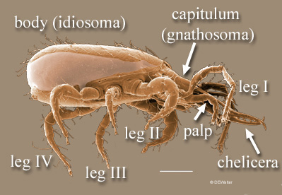 tagmata: capitulumgnathosoma:
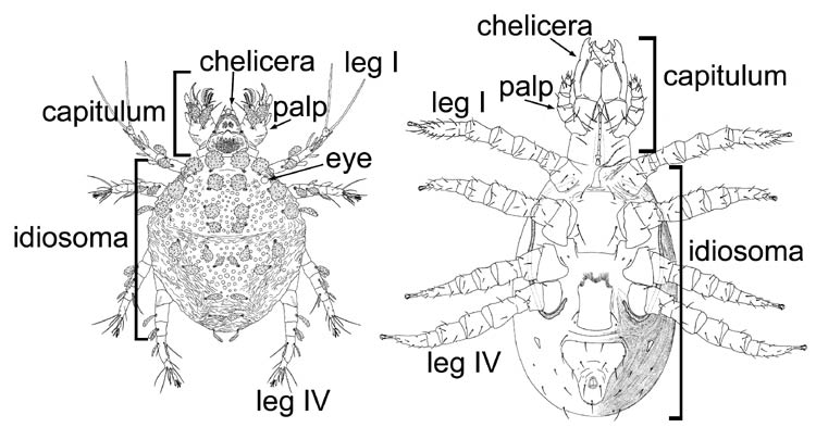
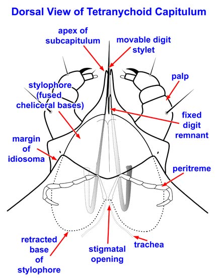
(= capitulum) the anteriormost part of a mite or ricinuleid, composed of the cheliceral and pedipalpal segments and separated from the body (idiosoma) by a ring of soft cuticle.
(=gnathosoma), idiosoma
Eyes: +/- 1–2 median ocelliocellus:
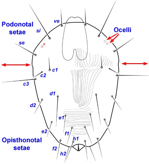
(pl. ocelli) a simple eye. Mites with eyes usually have one or two pairs of lateral ocelli, but some Opilioacarida have three pairs. Additionally, some acariform mites have one or two median ocelli on the underside of the naso.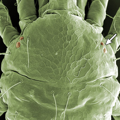 ; +/- 1–2 pairs lateral ocelliocellus:
(pl. ocelli) a simple eye. Mites with eyes usually have one or two pairs of lateral ocelli, but some Opilioacarida have three pairs. Additionally, some acariform mites have one or two median ocelli on the underside of the naso.
Antennae: absent
Mouthparts: 2–3 segmented chelicerae; pedipalps (palps) with 1–5 free segments; pedipalpal coxae fused into subcapitulumsubcapitulum:
(also infracapitulum) the venter of the capitulum; the ventral faces of the fused palpcoxae; apparently formed independently in the two superorders of mites.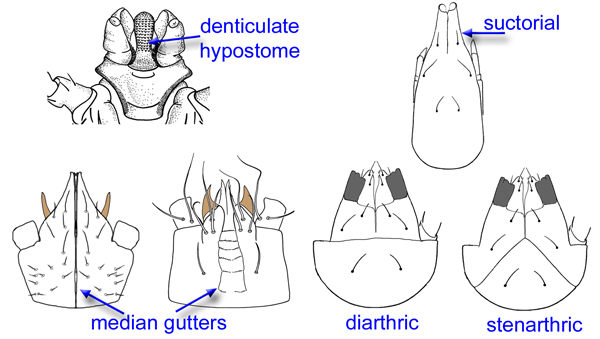
Legs: 1–4 pairs, typically 3 pairs in larvae, 4 pairs in nymphs and adults (rarely 2 pairs, 1 pair)
Distinguishing features: capitulumgnathosoma:
(= capitulum) the anteriormost part of a mite or ricinuleid, composed of the cheliceral and pedipalpal segments and separated from the body (idiosoma) by a ring of soft cuticle.
, loss of visible body segmentationsegmentation:
in mites distinct external segments have been lost but remnants of segmentation may be represented by hysterosomal folds or transverse arrays of setae and other cuticular sense organs. In theory, all chelicerates have a prosoma composed of 6 segments (cheliceral, pedipalpal, and four leg-bearing segments = body segments I-VI). Ventrally the positions of the prosomal segments can be identified by the insertions of their appendages, but dorsally they are obscured. The opisthosoma is thought to comprise an additional 12-13 segments (body segments VII-XVIII or XIX), but appears to be somewhat to much reduced in most mites, except possibly Opilioacarida. In early derivative Acariformes (e.g., many Endeostigmata), hysterosomal folds are thought to represent segmentation and in the Grandjean system are designated (from the sejugal furrow to the anus): C, D, E, F, H, PS AD, AN, PA. There is disagreement in the literature over the origin of 'segments' C and D. Adherents of Grandjean consider them to be opisthosomatic (with C probably representing a fusion of the pregenital [body segment VII] and genital [VIII] segments). Others believe that C and D are the dorsal regions of the last two prosomal segments that bear leggs III and IV (i.e., body segments V & VI).
Mites have the first two segments (bearing the chelicerae and palps) separated from the bodybody:
the idiosoma of mites. by flexible cuticle. This anterior region is called the capitulumgnathosoma:
(= capitulum) the anteriormost part of a mite or ricinuleid, composed of the cheliceral and pedipalpal segments and separated from the body (idiosoma) by a ring of soft cuticle.
('little head') or gnathosomagnathosoma:
(= capitulum) the anteriormost part of a mite or ricinuleid, composed of the cheliceral and pedipalpal segments and separated from the body (idiosoma) by a ring of soft cuticle.
('jaw body'). The remainder of the mite bodybody:
the idiosoma of mites. is called the idiosoma and lacks primary segmentationsegmentation:
in mites distinct external segments have been lost but remnants of segmentation may be represented by hysterosomal folds or transverse arrays of setae and other cuticular sense organs. In theory, all chelicerates have a prosoma composed of 6 segments (cheliceral, pedipalpal, and four leg-bearing segments = body segments I-VI). Ventrally the positions of the prosomal segments can be identified by the insertions of their appendages, but dorsally they are obscured. The opisthosoma is thought to comprise an additional 12-13 segments (body segments VII-XVIII or XIX), but appears to be somewhat to much reduced in most mites, except possibly Opilioacarida. In early derivative Acariformes (e.g., many Endeostigmata), hysterosomal folds are thought to represent segmentation and in the Grandjean system are designated (from the sejugal furrow to the anus): C, D, E, F, H, PS AD, AN, PA. There is disagreement in the literature over the origin of 'segments' C and D. Adherents of Grandjean consider them to be opisthosomatic (with C probably representing a fusion of the pregenital [body segment VII] and genital [VIII] segments). Others believe that C and D are the dorsal regions of the last two prosomal segments that bear leggs III and IV (i.e., body segments V & VI).
. Larval mites are usually hexapodhexapod:
with three pairs of legs (i.e. 6 legs), as in the larvae of mites or the larviform stages of others.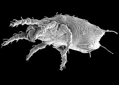 (6 legs); nymphs and adults usually octopod (8 legs).
"Heteromorphicheteromorphic:
having different morphological forms; referring either to different forms within a particular life stage (e.g., normal and heteromorphic deutonymphs in some Mesostigmata; protogynes vs. deutogynes in Eriophyoidea; heteromorphic vs. homeomorphic males in the Astigmata) or to a developmental stage that differs radically from other stages (e.g., the heteromorphic deutonymph or hypopus in the Astigmata).
" means having different morphological forms. It can refer either to different forms within a particular life stagestage:
a distinct developmental form, e.g., the egg, larval, nymphal and adult stages. Since mite instars are usually morphologically distinct, they are also stages (and see stase). Some authors, however, insist that instar should be apolysis to apolysis and stage ecdysis to ecdysis. Since apolysis can be a discontinuous process and, in any case, is difficult to determine, in practice the difference between a stage and an instar is abstract and of importance only if you have a contentious referee.
(e.g., normal and heteromorphicheteromorphic:
having different morphological forms; referring either to different forms within a particular life stage (e.g., normal and heteromorphic deutonymphs in some Mesostigmata; protogynes vs. deutogynes in Eriophyoidea; heteromorphic vs. homeomorphic males in the Astigmata) or to a developmental stage that differs radically from other stages (e.g., the heteromorphic deutonymph or hypopus in the Astigmata).
deutonymphs in some Mesostigmata; protogynes vs. deutogynes in Eriophyoidea; or heteromorphicheteromorphic:
having different morphological forms; referring either to different forms within a particular life stage (e.g., normal and heteromorphic deutonymphs in some Mesostigmata; protogynes vs. deutogynes in Eriophyoidea; heteromorphic vs. homeomorphic males in the Astigmata) or to a developmental stage that differs radically from other stages (e.g., the heteromorphic deutonymph or hypopus in the Astigmata).
vs. homeomorphic males in the Astigmata), or to a developmental stagestage:
a distinct developmental form, e.g., the egg, larval, nymphal and adult stages. Since mite instars are usually morphologically distinct, they are also stages (and see stase). Some authors, however, insist that instar should be apolysis to apolysis and stage ecdysis to ecdysis. Since apolysis can be a discontinuous process and, in any case, is difficult to determine, in practice the difference between a stage and an instar is abstract and of importance only if you have a contentious referee.
that differs radically from other stages (e.g., the heteromorphicheteromorphic:
having different morphological forms; referring either to different forms within a particular life stage (e.g., normal and heteromorphic deutonymphs in some Mesostigmata; protogynes vs. deutogynes in Eriophyoidea; heteromorphic vs. homeomorphic males in the Astigmata) or to a developmental stage that differs radically from other stages (e.g., the heteromorphic deutonymph or hypopus in the Astigmata).
deutonymph or hypopus in the Astigmata).
In addition to heteromorphicheteromorphic:
having different morphological forms; referring either to different forms within a particular life stage (e.g., normal and heteromorphic deutonymphs in some Mesostigmata; protogynes vs. deutogynes in Eriophyoidea; heteromorphic vs. homeomorphic males in the Astigmata) or to a developmental stage that differs radically from other stages (e.g., the heteromorphic deutonymph or hypopus in the Astigmata).
deutonymphs and females, which both serve as dispersal or survival stages, some mites, particularly in Astigmata, also exhibit heteromorphicheteromorphic:
having different morphological forms; referring either to different forms within a particular life stage (e.g., normal and heteromorphic deutonymphs in some Mesostigmata; protogynes vs. deutogynes in Eriophyoidea; heteromorphic vs. homeomorphic males in the Astigmata) or to a developmental stage that differs radically from other stages (e.g., the heteromorphic deutonymph or hypopus in the Astigmata).
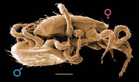
males. These males differ drastically from females, often possessing enlarged legs or chelicerae used for fighting. They typically engage in combat with each other or with homeomorphic males (those morphologically similar to females) but not with females or immatures. Heteromorphicheteromorphic:
having different morphological forms; referring either to different forms within a particular life stage (e.g., normal and heteromorphic deutonymphs in some Mesostigmata; protogynes vs. deutogynes in Eriophyoidea; heteromorphic vs. homeomorphic males in the Astigmata) or to a developmental stage that differs radically from other stages (e.g., the heteromorphic deutonymph or hypopus in the Astigmata).
males are produced under low population densities, where they always kill homeomorphic males, eventually allowing the strongest heteromorphicheteromorphic:
having different morphological forms; referring either to different forms within a particular life stage (e.g., normal and heteromorphic deutonymphs in some Mesostigmata; protogynes vs. deutogynes in Eriophyoidea; heteromorphic vs. homeomorphic males in the Astigmata) or to a developmental stage that differs radically from other stages (e.g., the heteromorphic deutonymph or hypopus in the Astigmata).
male to monopolize all females in the population. In contrast, heteromorphicheteromorphic:
having different morphological forms; referring either to different forms within a particular life stage (e.g., normal and heteromorphic deutonymphs in some Mesostigmata; protogynes vs. deutogynes in Eriophyoidea; heteromorphic vs. homeomorphic males in the Astigmata) or to a developmental stage that differs radically from other stages (e.g., the heteromorphic deutonymph or hypopus in the Astigmata).
males are typically not produced in large populations. A variation of this strategy occurs in the family Pyroglyphidae (Astigmata), where heteromorphicheteromorphic:
having different morphological forms; referring either to different forms within a particular life stage (e.g., normal and heteromorphic deutonymphs in some Mesostigmata; protogynes vs. deutogynes in Eriophyoidea; heteromorphic vs. homeomorphic males in the Astigmata) or to a developmental stage that differs radically from other stages (e.g., the heteromorphic deutonymph or hypopus in the Astigmata).
males use their enlarged legs during mating and postcopulatory guarding to prevent other males from mating with the same female.
In most cases, identification of Mesostigmata to family or lower can be accomplished only if the specimen is an adult female. The easiest way to determine the stagestage:
a distinct developmental form, e.g., the egg, larval, nymphal and adult stages. Since mite instars are usually morphologically distinct, they are also stages (and see stase). Some authors, however, insist that instar should be apolysis to apolysis and stage ecdysis to ecdysis. Since apolysis can be a discontinuous process and, in any case, is difficult to determine, in practice the difference between a stage and an instar is abstract and of importance only if you have a contentious referee.
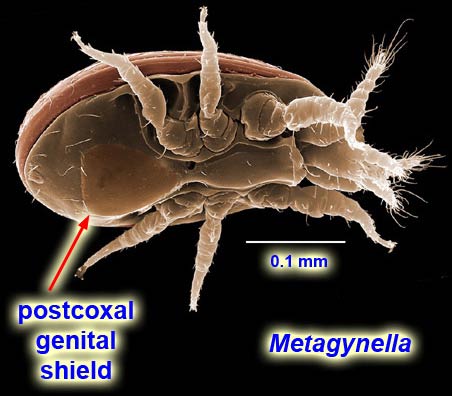
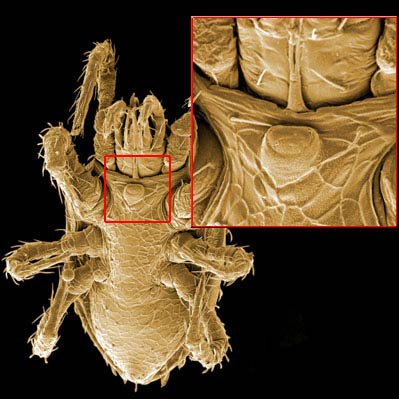
and sex of your specimen is to look at the intercoxal region. Adult females have a genital opening that is almost invariably in the intercoxal region (species of Metagynella (Fig. 1) are exceptions) and covered by a sclerotized shield which may be truncate posteriorly or continue onto the ventral regionventral region:
in Mesostigmata, the area between the genital and anal regions.
. Adult males and immature stages of both sexes have a continuous intercoxal shield that may be truncate or continue onto the ventral regionventral region:
in Mesostigmata, the area between the genital and anal regions.
or even incorporate the entire ventral regionventral region:
in Mesostigmata, the area between the genital and anal regions.
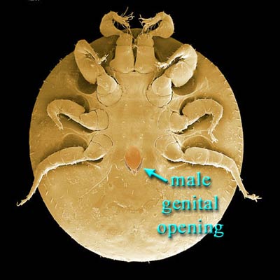
. Adult males have a subcircular opening in the intercoxal shield (sometimes called a sternogenital or sternitogenital shieldsternogenitalshield:
(also sternitogenital, sternitigenital) in male Mesostigmata, a shield covering the intercoxal region and bearing the genital opening.
) somewhere between the posteriorposterior:
the back part of the body or towards that region in comparison, e.g., 'posterior to'.
margins of coxae IV (Fig. 2) and the basebase:
the usually columnar basal part of the tritosternum; sometimes expanded and rectangular or otherwise modified; the most basal part of any structure.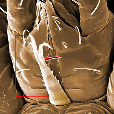 of the tritosternumtritosternum:
the sternum of the 3rd body segment (between legs I); produced as a biflagellate structure in Mesostigmata, although sometimes the flagellae (laciniae) are partially or completely fused. 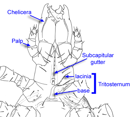 (Fig. 3). Immature stages (larva, protonymphprotonymph:
the first nymphal stage or instar, usually octopod.
, deutonymphdeutonymph:
(also deuteronymph) the second nymphal stage or instar.
) of Mesostigmata have no genital opening.
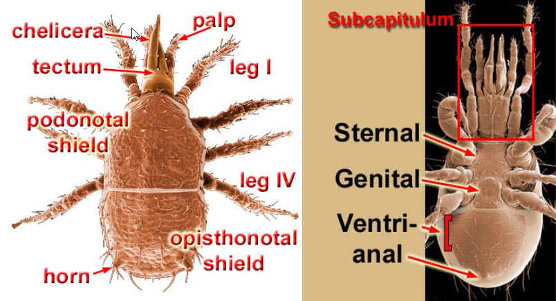
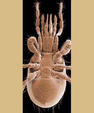
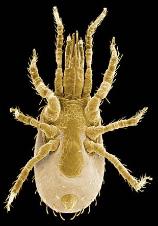
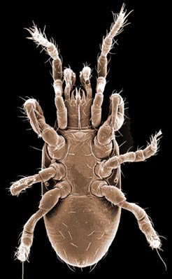
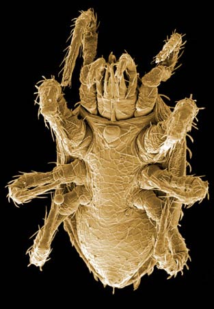
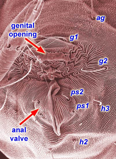
Female mesostigmatans are easily recognized by their genital opening, usually found between coxacoxa:
the basal segment of the leg, articulating with (Parasitiformes) or fused to (Acariformes) the body wall.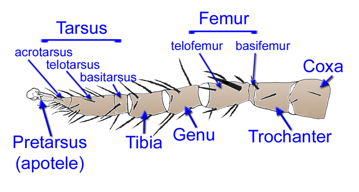 IV and covered by one or more genital shields (Fig. 4). Although called 'genital' (or epigynialepigynal:
(also epigynial) of or relating to the female genital opening or a shield protecting it.
, epigynalepigynal:
(also epigynial) of or relating to the female genital opening or a shield protecting it.
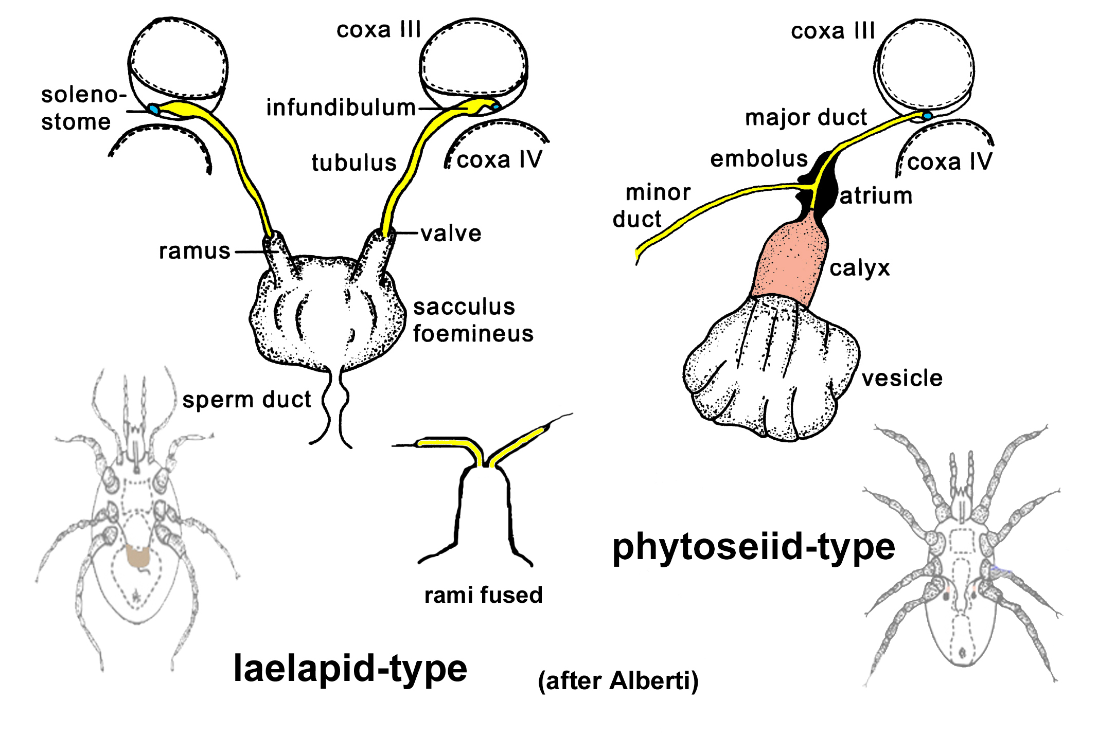
), the shield protects only the ovipore in mesostigmatans with a secondary sperm transfer system (Dermanyssiae; Fig. 5). Females lay eggs relatively large in comparison to their bodybody:
the idiosoma of mites. size, so the genital opening and its shield are relatively large, and may be extended posteriorly to incorporate ventralventral:
relating to the lower or under side; opposed to dorsal.
or ventrianal elements.
The genital shieldgenital shield:
a shield or shields covering the genital opening; in female mongynaspine Mesostigmata this shield is usually called the epigynal (epigynial) shield.
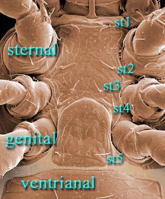
often bears the 5th pair of sternal setaesternal setae:
in Mesostigmata, the five pairs of setae in the intercoxal region designated st1-5; st1-3 are present in the larva and usually are borne on a sternal shield in the adult female; st4, the metasternal setae, are added in the deutonymph, often are borne on metasternal platelets, and sometimes on the sternal shield; st5, the genital setae, are added in the protonymph and usually borne on or are inserted laterad the epigynal shield in the adult female.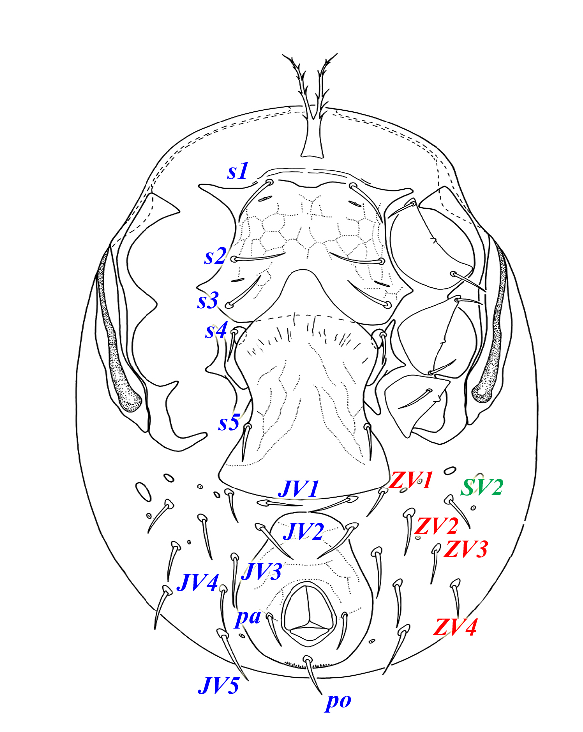 (st5), but may be nude and variously formed. Adult females also usually have one or more shields anterior to the genital shield(s) called sternal shields, and these usually sport one or more of sternal setaesternal setae:
in Mesostigmata, the five pairs of setae in the intercoxal region designated st1-5; st1-3 are present in the larva and usually are borne on a sternal shield in the adult female; st4, the metasternal setae, are added in the deutonymph, often are borne on metasternal platelets, and sometimes on the sternal shield; st5, the genital setae, are added in the protonymph and usually borne on or are inserted laterad the epigynal shield in the adult female. st1–4. Other shields cover the ventralventral:
relating to the lower or under side; opposed to dorsal.
and anal regions. The female ologamasid mite in Figures 6 and 7 has a large ventrianal shieldventrianal shield:
in Mesostigmata, a ventral shield bearing the anal opening, circum anal setae, and one or more pairs of ventral setae or pores (lyrifissures) [see anal shield]; maybe rather narrow or very broad and covering most of the gaster.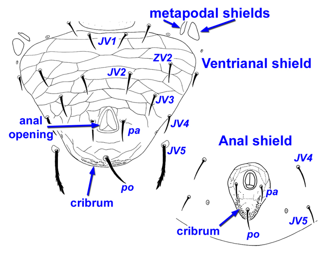 , while the female laelapid mite in Figures 8 and 9 has a small anal shield.
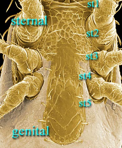
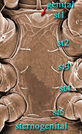
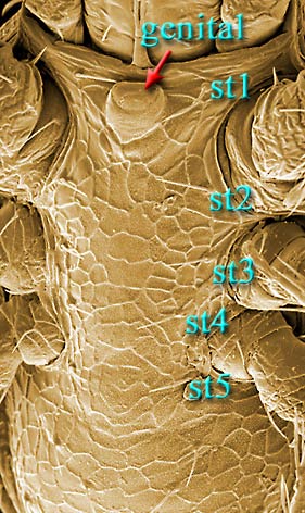
Males, however, produce relatively small sperm packets (spermatophores) and have much smaller genital openings. Although these openings also may lie between coxae IV, they are more typically towards the anterior end of the intercoxal region and often at the basebase:
the usually columnar basal part of the tritosternum; sometimes expanded and rectangular or otherwise modified; the most basal part of any structure. of the tritosternumtritosternum:
the sternum of the 3rd body segment (between legs I); produced as a biflagellate structure in Mesostigmata, although sometimes the flagellae (laciniae) are partially or completely fused. (see details in Figs. 10–13). In the male ologamasid mite (Figs. 10, 11), the genital opening is in a sternogenital (also sternitogenital) shield that bears 5 pairs of setae (st1–5). In the male laelapid mite (Figs. 12, 13), however, the sternogenital shield is fused to the ventrianal shieldventrianal shield:
in Mesostigmata, a ventral shield bearing the anal opening, circum anal setae, and one or more pairs of ventral setae or pores (lyrifissures) [see anal shield]; maybe rather narrow or very broad and covering most of the gaster. and other elements to form a ventral shieldventral shield:
in Mesostigmata, any shield or shields in the ventral region; often fused with the anal shield to form a ventrianal shield. [Back to Top]
or plate. Males also often, but not always, have modifications to their chelicerae for transferring sperm, e.g., spermatodactyls or spermatotremes.
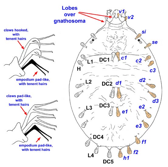
Find more anatomy drawings and a morphological diagnosis on the Bryobiinae fact sheet.
Find more images and a morphological diagnosis on the Tetranychinae fact sheet.
Author: David E. Walter
Content last updated Sept. 2006, unless indicated otherwise
Lucid key player updated Sept. 2025
idtools.org