Since bee-associated mites come from a variety of diverse lineages (see tool scope and classification), it is difficult to provide an exhaustive morphological account. Please consult Krantz & Walter (2009)) for additional details. Here we offer a short introduction to mite morphology, focusing on diagnostic characters for the three principal groups that include bee-associated mites: Mesostigmata, Astigmata, and Prostigmata. In the comparison table below, an asterisk indicates a defining trait for that group; if a mite bears that trait, it is undoubtedly a member of that group, and checking other characters is not necessary. Other rarely used diagnostic traits, such as those used to distinguish two or a few mite genera are pointed out in fact sheets and key images.
See the "Setae, solenidiasolenidion:
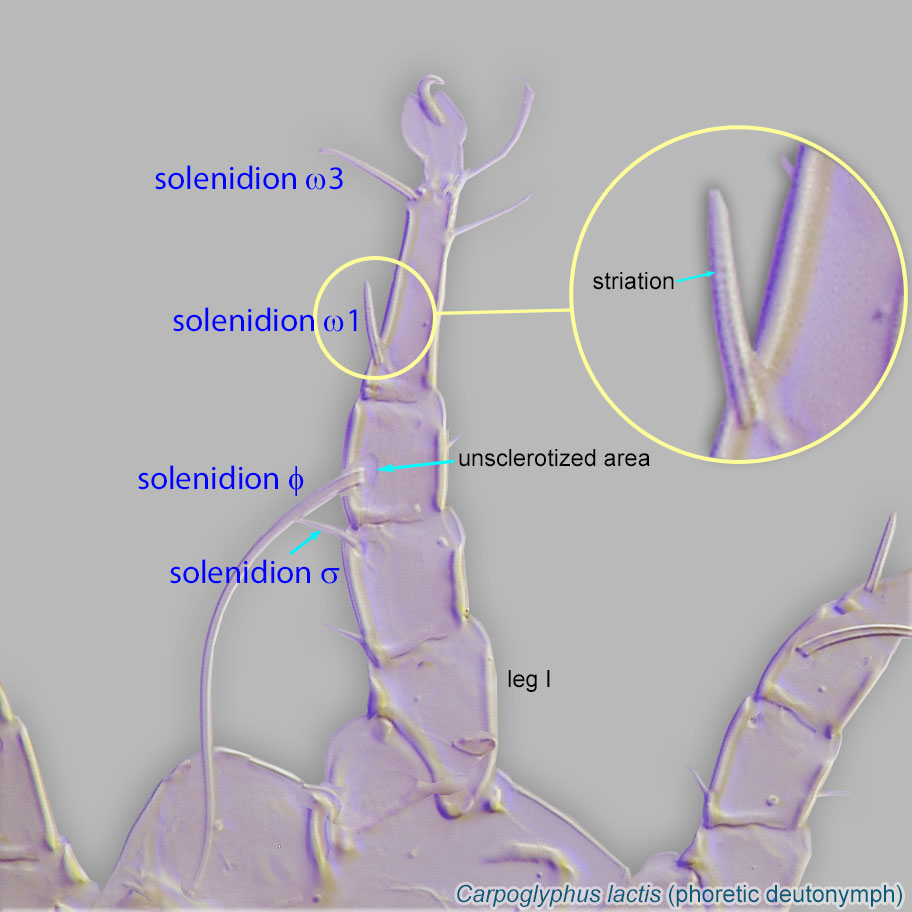
Thin-walled, terminally rounded or pointed filiform or peglike structure that is not birefringent in polarized light (unlike common setae in Acariformes). Often appears striated because of its internal structure. Found on the palpal tarsus on the gnathosoma and may also occur on the tarsus and tibia, less frequently on the genu, and occasionally on the femur of legs I-IV. In Acariformes, leg solenidia often arise from unsclerotized areas.
 , & suckers" tab for more details on using these features for mite identification.
| Mesostigmata | Astigmata | Prostigmata | |
|---|---|---|---|
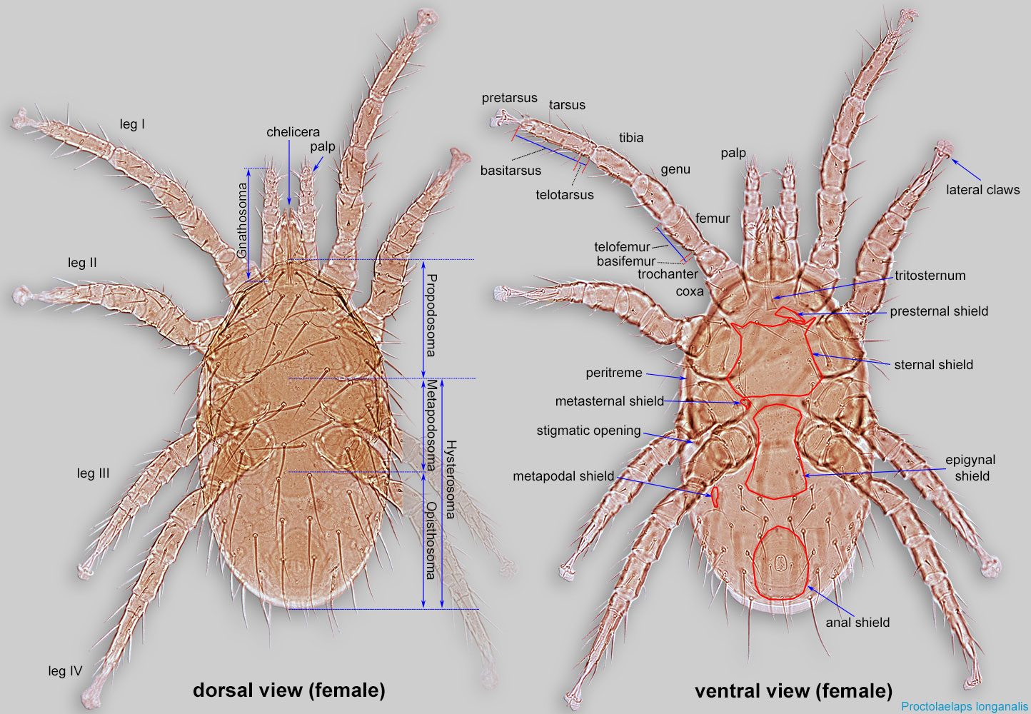
| tritosternumtritosternum: In Mesostigmata, a biflagellate structure situated on the ventral side of the body, posterior to the gnathosoma and anterior to the sternal shield. Sometimes the flagellae (laciniae) are partially or completely fused. 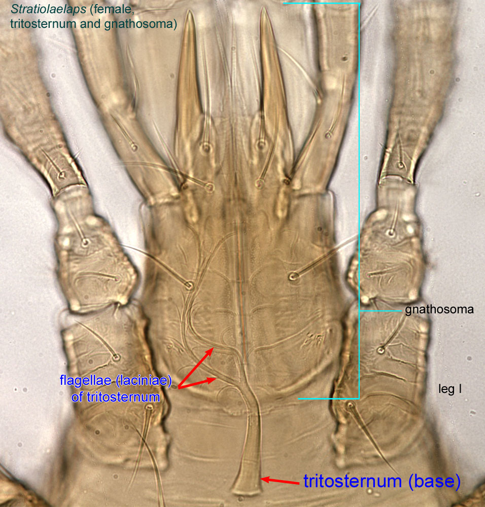 |
*present (Fig. 1) | absent | absent |
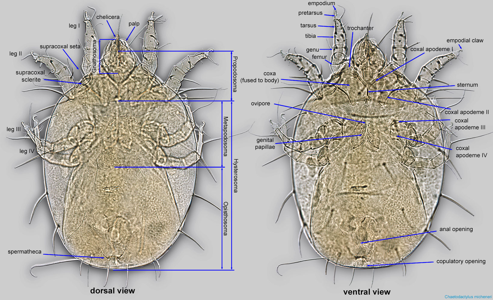
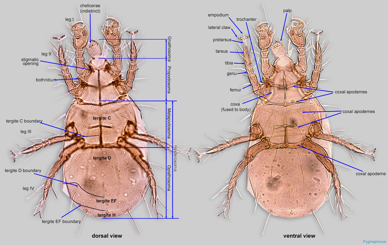
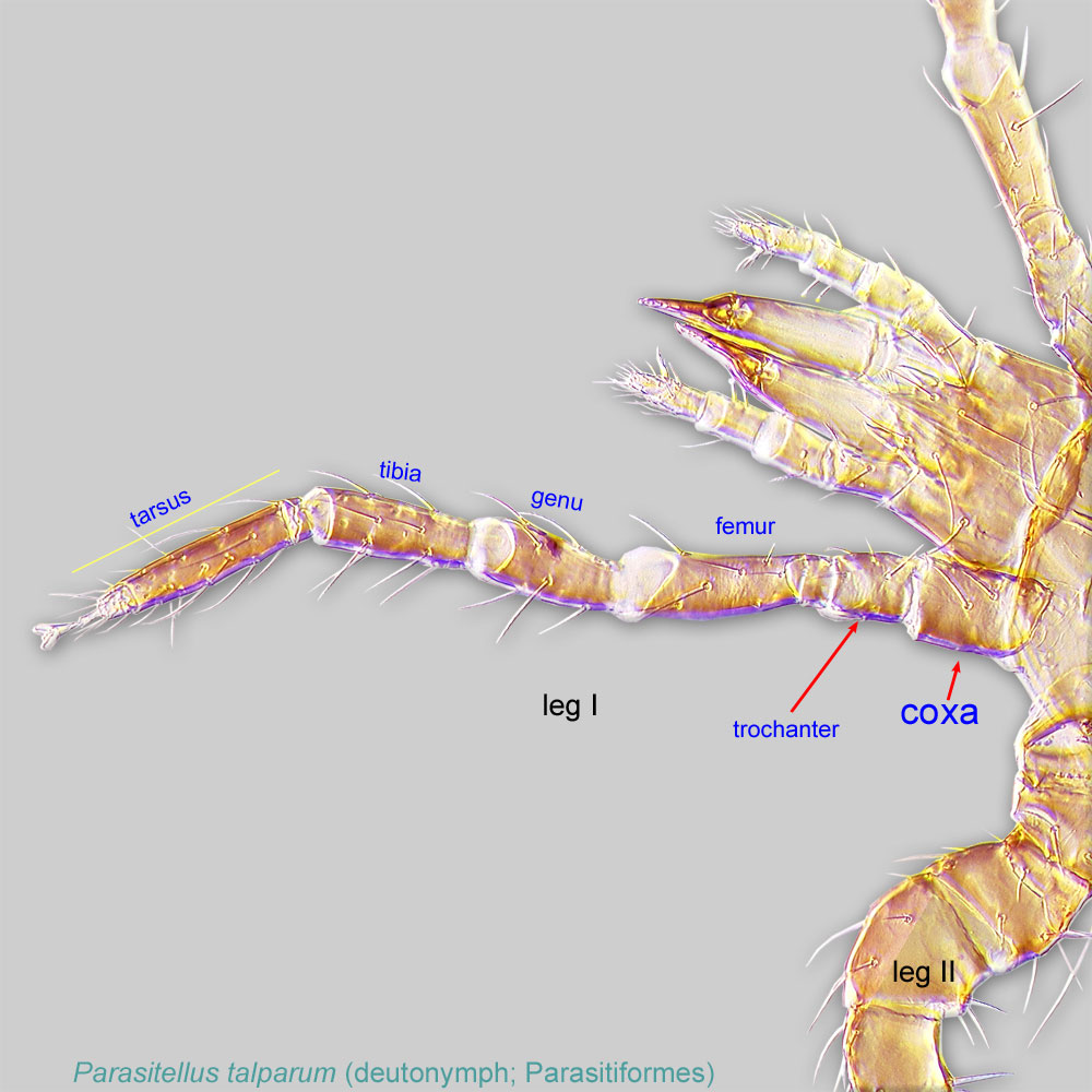
| leg coxae | *free, not fused to body (Fig. 1) | fused to body, form distinct coxal apodemes (Fig. 4) | fused to body, form distinct coxal apodemes (fig. 7) |
| stigmatic openingsstigmatic opening: An opening that connects to a well-developed tracheal system (exceptions exist when tracheal system and stigmatal openings are disassociated). Situated at or on the cheliceral bases, anterolaterally on the propodosoma, or lateral sides of the body. Also known as stigmatal opening or stigma (pl. stigmata). 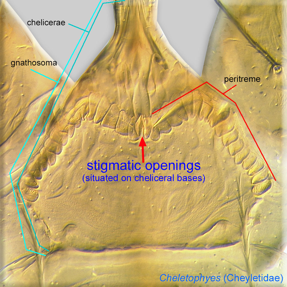 |
*present on mid sides of the body, posterior to leg II or III insertions (Fig. 1) | *absent | *present, either near associated with the chelicerae or on the dorsal propodosomapropodosoma: Anterior part of idiosoma, in front of sejugal furrow. 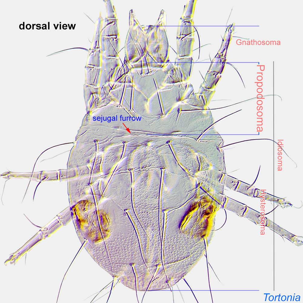 (Fig. 7) (Fig. 7) |
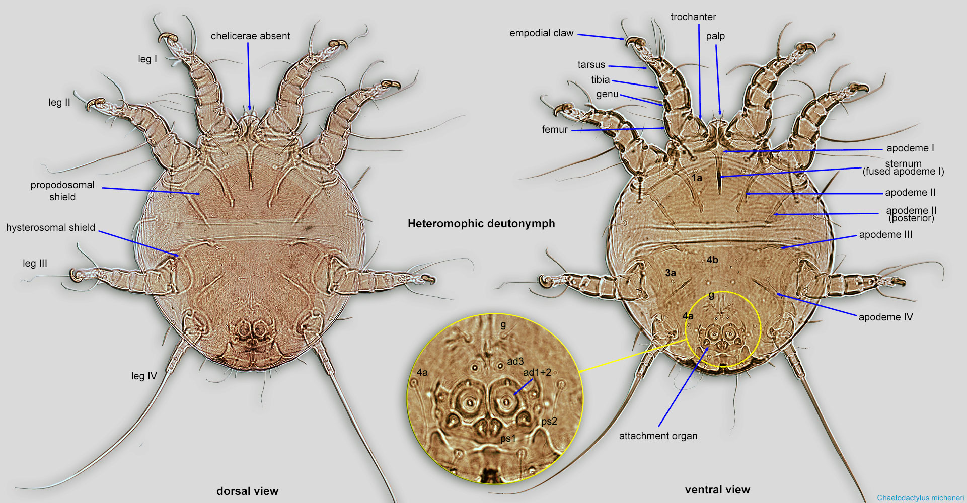
| empodial claws; lateral claws | empodial claws absent from all legs, lateral claws usually present, at least on some legs | *only empodial clawempodial claw: Claw-like, membranous, or pad-like structure of setal origin. Present only on the pretarsus in Acariformes. In Astigmata, it is the only claw on the pretarsus and often referred to simply as the claw. In the remaining Acariformes, may be accomanied by two lateral claws. Also known as empodium, pretarsal empodium, or central claw. 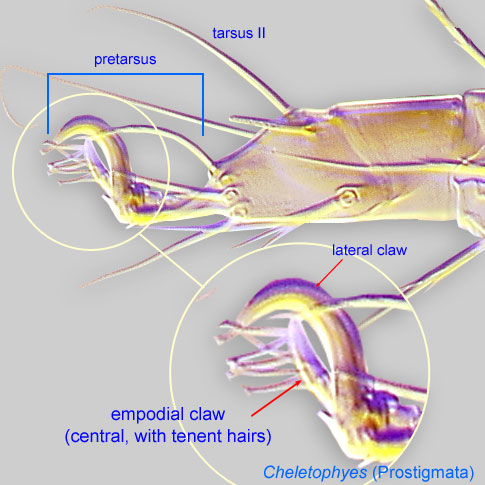 is present (Figs. 4, and 6); lateral claws are absent from all legs is present (Figs. 4, and 6); lateral claws are absent from all legs |
lateral and empodial claws are usually present, at least on some legs |
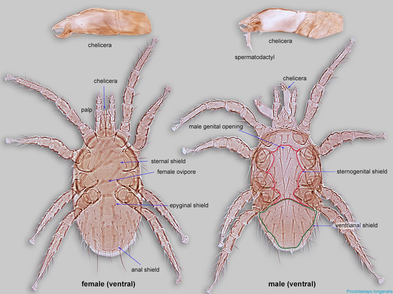
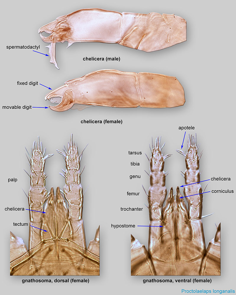
| male aedeagus | absent; some have spermatodactylspermatodactyl: The "sperm finger" on the chelicerae of male Dermanyssina (Parasitiformes) used to transfer sperm to the secondary sperm system in the female. Has various forms, from simple finger-like processes to very long, contorted structures. 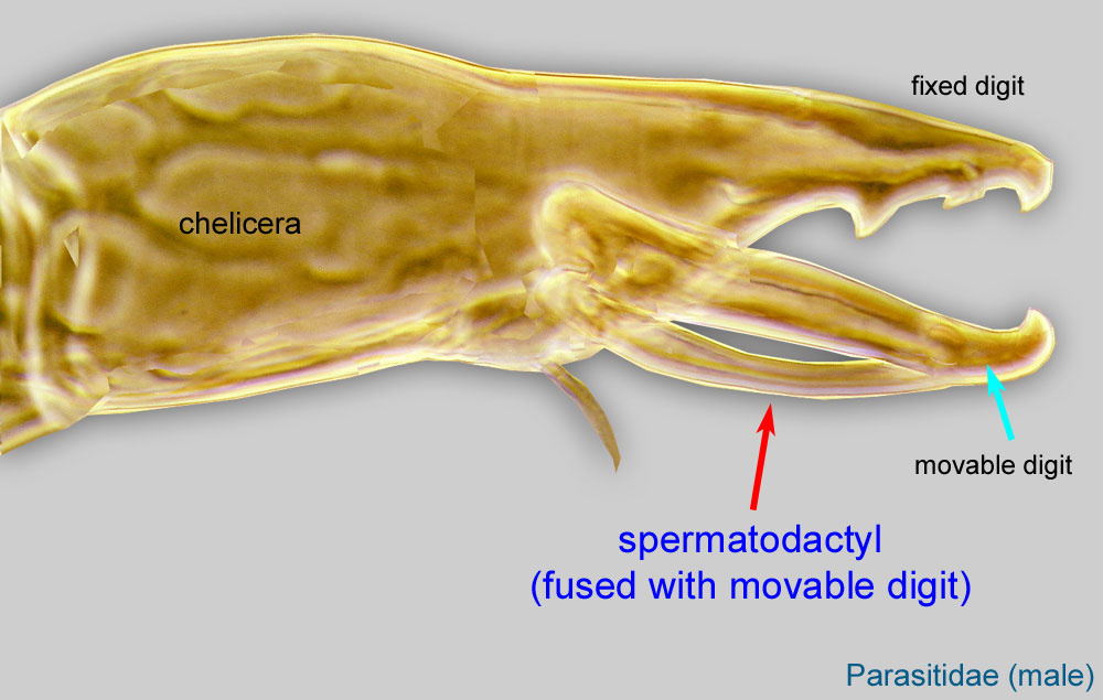 instead (Figs. 2, 3) instead (Figs. 2, 3) |
present (Fig. 5) | present or absent |
| dispersal stage/non-dispersal stage | dispersal stages (fertilized female or deutonymphsdeutonymph: Ontogenetic stage between protonymph and tritonymph (or adult, if tritonymph is absent). See <a href="index.cfm?pageID=1720">Life stages page</a> for more details. 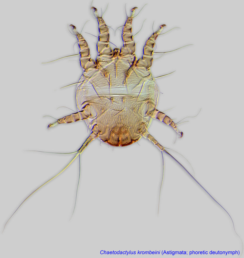 ) are not substantially different from non-dispersal stages ) are not substantially different from non-dispersal stages |
*dispersal stage (phoretic deutonymphsdeutonymph: Ontogenetic stage between protonymph and tritonymph (or adult, if tritonymph is absent). See <a href="index.cfm?pageID=1720">Life stages page</a> for more details. ) substantially different from non-dispersal stages (Fig. 6) |
dispersal stage is not substantially different from non-dispersal stages |
| attachment organattachment organ: Complex unpaired structure in phoretic deutonymphs of Astigmata situated on the posteroventral end of the body that serves for attachment to the host during phoresy. In deutonymphs phoretic on insects, attachment organ consists of a vestigial anal opening and two types of attachment elements: true suckers that create negative pressure and conoids that create adhesive forces. Not to be confused with pedicel. 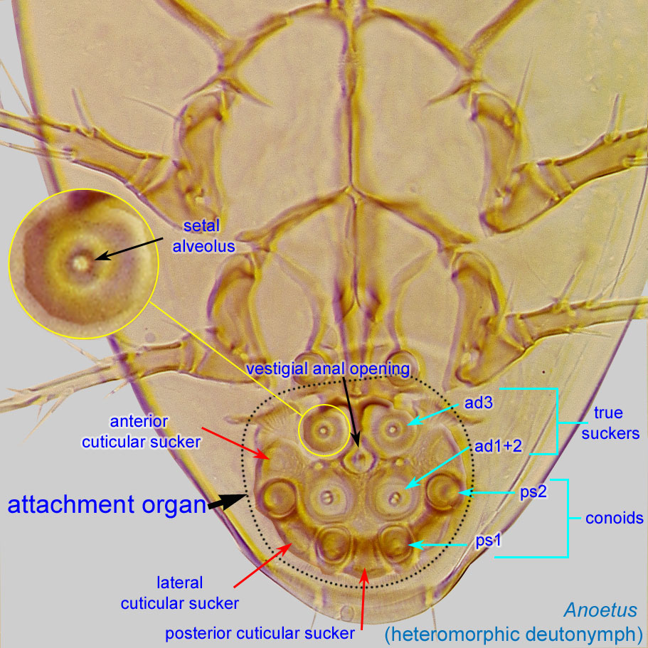 |
absent | present (Fig. 6) | absent |
| chelicerae in dispersal stage | present | absent (Fig. 6) | developed but sometimes indistinct |
Coxal setae are paired setae situated on or near coxaecoxa:
In Parasitiformes, most basal leg segment (or podomere) forming a joint with the body. Areas delimited by coxal apodemes are called coxal fields in Astigmata or coxisternal plates in Prostigmata.
 .
.
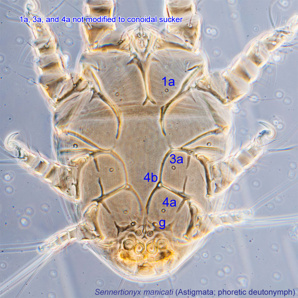
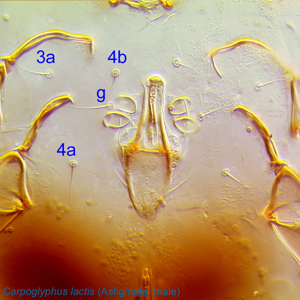
coxal seta 1a: situated on coxaecoxa:
In Parasitiformes, most basal leg segment (or podomere) forming a joint with the body. Areas delimited by coxal apodemes are called coxal fields in Astigmata or coxisternal plates in Prostigmata.
I. In Astigmata present in all active developmental stages. In phoreticphoretic:
Pertaining to phoresy; using another organism (i.e., a host) for dispersal to new habitats. Phoresy can be distinguished from parasitism because feeding typically does not occur during phoresy.
deutonymphsdeutonymph:
Ontogenetic stage between protonymph and tritonymph (or adult, if tritonymph is absent). See <a href="index.cfm?pageID=1720">Life stages page</a> for more details. of Astigmata, can be modified to a conoid or be vestigial or absent.
coxal seta 3a: situated on coxaecoxa:
In Parasitiformes, most basal leg segment (or podomere) forming a joint with the body. Areas delimited by coxal apodemes are called coxal fields in Astigmata or coxisternal plates in Prostigmata.
III. In Astigmata present in all active developmental stages. In phoreticphoretic:
Pertaining to phoresy; using another organism (i.e., a host) for dispersal to new habitats. Phoresy can be distinguished from parasitism because feeding typically does not occur during phoresy.
deutonymphsdeutonymph:
Ontogenetic stage between protonymph and tritonymph (or adult, if tritonymph is absent). See <a href="index.cfm?pageID=1720">Life stages page</a> for more details. of Astigmata, can be modified to a conoid or be vestigial or absent.
coxal seta 4a: situated on coxaecoxa:
In Parasitiformes, most basal leg segment (or podomere) forming a joint with the body. Areas delimited by coxal apodemes are called coxal fields in Astigmata or coxisternal plates in Prostigmata.
IV. In Astigmata appear at the stage of deutonymphdeutonymph:
Ontogenetic stage between protonymph and tritonymph (or adult, if tritonymph is absent). See <a href="index.cfm?pageID=1720">Life stages page</a> for more details.. In phoreticphoretic:
Pertaining to phoresy; using another organism (i.e., a host) for dispersal to new habitats. Phoresy can be distinguished from parasitism because feeding typically does not occur during phoresy.
deutonymphsdeutonymph:
Ontogenetic stage between protonymph and tritonymph (or adult, if tritonymph is absent). See <a href="index.cfm?pageID=1720">Life stages page</a> for more details. of Astigmata, can be modified to a conoid or be vestigial or absent.
coxal seta 4b: usually situated between setae 3a and anterior to setae 4a or occasionally absent. In Astigmata appear at the stage of deutonymphdeutonymph:
Ontogenetic stage between protonymph and tritonymph (or adult, if tritonymph is absent). See <a href="index.cfm?pageID=1720">Life stages page</a> for more details..
genital seta g: Paired setae associated with the genital region. In Astigmata appear at the protonymphprotonymph:
Ontogenetic stage between larva and deutonymph. See <a href="index.cfm?pageID=1720">Life stages page</a> for more details.
stage.
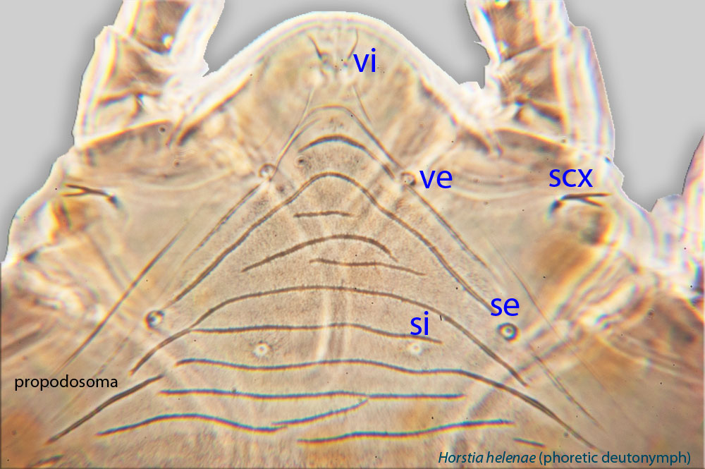
external scapular seta: Paired prodorsalprodorsal:
Pertaining to the prodorsum.
setae se in Astigmata (also known as sce; in Oribatida known as ex).
external vertical seta: Paired prodorsalprodorsal:
Pertaining to the prodorsum.
setae ve in Astigmata (in Oribatida known as le); sometimes absent.
internal scapular seta: Paired prodorsalprodorsal:
Pertaining to the prodorsum.
setae si in Astigmata (also known as sce; in Oribatida known as in).
internal vertical seta: Paired prodorsalprodorsal:
Pertaining to the prodorsum.
setae vi in Astigmata (in Oribatida known as ro).
supracoxal seta: Paired, often modified dorsal coxal seta (scx) in Astigmata associated with the supracoxal glandsupracoxal gland:
Paired glands serving for maintenance of water balance in Astigmata. These glands are usually invisible on typical microscopic preparations. However, their presence can be detected by the well-sclerotized supracoxal gland openings, which are situated on supracoxal sclerites.
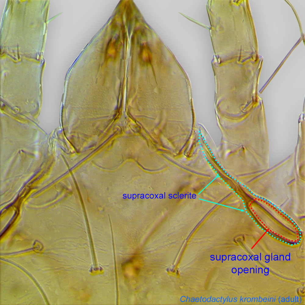 opening. Situated on the lateral propodosomapropodosoma:
Anterior part of idiosoma, in front of sejugal furrow.
(because coxacoxa:
In Parasitiformes, most basal leg segment (or podomere) forming a joint with the body. Areas delimited by coxal apodemes are called coxal fields in Astigmata or coxisternal plates in Prostigmata.
are fused to the body in Acariformes). Also known as elc I.
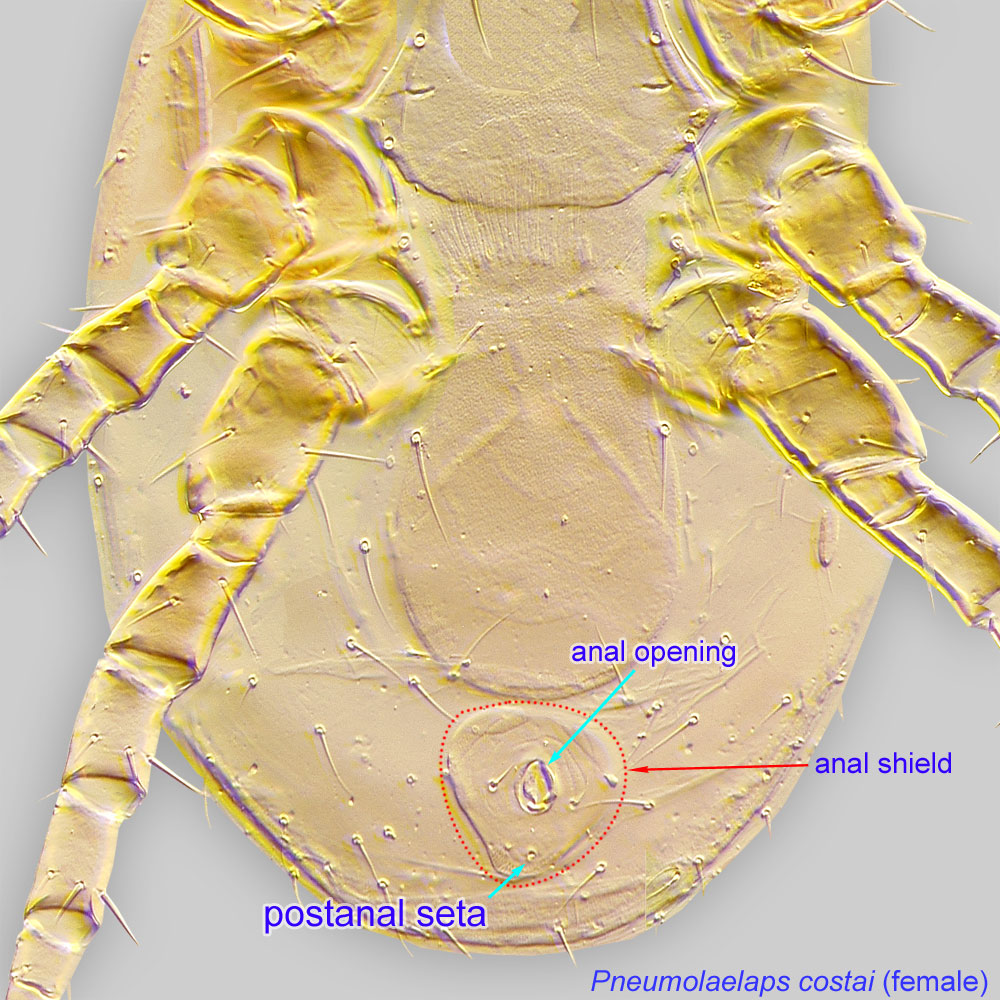
postanal seta: Unpaired median seta inserted posterior to the anal opening in the Mesostigmata (Parasitiformes); usually on the anal or ventrianal shieldventrianal shield:
In Mesostigmata, a ventral shield bearing the anal opening, circumanal (postanal and adanal) setae, and one or more pairs of ventral setae or pores (lyrifissures) (see anal shield); may be narrow or very broad and covering most of the gaster.
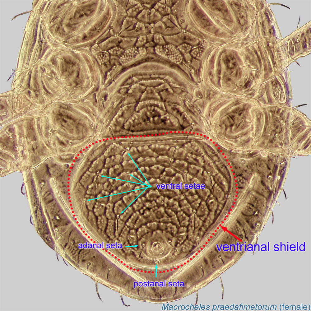 .
.
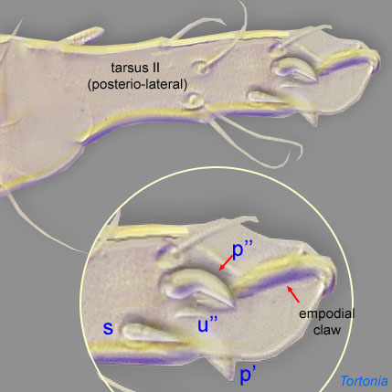
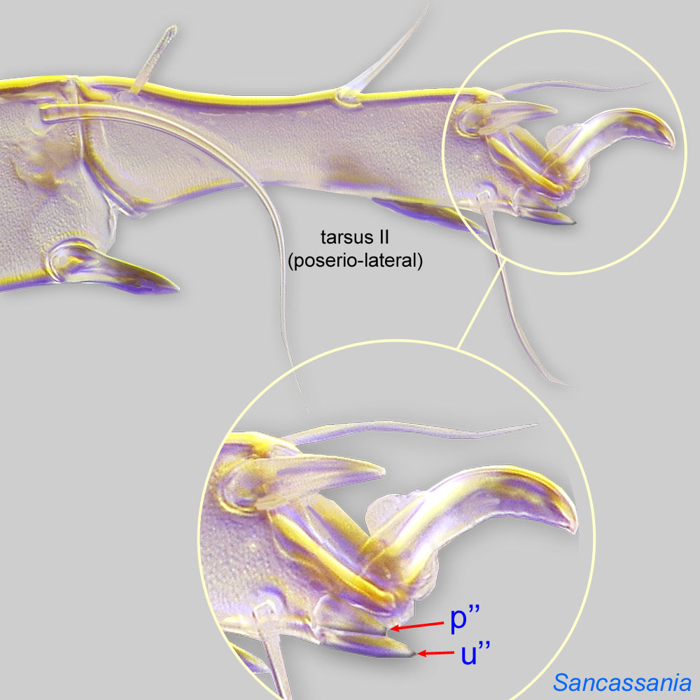
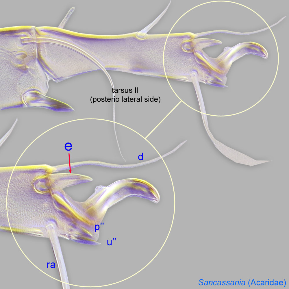
In the Acariformes, in the ancestral complement of five setae surrounding the empodial clawempodial claw:
Claw-like, membranous, or pad-like structure of setal origin. Present only on the pretarsus in Acariformes. In Astigmata, it is the only claw on the pretarsus and often referred to simply as the claw. In the remaining Acariformes, may be accomanied by two lateral claws. Also known as empodium, pretarsal empodium, or central claw.
ventrally, one is unpaired and the most ventral (s), and four others are paired (u' and u'' and p' and p''). Relative to each other, the unguinal setae u are more ventral and the proral setae p are more dorsal.
proral seta p or proral p: Paired disto-ventral setae (p' and p'') on tarsitarsus:
Terminal segment (also known as podomere or palpomere) of legs or palps. In Parasitoformes it can be subdivided into telotarsus and basitarsus.
 I-IV of Acariformes.
subunguinal seta s: Unpaired disto-ventral seta on tarsitarsus:
Terminal segment (also known as podomere or palpomere) of legs or palps. In Parasitoformes it can be subdivided into telotarsus and basitarsus.
I-IV of Acariformes.
unguinal seta u: Paired disto-venrtal setae u' and u'' on tarsitarsus:
Terminal segment (also known as podomere or palpomere) of legs or palps. In Parasitoformes it can be subdivided into telotarsus and basitarsus.
I-IV of Acariformes.
tarsal seta aa: On tarsustarsus:
Terminal segment (also known as podomere or palpomere) of legs or palps. In Parasitoformes it can be subdivided into telotarsus and basitarsus.
I in some Astigmata. Usually situated close to solenidionsolenidion:
Thin-walled, terminally rounded or pointed filiform or peglike structure that is not birefringent in polarized light (unlike common setae in Acariformes). Often appears striated because of its internal structure. Found on the palpal tarsus on the gnathosoma and may also occur on the tarsus and tibia, less frequently on the genu, and occasionally on the femur of legs I-IV. In Acariformes, leg solenidia often arise from unsclerotized areas.
ω2 in basal part of tarsustarsus:
Terminal segment (also known as podomere or palpomere) of legs or palps. In Parasitoformes it can be subdivided into telotarsus and basitarsus.
. Absent from tarsustarsus:
Terminal segment (also known as podomere or palpomere) of legs or palps. In Parasitoformes it can be subdivided into telotarsus and basitarsus.
II, hence can be easily identified by comparison of tarsitarsus:
Terminal segment (also known as podomere or palpomere) of legs or palps. In Parasitoformes it can be subdivided into telotarsus and basitarsus.
I and II setations. Homologous to anterolateral setae a'' of Oribatida. (Not illustrated.)
tarsal seta ba: On tarsitarsus:
Terminal segment (also known as podomere or palpomere) of legs or palps. In Parasitoformes it can be subdivided into telotarsus and basitarsus.
I-II in many Astigmata. Usually situated in the mid dorso-anterior part of tarsustarsus:
Terminal segment (also known as podomere or palpomere) of legs or palps. In Parasitoformes it can be subdivided into telotarsus and basitarsus.
. Many Acaridae lack ba from tarsustarsus:
Terminal segment (also known as podomere or palpomere) of legs or palps. In Parasitoformes it can be subdivided into telotarsus and basitarsus.
I in phoreticphoretic:
Pertaining to phoresy; using another organism (i.e., a host) for dispersal to new habitats. Phoresy can be distinguished from parasitism because feeding typically does not occur during phoresy.
deutonymphsdeutonymph:
Ontogenetic stage between protonymph and tritonymph (or adult, if tritonymph is absent). See <a href="index.cfm?pageID=1720">Life stages page</a> for more details., while this seta is present in adults and other feeding stages (in contrast, setae ba II uniformly present on tarsitarsus:
Terminal segment (also known as podomere or palpomere) of legs or palps. In Parasitoformes it can be subdivided into telotarsus and basitarsus.
II in all stages of these taxa). Homologous to fastigial setae ft' of Oribatida. (Not illustrated.)
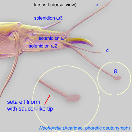
tarsal seta e: Posterodistal seta on tarsitarsus:
Terminal segment (also known as podomere or palpomere) of legs or palps. In Parasitoformes it can be subdivided into telotarsus and basitarsus.
I-IV of Astigmata. On tarsitarsus:
Terminal segment (also known as podomere or palpomere) of legs or palps. In Parasitoformes it can be subdivided into telotarsus and basitarsus.
I-II of phoreticphoretic:
Pertaining to phoresy; using another organism (i.e., a host) for dispersal to new habitats. Phoresy can be distinguished from parasitism because feeding typically does not occur during phoresy.
deutonymphsdeutonymph:
Ontogenetic stage between protonymph and tritonymph (or adult, if tritonymph is absent). See <a href="index.cfm?pageID=1720">Life stages page</a> for more details. of Acaridae and related lineages (Astigmata), these setae have a characteristic 'saucer' at the tip. Homologous to tectal seta tc'' of Oribatida.
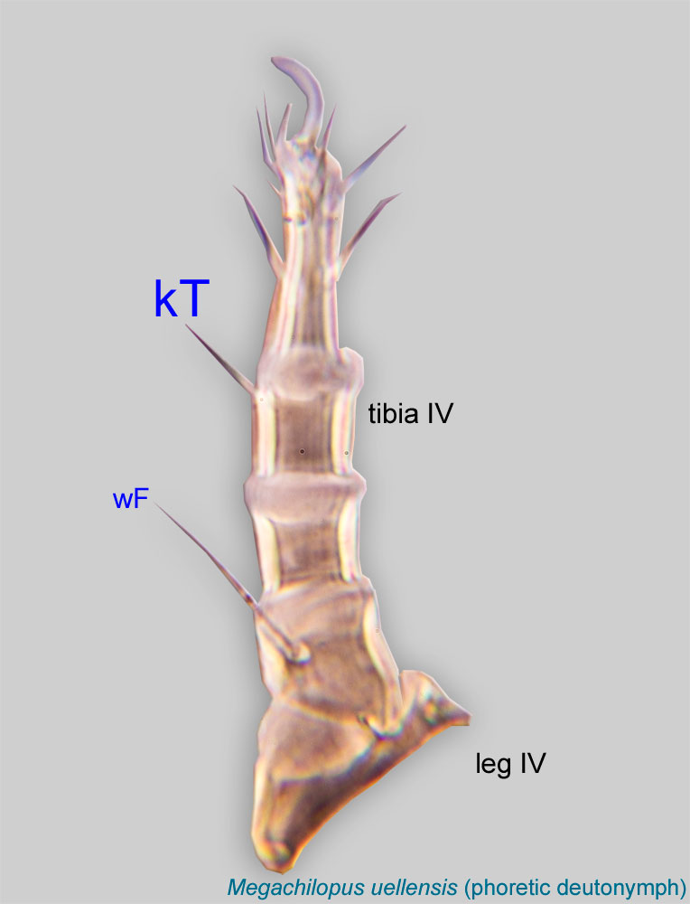
tibial seta gT: Anterior seta on tibiaetibia:
Leg or palp segment (also known as podomere or palpomere) between tarsus and genu.
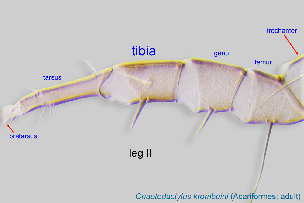 I-II of Astigmata. Homologous to seta v' of Oribatida. (Not illustrated.)
tibial seta hT: Posterior seta on tibiaetibia:
Leg or palp segment (also known as podomere or palpomere) between tarsus and genu.
I-II of Astigmata. Homologous to seta c'' of Oribatida. (Not illustrated.)
tibial seta kT: Anterior seta on tibiaetibia:
Leg or palp segment (also known as podomere or palpomere) between tarsus and genu.
III-IV of Astigmata. Homologous to seta v' of Oribatida.
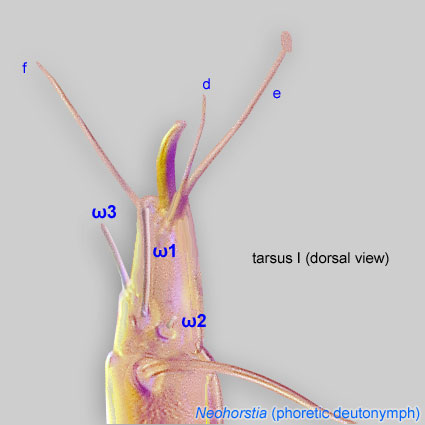
Solenidia ω1, ω2, and ω3 are found on tarsitarsus:
Terminal segment (also known as podomere or palpomere) of legs or palps. In Parasitoformes it can be subdivided into telotarsus and basitarsus.
in many Acariformes.
Solenidia ω1, ω2, and ω3 are found on tarsitarsus:
Terminal segment (also known as podomere or palpomere) of legs or palps. In Parasitoformes it can be subdivided into telotarsus and basitarsus.
in many Acariformes.
solenidion solenidion:
Thin-walled, terminally rounded or pointed filiform or peglike structure that is not birefringent in polarized light (unlike common setae in Acariformes). Often appears striated because of its internal structure. Found on the palpal tarsus on the gnathosoma and may also occur on the tarsus and tibia, less frequently on the genu, and occasionally on the femur of legs I-IV. In Acariformes, leg solenidia often arise from unsclerotized areas.
ω1: In Astigmata, usually situated in the dorso-basal part of tarsitarsus:
Terminal segment (also known as podomere or palpomere) of legs or palps. In Parasitoformes it can be subdivided into telotarsus and basitarsus.
I-II (absent from tarsitarsus:
Terminal segment (also known as podomere or palpomere) of legs or palps. In Parasitoformes it can be subdivided into telotarsus and basitarsus.
III-IV) and typically has a club-shaped, or rounded terminal end. Invariably present in all developmental stages.
solenidion solenidion:
Thin-walled, terminally rounded or pointed filiform or peglike structure that is not birefringent in polarized light (unlike common setae in Acariformes). Often appears striated because of its internal structure. Found on the palpal tarsus on the gnathosoma and may also occur on the tarsus and tibia, less frequently on the genu, and occasionally on the femur of legs I-IV. In Acariformes, leg solenidia often arise from unsclerotized areas.
ω2: In Astigmata, usually situated in the basal, posterolateral part of tarsustarsus:
Terminal segment (also known as podomere or palpomere) of legs or palps. In Parasitoformes it can be subdivided into telotarsus and basitarsus.
I (and absent from all other tarsitarsus:
Terminal segment (also known as podomere or palpomere) of legs or palps. In Parasitoformes it can be subdivided into telotarsus and basitarsus.
), often close to setae aa. Often is much shorter than ω1 and has a rounded tip. Present in protonymphsprotonymph:
Ontogenetic stage between larva and deutonymph. See <a href="index.cfm?pageID=1720">Life stages page</a> for more details.
to adults and absent in larvae.
solenidion solenidion:
Thin-walled, terminally rounded or pointed filiform or peglike structure that is not birefringent in polarized light (unlike common setae in Acariformes). Often appears striated because of its internal structure. Found on the palpal tarsus on the gnathosoma and may also occur on the tarsus and tibia, less frequently on the genu, and occasionally on the femur of legs I-IV. In Acariformes, leg solenidia often arise from unsclerotized areas.
ω3: In Astigmata, usually situated in the anterio-lateral part of tarsustarsus:
Terminal segment (also known as podomere or palpomere) of legs or palps. In Parasitoformes it can be subdivided into telotarsus and basitarsus.
I (and absent from all other tarsitarsus:
Terminal segment (also known as podomere or palpomere) of legs or palps. In Parasitoformes it can be subdivided into telotarsus and basitarsus.
). In phoreticphoretic:
Pertaining to phoresy; using another organism (i.e., a host) for dispersal to new habitats. Phoresy can be distinguished from parasitism because feeding typically does not occur during phoresy.
deutonymphsdeutonymph:
Ontogenetic stage between protonymph and tritonymph (or adult, if tritonymph is absent). See <a href="index.cfm?pageID=1720">Life stages page</a> for more details. usually situated in the basal part of tarsustarsus:
Terminal segment (also known as podomere or palpomere) of legs or palps. In Parasitoformes it can be subdivided into telotarsus and basitarsus.
(sometimes close to ω1), while in adults and other feeding stages it is terminal. Present in deutonymphsdeutonymph:
Ontogenetic stage between protonymph and tritonymph (or adult, if tritonymph is absent). See <a href="index.cfm?pageID=1720">Life stages page</a> for more details. to adults and absent in larvae and protonymphsprotonymph:
Ontogenetic stage between larva and deutonymph. See <a href="index.cfm?pageID=1720">Life stages page</a> for more details.
.
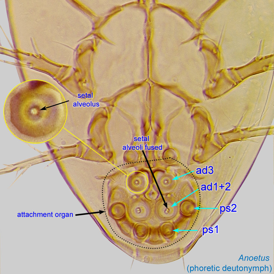
Suckers ad1+2 and ad3 and conoids ps1 and ps2 are paired suckers of setal origin located on the attachment organattachment organ:
Complex unpaired structure in phoretic deutonymphs of Astigmata situated on the posteroventral end of the body that serves for attachment to the host during phoresy. In deutonymphs phoretic on insects, attachment organ consists of a vestigial anal opening and two types of attachment elements: true suckers that create negative pressure and conoids that create adhesive forces. Not to be confused with pedicel.
of phoreticphoretic:
Pertaining to phoresy; using another organism (i.e., a host) for dispersal to new habitats. Phoresy can be distinguished from parasitism because feeding typically does not occur during phoresy.
deutonymphsdeutonymph:
Ontogenetic stage between protonymph and tritonymph (or adult, if tritonymph is absent). See <a href="index.cfm?pageID=1720">Life stages page</a> for more details. of Astigmata. Suckers function as attachment structures by creating negative pressure; they are modified from setal alveoli, vestiges of which are visible in the center of the sucker. In contrast, conoids are attachment elements that create adhesive forces; they are modified setae arising from well developed alveoli.
sucker ad1+2: Paired true suckers (from adanal setae ad1 and ad2 fused together) typically situated centrally on the attachment organattachment organ:
Complex unpaired structure in phoretic deutonymphs of Astigmata situated on the posteroventral end of the body that serves for attachment to the host during phoresy. In deutonymphs phoretic on insects, attachment organ consists of a vestigial anal opening and two types of attachment elements: true suckers that create negative pressure and conoids that create adhesive forces. Not to be confused with pedicel.
. Each sucker has two setal alveoli (often fused into a single alveolus).
sucker ad3: Paired true suckers (from adanal setae ad1 and ad2 fused together) typically situated anteriorly on the attachment organattachment organ:
Complex unpaired structure in phoretic deutonymphs of Astigmata situated on the posteroventral end of the body that serves for attachment to the host during phoresy. In deutonymphs phoretic on insects, attachment organ consists of a vestigial anal opening and two types of attachment elements: true suckers that create negative pressure and conoids that create adhesive forces. Not to be confused with pedicel.
. Each sucker has a setal alveolus.
conoid ps1: Paired conoids originating from pseudanal setae 1 (ps1) typically situated posterolaterally on the attachment organattachment organ:
Complex unpaired structure in phoretic deutonymphs of Astigmata situated on the posteroventral end of the body that serves for attachment to the host during phoresy. In deutonymphs phoretic on insects, attachment organ consists of a vestigial anal opening and two types of attachment elements: true suckers that create negative pressure and conoids that create adhesive forces. Not to be confused with pedicel.
. Lacks setal alveoli.
conoid ps2: Paired conoids originating from pseudanal setae 2 (ps2) typically situated medio-laterally on the attachment organattachment organ:
Complex unpaired structure in phoretic deutonymphs of Astigmata situated on the posteroventral end of the body that serves for attachment to the host during phoresy. In deutonymphs phoretic on insects, attachment organ consists of a vestigial anal opening and two types of attachment elements: true suckers that create negative pressure and conoids that create adhesive forces. Not to be confused with pedicel.
. Lacks setal alveoli.
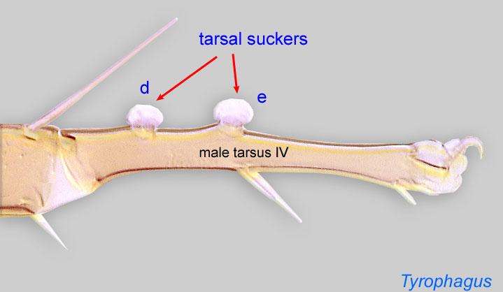
tarsal sucker: In some genera of Astigmata, tarsal setae e (=tc'') and d (=ft'') that are modified to suckers on tarsustarsus:
Terminal segment (also known as podomere or palpomere) of legs or palps. In Parasitoformes it can be subdivided into telotarsus and basitarsus.
IV of the male. Used for holding female during copulation. Otherwise, these setae are filiform and not modified.
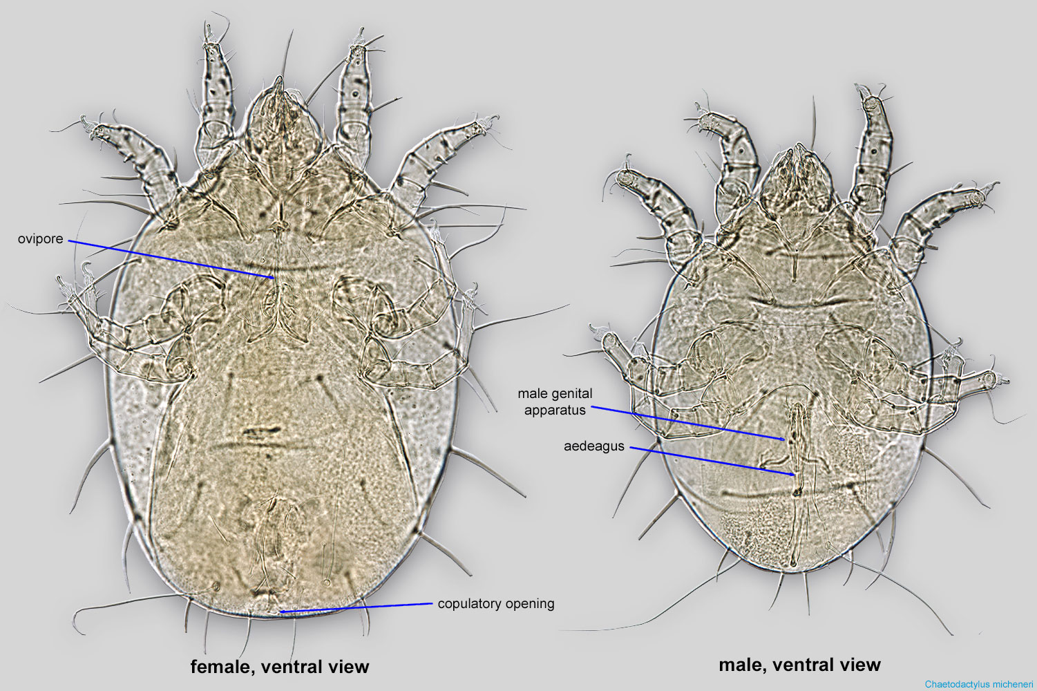
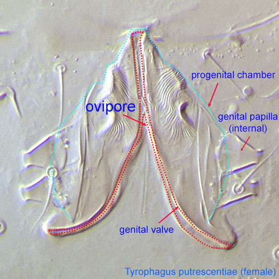
Adult female mites have an oviporeovipore:
Opening of female which serves as an outlet for oviposition. In Astigmata and non-astigmatan Oribatida, situated between genital valves. In Astigmata, not to be confused with the copulatory opening (which serves for sperm intake).
 , while adult males have an aedeagusaedeagus:
An external organ of a male arthropod that is specialized to deliver sperm during copulation.
(Astigmata [Fig. 2] and some Prostigmata; see Overview), a gonopodegonopode:
An appendage specialized to transfer sperm, such as the modified male chelicera in Mesostigmata.
(Mesostigmata [Fig. 1]), or a spermatopositorspermatopositor:
A small male organ for depositing spermatophores.
(Oribatida and some Prostigmata).
Developing eggs can be observed inside females. Eggs are usually large compared to the female body and are easily noticeable. In addition to the oviporeovipore:
Opening of female which serves as an outlet for oviposition. In Astigmata and non-astigmatan Oribatida, situated between genital valves. In Astigmata, not to be confused with the copulatory opening (which serves for sperm intake).
, mite females in Astigmata and Mesostigmata may have a copulatory opening. These openings serve for insemination, are usually very small (Fig. 2), and their position varies in different groups of mites.
Sex is usually apparent only in adult mites. Locustacarus is an exception. In this species, the larval stage is represented by female larvae (called "larviform females"), which can copulate but lay eggs after molting, and larviform males, which do not molt further. Sometimes, despite lacking functional genital structures, immature stages can be sexed based on differences in shape and size, since males are usually smaller.
Authors: P. Klimov, B. OConnor, R. Ochoa, G. Bauchan, A. Redford, J. Scher
Last updated October 2016
tool images at ITP Node
idtools.org