scavenger or mutualist; feeds on various microorganisms in bee nests
Histiostoma Kramer, 1876
Superorder Acariformes » Order Sarcoptiformes » Suborder Oribatida » Infraorder Desmonomata » Hyporder Astigmata » Family Histiostomatidae » Genus Histiostoma
Histiostoma pectineum Kramer, 1876 (=Hypopus feroniarum Dufour, 1839)
Phyllostoma Kramer, 1876 (nom. preocc.); Zschachia Oudemans, 1929. In literature, the name Histiostoma has been used to group species now assigned to genera Histiostoma and Anoetus.
Phoretic phoretic:
Pertaining to phoresy; using another organism (i.e., a host) for dispersal to new habitats. Phoresy can be distinguished from parasitism because feeding typically does not occur during phoresy.
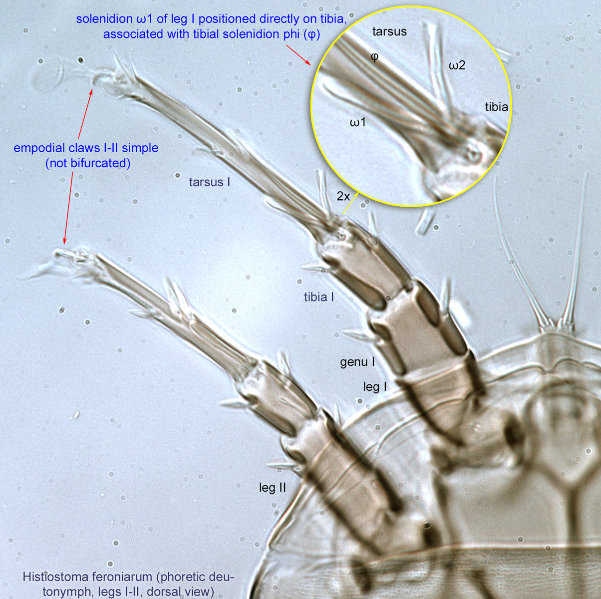
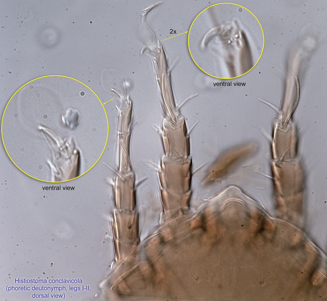
deutonymph: Solenidionsolenidion:
Thin-walled, terminally rounded or pointed filiform or peglike structure that is not birefringent in polarized light (unlike common setae in Acariformes). Often appears striated because of its internal structure. Found on the palpal tarsus on the gnathosoma and may also occur on the tarsus and tibia, less frequently on the genu, and occasionally on the femur of legs I-IV. In Acariformes, leg solenidia often arise from unsclerotized areas.
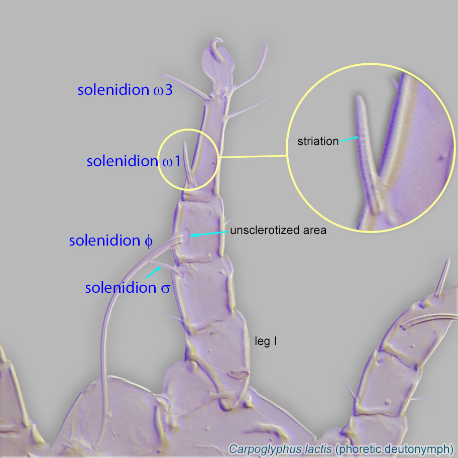 ω1 of leg I positioned directly on tibiatibia:
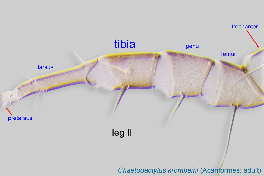
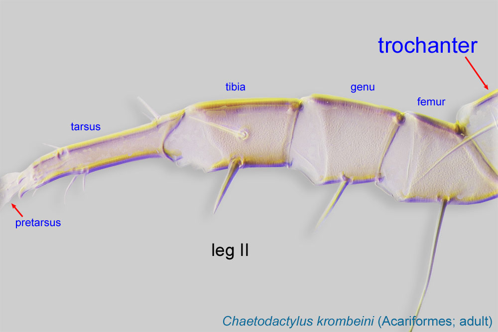
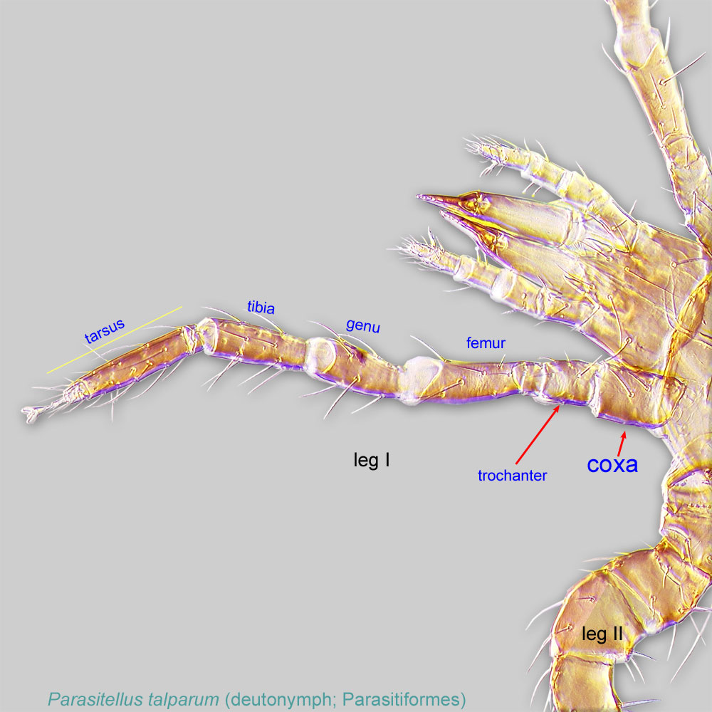
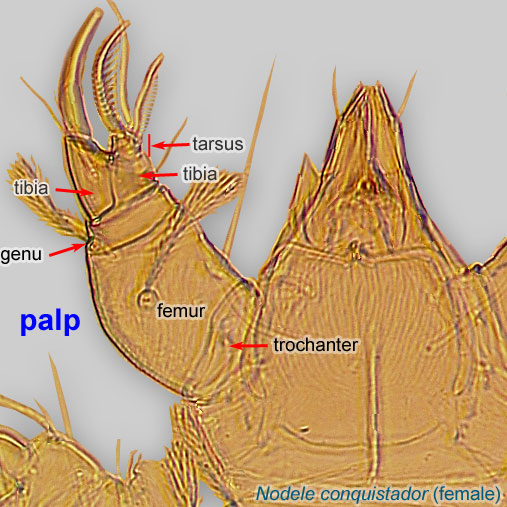
Leg or palp segment (also known as podomere or palpomere) between tarsus and genu.
 , associated with tibial solenidionsolenidion:
Thin-walled, terminally rounded or pointed filiform or peglike structure that is not birefringent in polarized light (unlike common setae in Acariformes). Often appears striated because of its internal structure. Found on the palpal tarsus on the gnathosoma and may also occur on the tarsus and tibia, less frequently on the genu, and occasionally on the femur of legs I-IV. In Acariformes, leg solenidia often arise from unsclerotized areas.
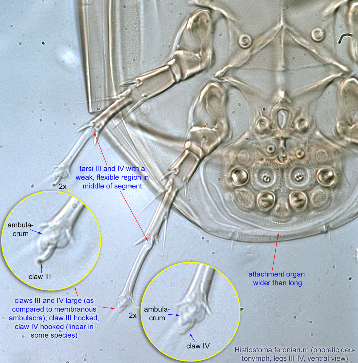
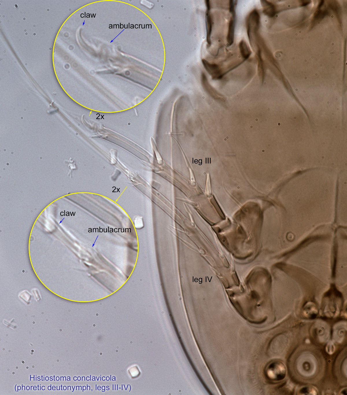
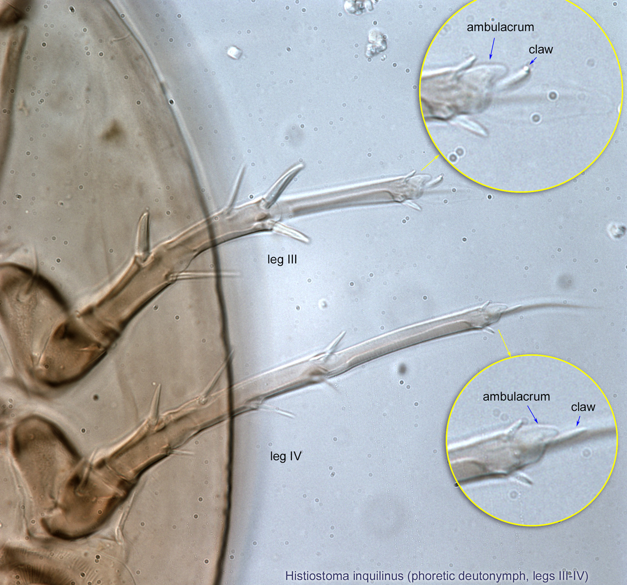
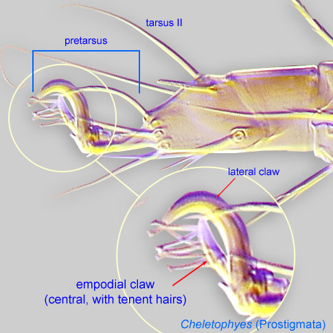
phi (φ) (Fig.4); claws III and IV large (as compared to membranous ambulacraambulacrum:
The claws and empodium of the apotele or pretarsus.
); claw III hooked; and claw IV hooked (Fig. 6) or linear (Figs. 22, 24).
Phoretic phoretic:
Pertaining to phoresy; using another organism (i.e., a host) for dispersal to new habitats. Phoresy can be distinguished from parasitism because feeding typically does not occur during phoresy.
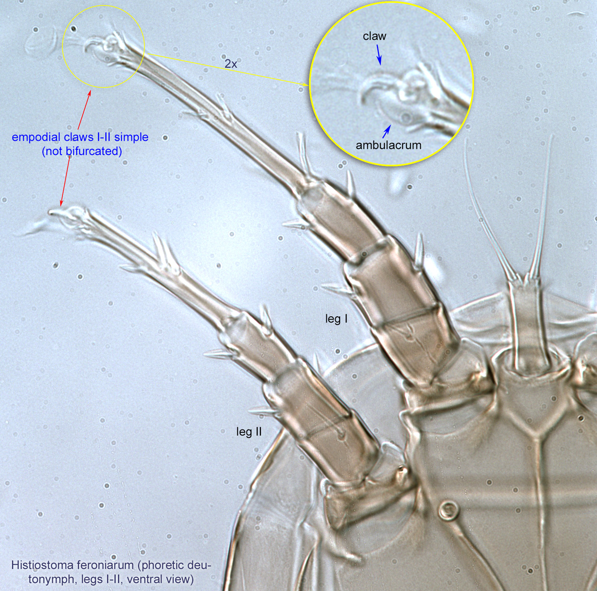
deutonymph: Empodial clawsEmpodial claw:
Claw-like, membranous, or pad-like structure of setal origin. Present only on the pretarsus in Acariformes. In Astigmata, it is the only claw on the pretarsus and often referred to simply as the claw. In the remaining Acariformes, may be accomanied by two lateral claws. Also known as empodium, pretarsal empodium, or central claw.
 I-IV simplesimple:
Of claws or setae; not modified or not bi- or trifurcate at tip.
(not bifurcated) (Fig. 4, 5). Tarsustarsus:
Terminal segment (also known as podomere or palpomere) of legs or palps. In Parasitoformes it can be subdivided into telotarsus and basitarsus.
 III and IV with a weak, flexible region in middle of segment (Fig. 6). Pretarsuspretarsus:
Terminal leg or palpal segment distal to tarsus.
III and IV with empodial clawsempodial claw:
Claw-like, membranous, or pad-like structure of setal origin. Present only on the pretarsus in Acariformes. In Astigmata, it is the only claw on the pretarsus and often referred to simply as the claw. In the remaining Acariformes, may be accomanied by two lateral claws. Also known as empodium, pretarsal empodium, or central claw.
(Fig. 6). TrochantersTrochanter:
Leg or palp segment (also known as podomere or palpomere) between femur and coxa. In Acariformes this is the most basal movable leg segment (or podomere) forming a joint with the body.
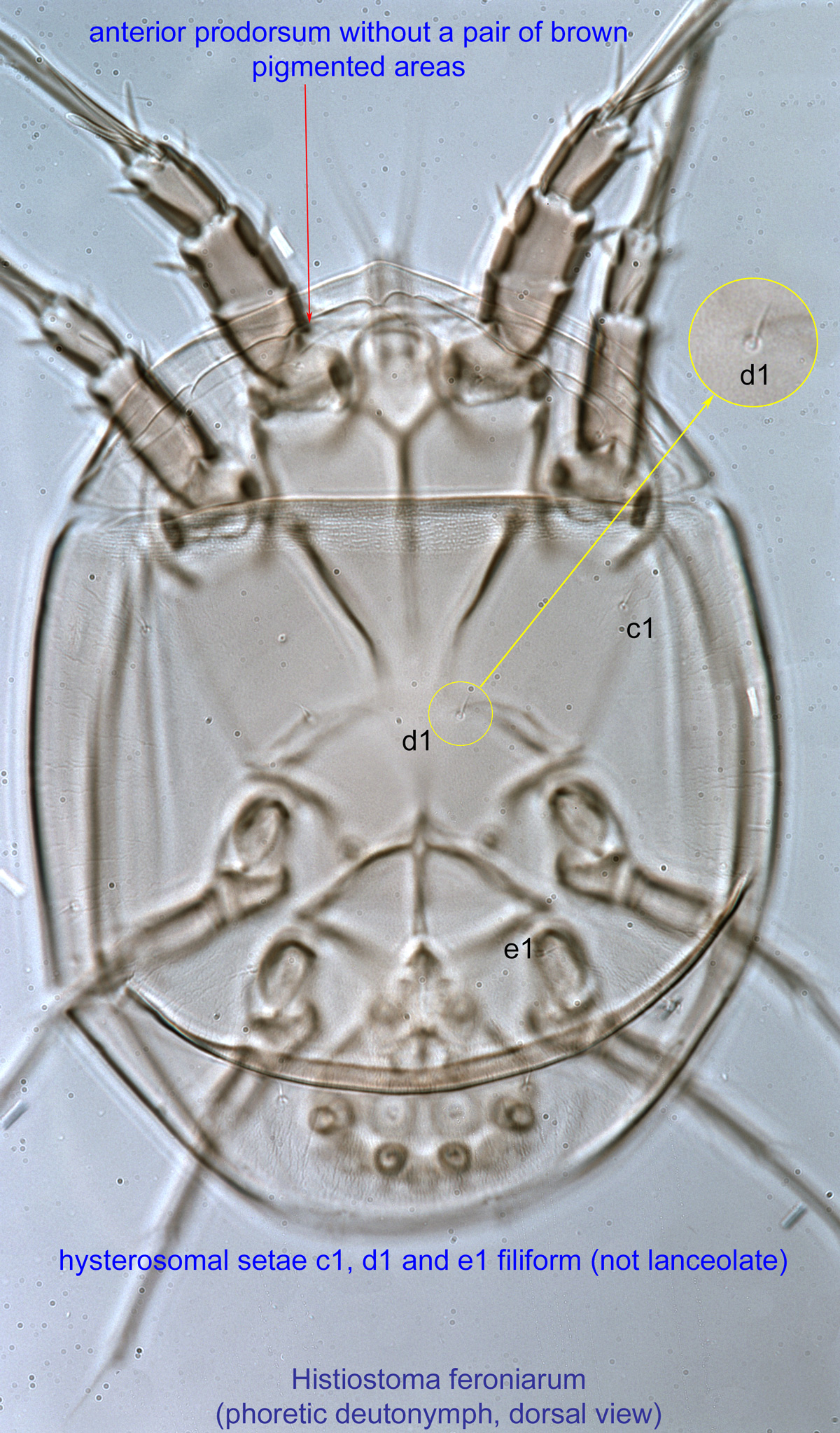
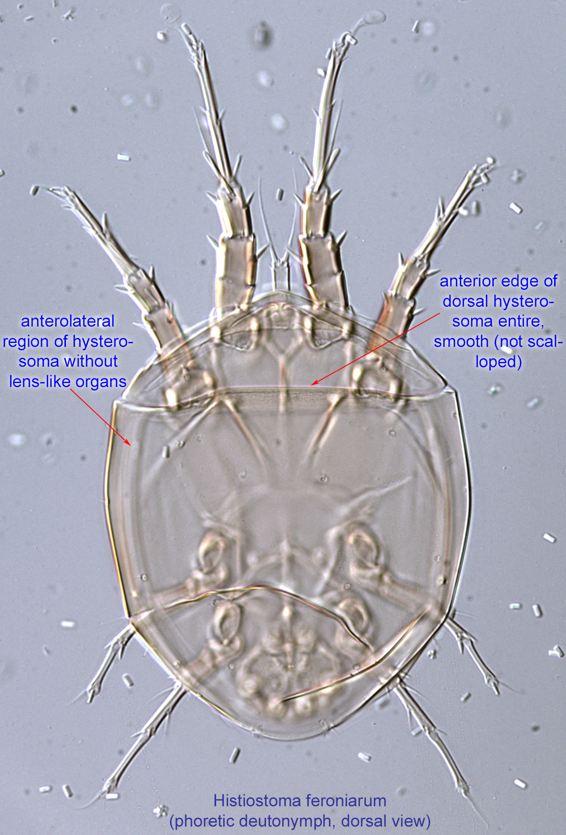
 I-II without setae. Anterior prodorsumprodorsum:
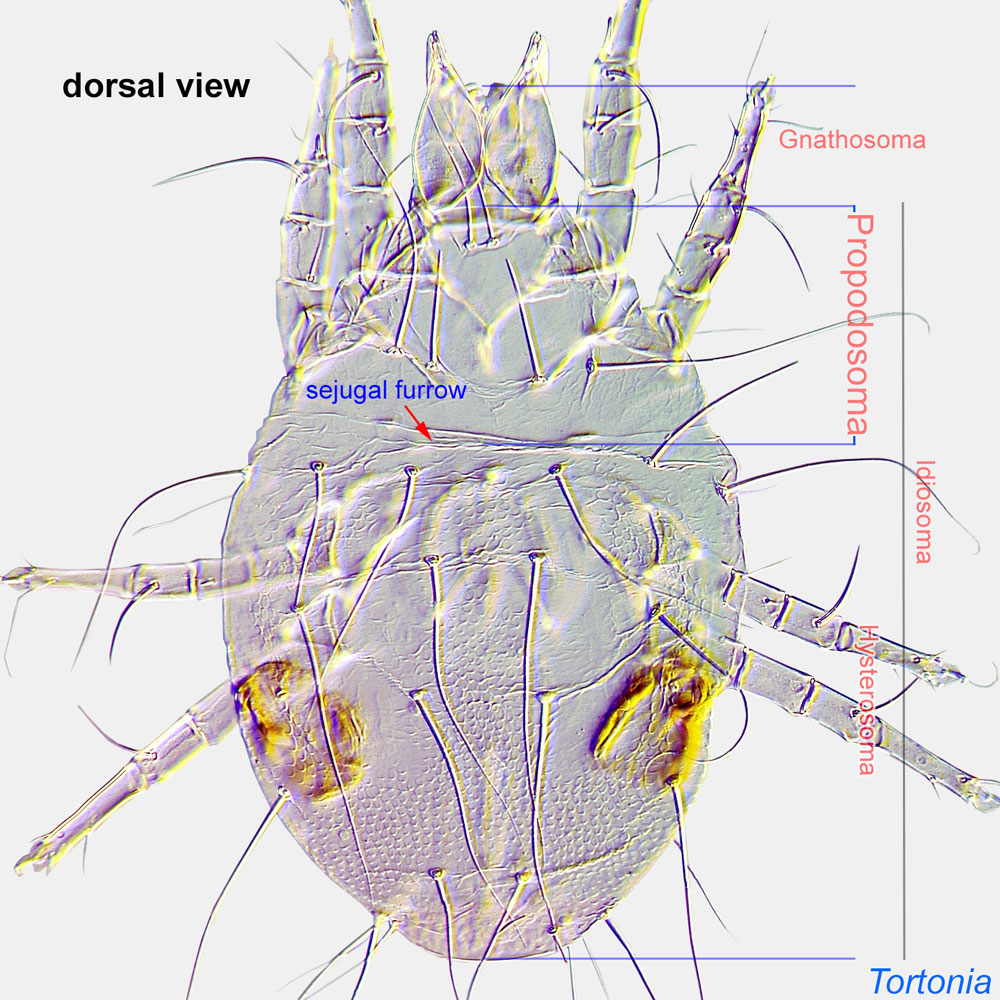
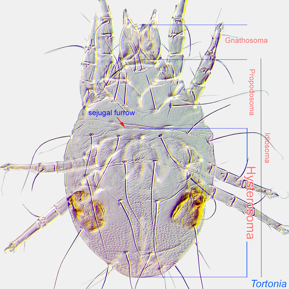
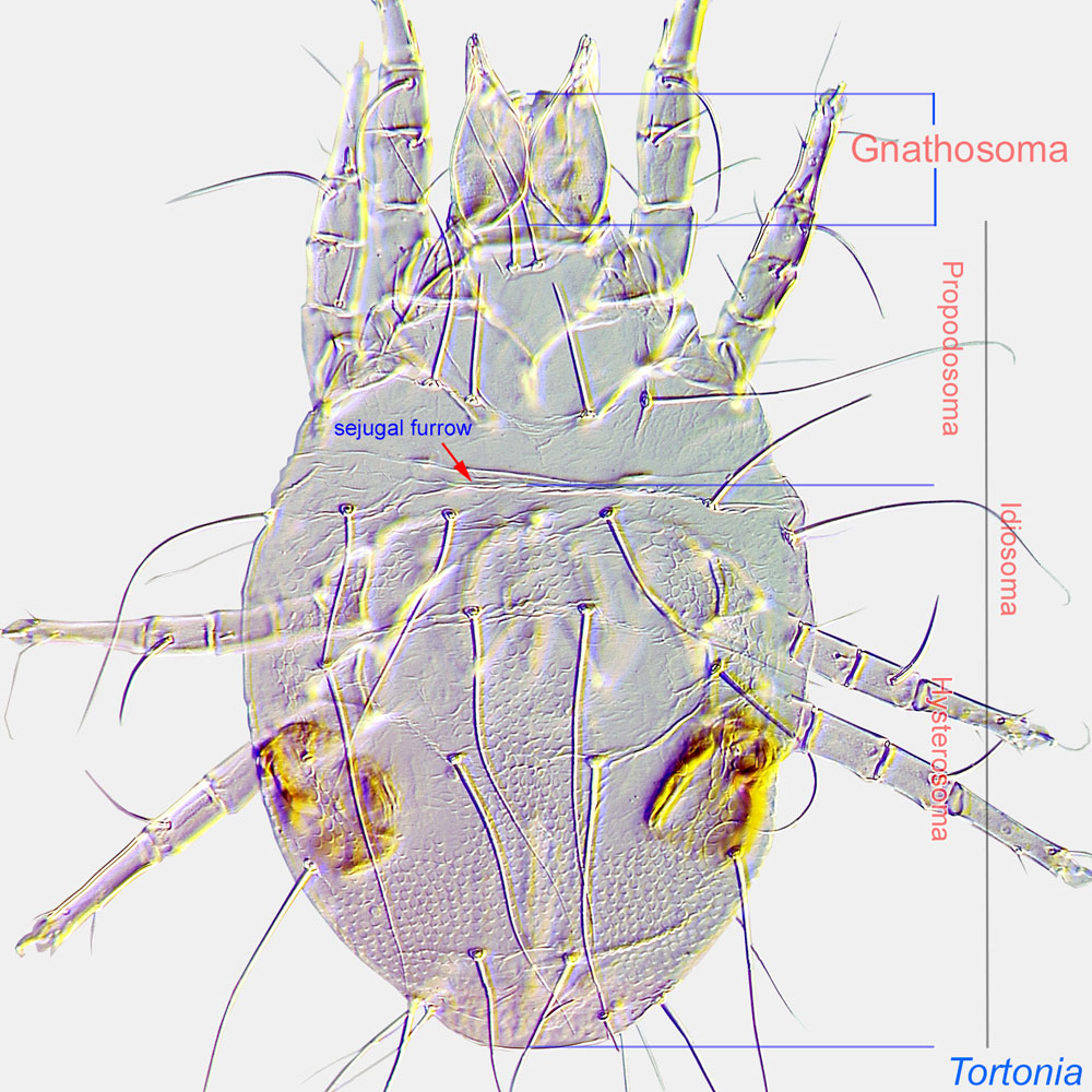
Dorsal surface of propodosoma.
 without a pair of brown pigmented areas (Fig. 1). Anterior edge of dorsal hysterosomahysterosoma:
Division of body posterior to the sejugal furrow, bearing legs III and IV.
 entire, smooth (not scalloped) (Fig. 3). Anterolateral region of hysterosomahysterosoma:
Division of body posterior to the sejugal furrow, bearing legs III and IV.
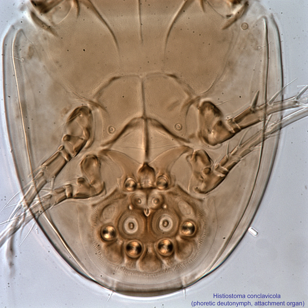
without lens-like organs (Fig. 3). Hysterosomal setae c1, d1, and e1 filiform (not lanceolate) (Fig. 1). Attachment organattachment organ:
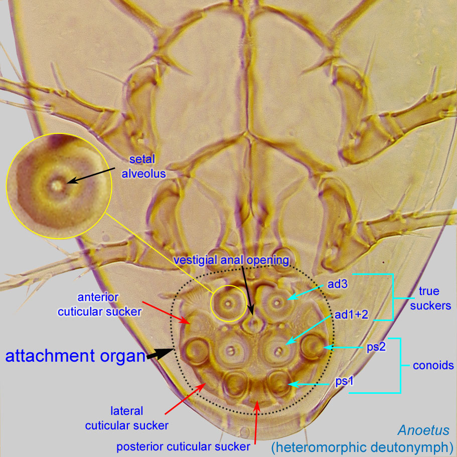
Complex unpaired structure in phoretic deutonymphs of Astigmata situated on the posteroventral end of the body that serves for attachment to the host during phoresy. In deutonymphs phoretic on insects, attachment organ consists of a vestigial anal opening and two types of attachment elements: true suckers that create negative pressure and conoids that create adhesive forces. Not to be confused with pedicel.
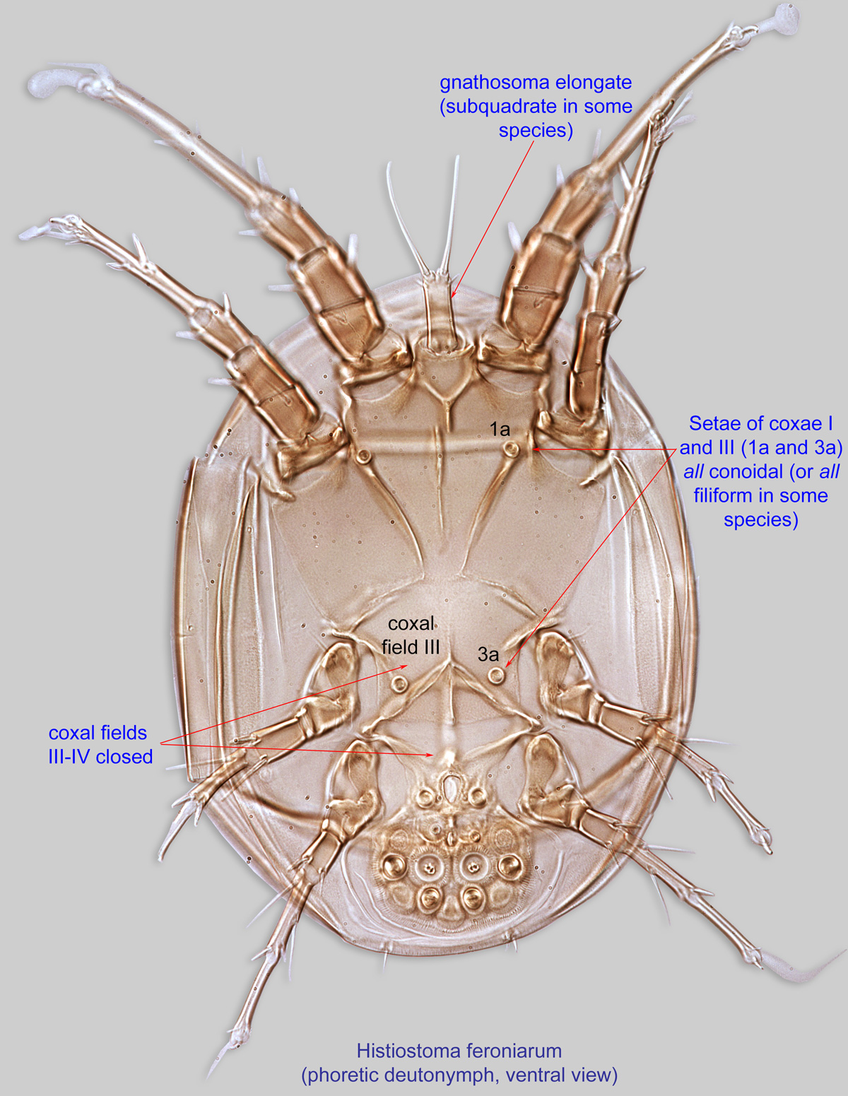
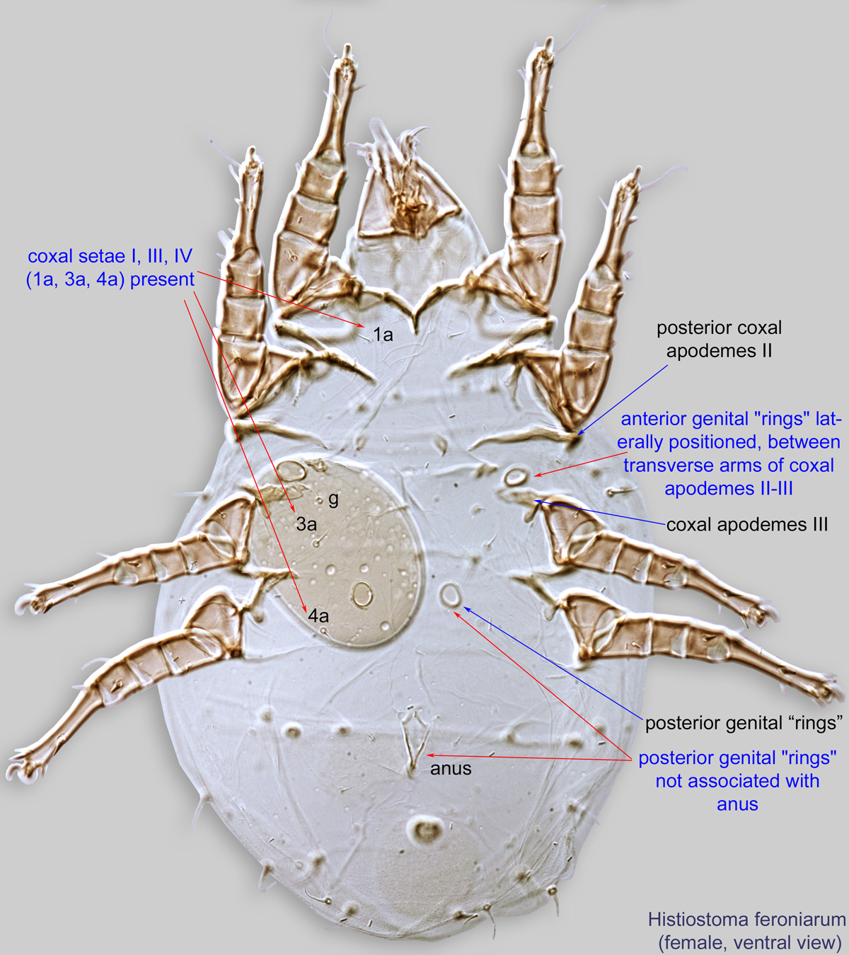
 wider than long (Fig. 6). Coxal fields III-IV closed (Fig. 2). Setae of coxaecoxa:
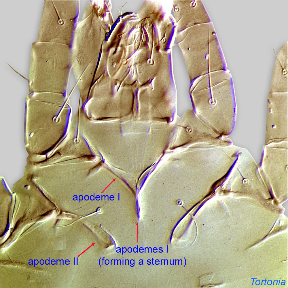
In Parasitiformes, most basal leg segment (or podomere) forming a joint with the body. Areas delimited by coxal apodemes are called coxal fields in Astigmata or coxisternal plates in Prostigmata.
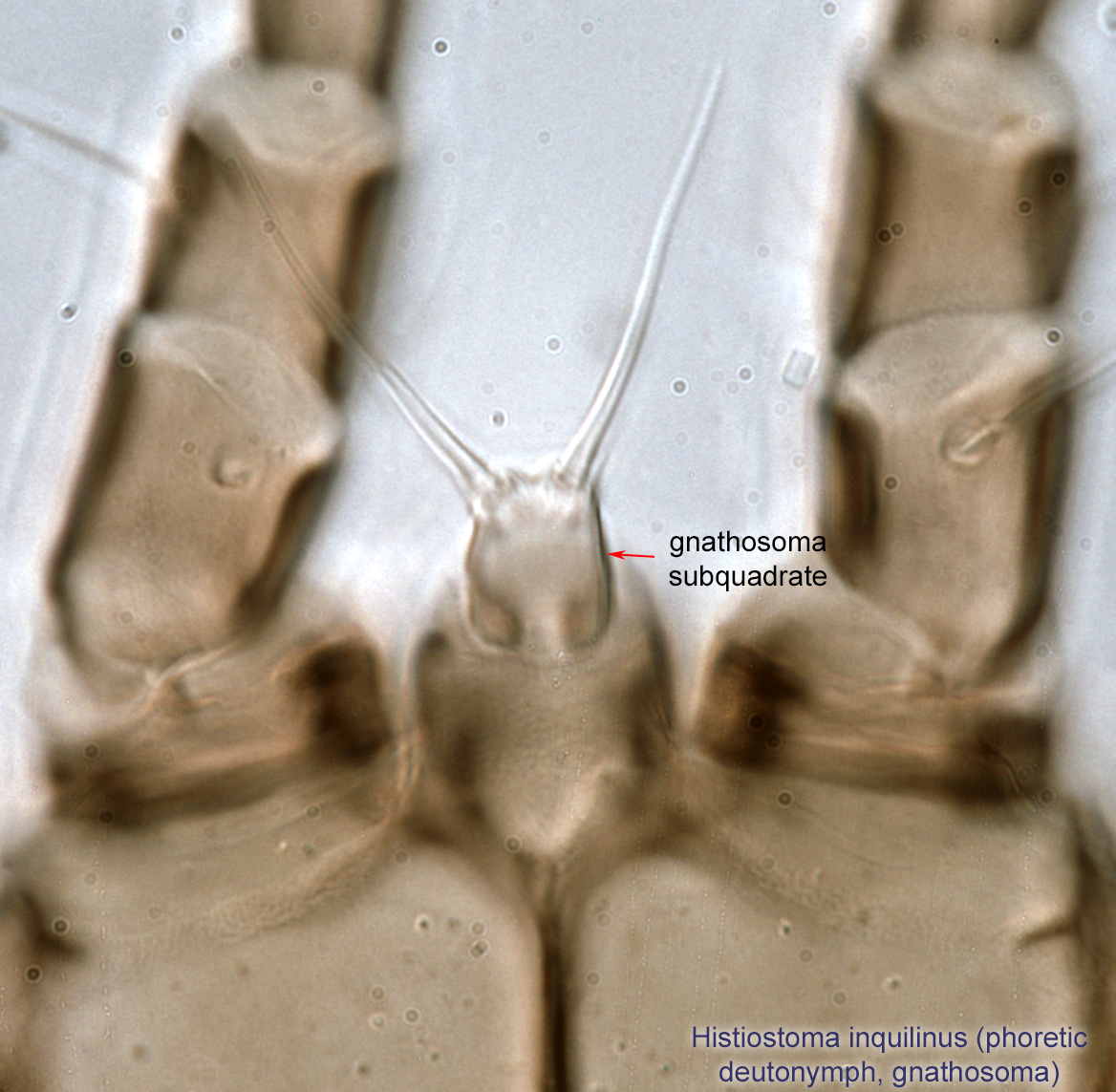
 I and III (1a and 3a) either all filiform or all conoidal (Fig. 2). Gnathosomagnathosoma:
Division of body anterior to the propodosoma bearing two pairs of appendages (palps and chelicerae).
 elongate (Fig. 2) or subquadrate (Fig. 23).
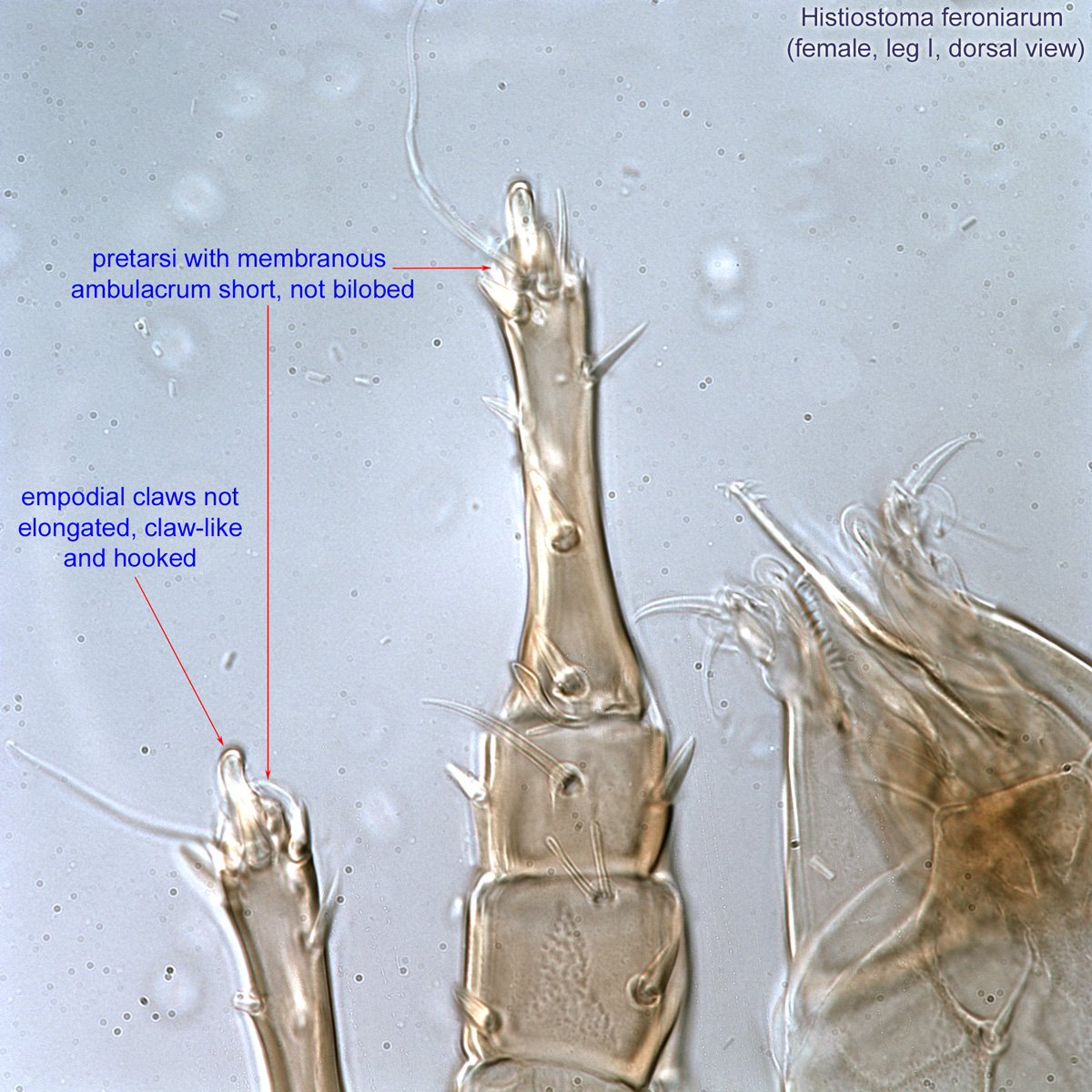
Female: PretarsiPretarsus:
Terminal leg or palpal segment distal to tarsus.
with membranous ambulacrumambulacrum:
The claws and empodium of the apotele or pretarsus.
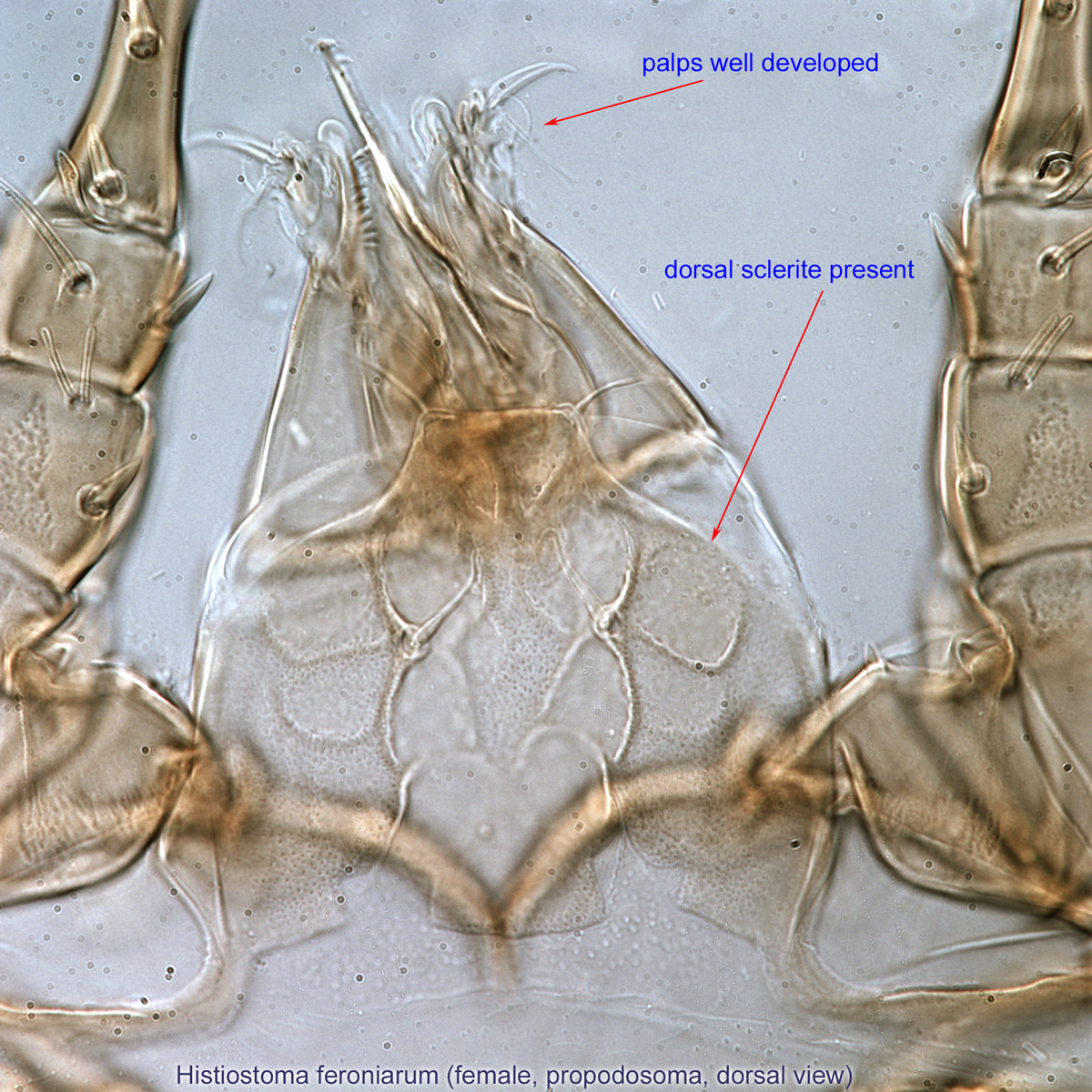
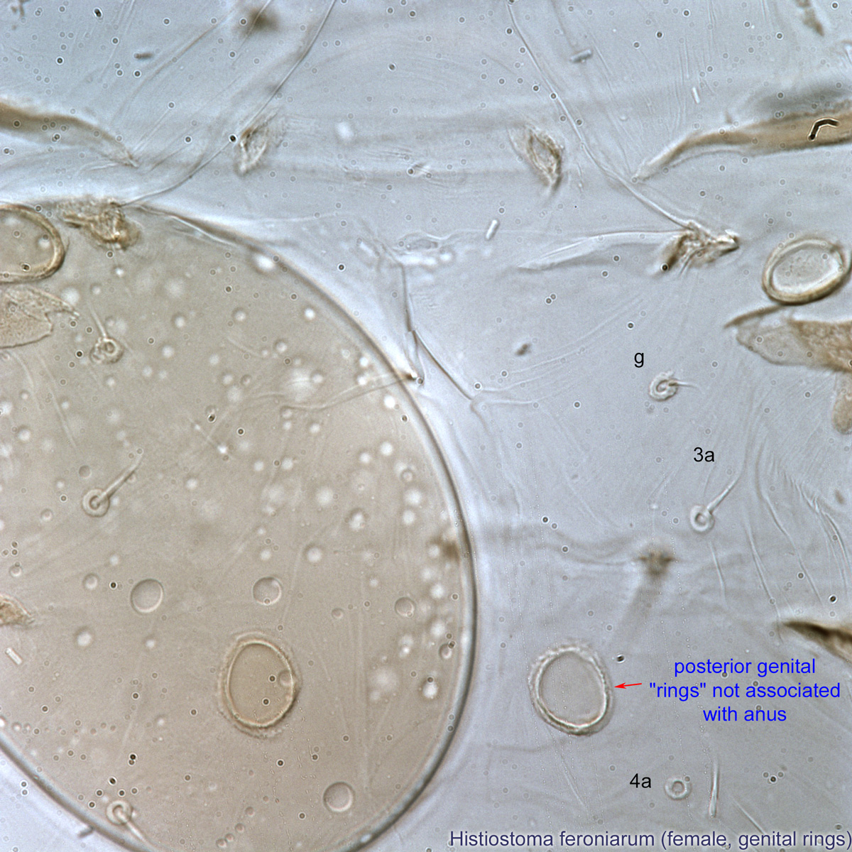
short, not bilobed (Fig. 11). Posterior genital "rings" not associated with the anus (Figs. 8, 10). Coxal setae I, III, IV present (Fig. 8). PalpsPalp:
Second (after chelicera) paired appendage of the gnathosoma. Has a sensory function, but may be variously modified for other functions (e.g., raptorial, attachment to host, or filtering).
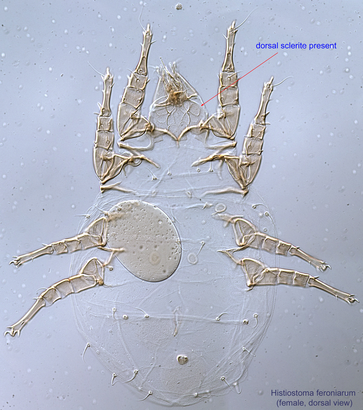
 well developed (Fig. 9). Dorsal scleritesclerite:
A component section of an exoskeleton; a plate forming the skeleton of an arthropod.
present (Figs. 7, 9). Anterior genital "rings" laterally positioned between transverse arms of coxal apodemesapodeme:
Internal sclerite that serves as an attachment site for muscles. Most commonly used (as "coxal apodeme") to describe elements of coxae fused to the ventral body in Acariformes (coxae are free and not fused to the body in Parasitiformes), and may be variously referred to as ventral, sternal, anterior, or posterior.
 II-III (Fig. 8). Empodial clawsEmpodial claw:
Claw-like, membranous, or pad-like structure of setal origin. Present only on the pretarsus in Acariformes. In Astigmata, it is the only claw on the pretarsus and often referred to simply as the claw. In the remaining Acariformes, may be accomanied by two lateral claws. Also known as empodium, pretarsal empodium, or central claw.
not elongated, claw-like and hooked (Fig. 11).
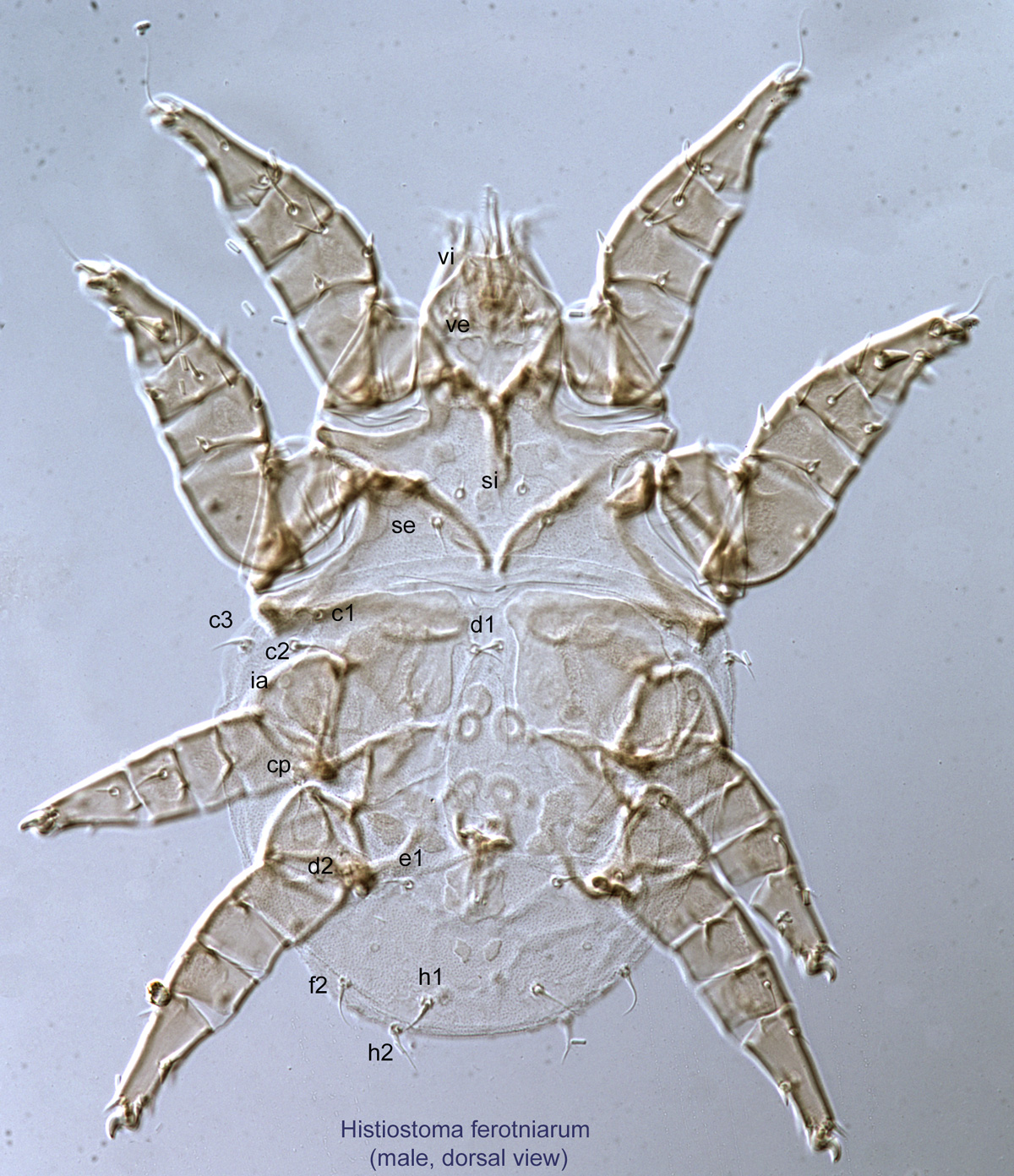
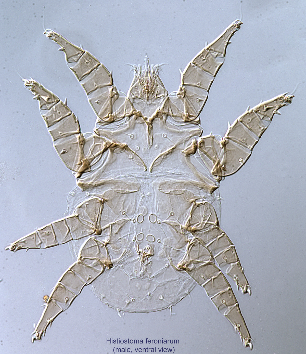
Male: Paranal suckers absent (Fig. 15). Aedeagusaedeagus:
An external organ of a male arthropod that is specialized to deliver sperm during copulation.
thin, short and posteriorly directed (Fig. 15). With two pairs of genital rings (Fig. 15). Mouthparts and setation normal (Fig. 14). External vertical setae short, not extending over the gnathosomagnathosoma:
Division of body anterior to the propodosoma bearing two pairs of appendages (palps and chelicerae).
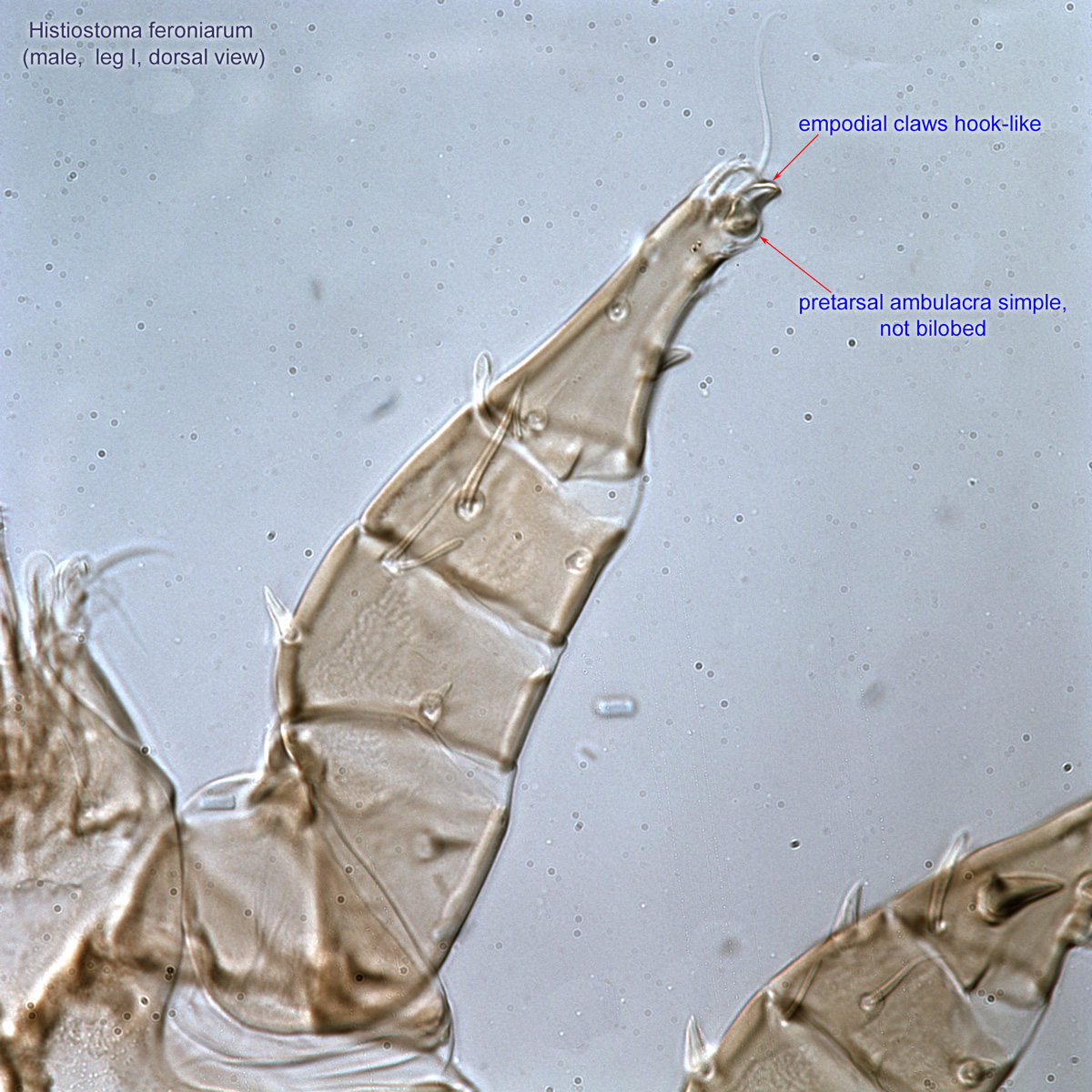
(Fig. 14). Dorsal setae filiform (not heavily barbed or blade-like) (Fig. 14). Pretarsal ambulacraambulacrum:
The claws and empodium of the apotele or pretarsus.
simple, not bilobed, empodial clawsempodial claw:
Claw-like, membranous, or pad-like structure of setal origin. Present only on the pretarsus in Acariformes. In Astigmata, it is the only claw on the pretarsus and often referred to simply as the claw. In the remaining Acariformes, may be accomanied by two lateral claws. Also known as empodium, pretarsal empodium, or central claw.
hook-like (Fig. 16). Body form and sclerotization variable; if propodosomapropodosoma:
Anterior part of idiosoma, in front of sejugal furrow.
and opisthosomaopisthosoma:
Body division posterior to legs IV; usually there is no distinct boundary delimiting this part of idiosoma.
each covered by a reticulate scleritesclerite:
A component section of an exoskeleton; a plate forming the skeleton of an arthropod.
, then setae d1 far posterior to cp. Setae h1 usually ventral in position.
This is a large genus with more than 200 described species. A dichotomous key not available. Most species have been described based on phoreticphoretic:
Pertaining to phoresy; using another organism (i.e., a host) for dispersal to new habitats. Phoresy can be distinguished from parasitism because feeding typically does not occur during phoresy.
deutonymphsdeutonymph:
Ontogenetic stage between protonymph and tritonymph (or adult, if tritonymph is absent). See <a href="index.cfm?pageID=1720">Life stages page</a> for more details.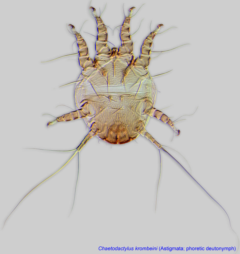 . Older, possibly out-dated keys to species (adults and phoreticphoretic:
Pertaining to phoresy; using another organism (i.e., a host) for dispersal to new habitats. Phoresy can be distinguished from parasitism because feeding typically does not occur during phoresy.
deutonymphsdeutonymph:
Ontogenetic stage between protonymph and tritonymph (or adult, if tritonymph is absent). See <a href="index.cfm?pageID=1720">Life stages page</a> for more details.) are available in Hughes and Jackson, 1958Hughes and Jackson, 1958:
Hughes, R. D. amp; C. G. Jackson. 1958. A review of the Anoetidae (Acari). Virginia Journal of Science. 8: 5-198.; Mahunka, 1970Mahunka, 1970:
Mahunka, S. 1970. Atkák V. - Acari V. Magyarország Állatvilága (Fauna Hungariae 101).18: 1-76.; Scheucher, 1957Scheucher, 1957:
Scheucher, R. 1957. Systematik und Ökologie der deutschen Anoetinen. In Beiträge zur Systematik und Ökologie mitteleuropäischer Acarina. Vol. 1, ed. S. H.-J., 233-384.; Sevastyanov, 1975Sevastyanov, 1975:
Sevastyanov, V. D. 1975. [Family Anoetidae]. In Opredelitel' obitayushchikh v pochve kleshchey. Sarcoptiformes. [Identification key to soil inhabiting mites. Sarcoptiformes] ed. M. S. Gilyarov, 382-416. Moscow: Nauka Publ..
Phoretic phoretic:
Pertaining to phoresy; using another organism (i.e., a host) for dispersal to new habitats. Phoresy can be distinguished from parasitism because feeding typically does not occur during phoresy.
deutonymphsdeutonymph:
Ontogenetic stage between protonymph and tritonymph (or adult, if tritonymph is absent). See <a href="index.cfm?pageID=1720">Life stages page</a> for more details. of Histiostoma are very similar to those of Anoetus. In Histiostoma claws III-IV are large (as compared to width of membranous ambulacraambulacrum:
The claws and empodium of the apotele or pretarsus.
), claw III is claw-like, claw IV either claw-like or linear; gnathosomagnathosoma:
Division of body anterior to the propodosoma bearing two pairs of appendages (palps and chelicerae).
is subquadrate or elongated. In Anoetus, claws III - IV are very small (as compared to width of membranous ambulacraambulacrum:
The claws and empodium of the apotele or pretarsus.
), thin, and linear; gnathosomagnathosoma:
Division of body anterior to the propodosoma bearing two pairs of appendages (palps and chelicerae).
is subquadrate.
Worldwide, including subantarctic islands. Species found in associations with bees have been recorded from the Nearctic, Palaearctic, Oriental, and African (including Madagascar) regions.
Among bees, this genus has been found in associations with Heriades, Xenoglossa, Apis, and Xylocopa.
facultative facultative:
can complete entire life cycle without bees or their close relative, wasps
in Histiostoma feroniarum and Histiostoma polypori
permanent permanent:
associated exclusively with bees or their close relative, wasps; cannot live without these hosts
in Histiostoma conclavicola, Histiostoma heriades, and Histiostoma inquilinus
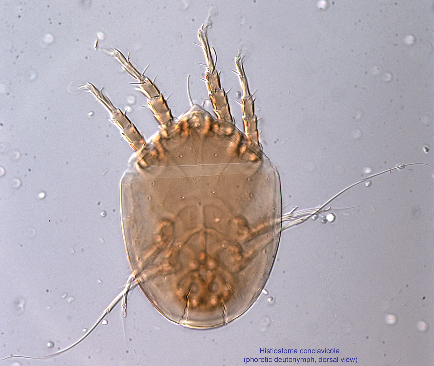
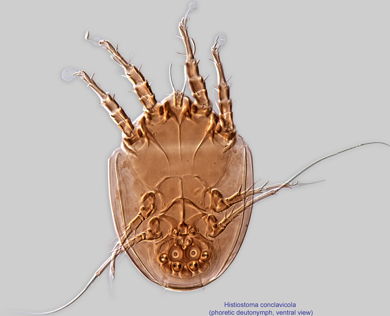
Bee specialists Histiostoma conclavicola, Histiostoma heriades, and Histiostoma inquilinus
disperse on adult bees. They are sometimes found in acarinariaacarinarium:Habitat generalist Histiostoma feroniarum
disperse on any suitable host, such as various insects that can enter beehives, rather than on adult bees.Earwig specialist Histiostoma polypori
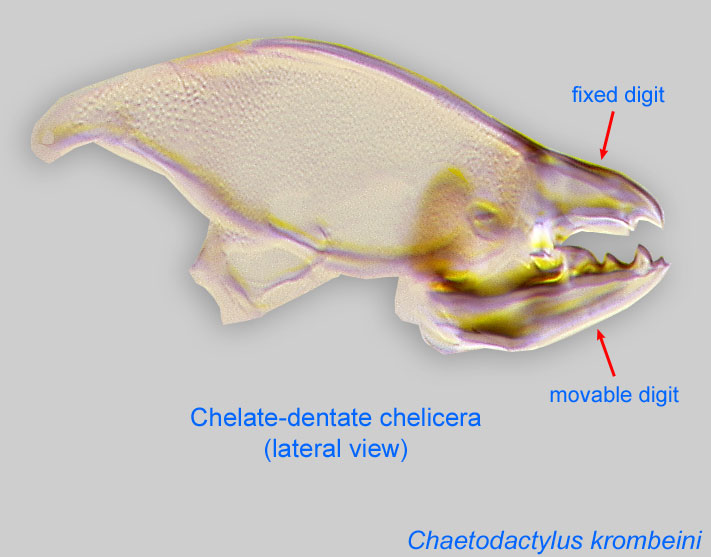
attach to and disperse on earwigs and can enter beehives with these hosts.These mites are common and live in a wide variety of terrestrial and semiaquatic freshwater habitats, where they primarily feed on various microorganisms by filtering them from the substrate using their modified, brush like cheliceraechelicera:
Anterior, paired appendage of the body. Primary organ for food acquisition, adapted for chewing, piercing, tearing, sucking, or filtering.
 . Some species are habitat generalists, though several show some degree of specialization to a particular host, such as bark beetles, scarab beetles, ants, earwigs, or bees.
Three species are bee specialists, found only on adult bees as phoreticphoretic:
Pertaining to phoresy; using another organism (i.e., a host) for dispersal to new habitats. Phoresy can be distinguished from parasitism because feeding typically does not occur during phoresy.
deutonymphs:deutonymph:
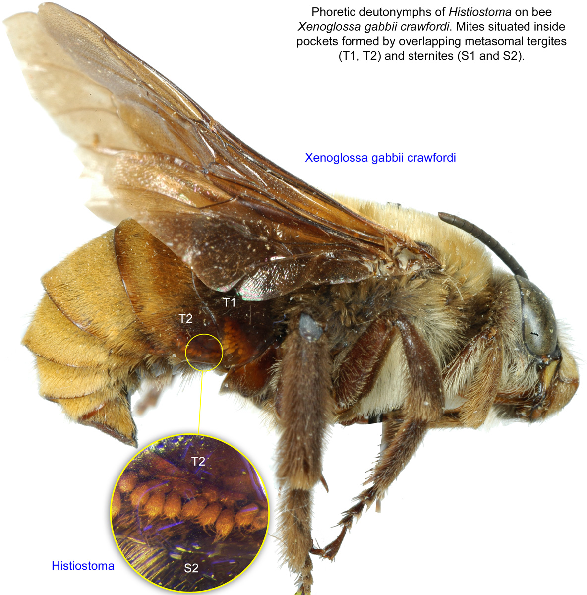
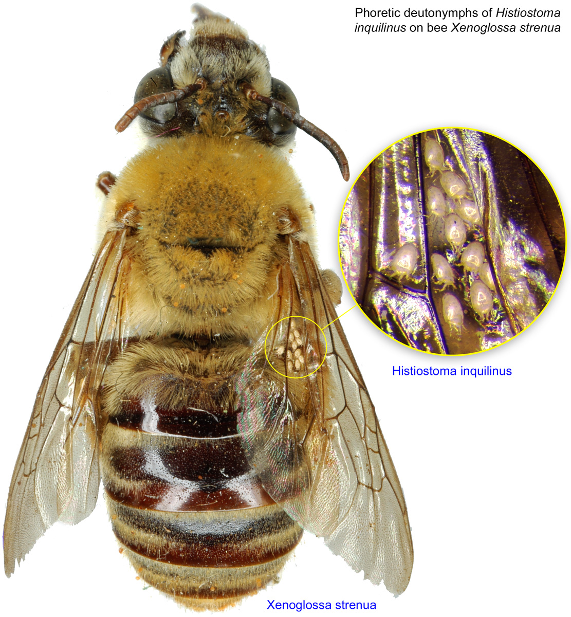
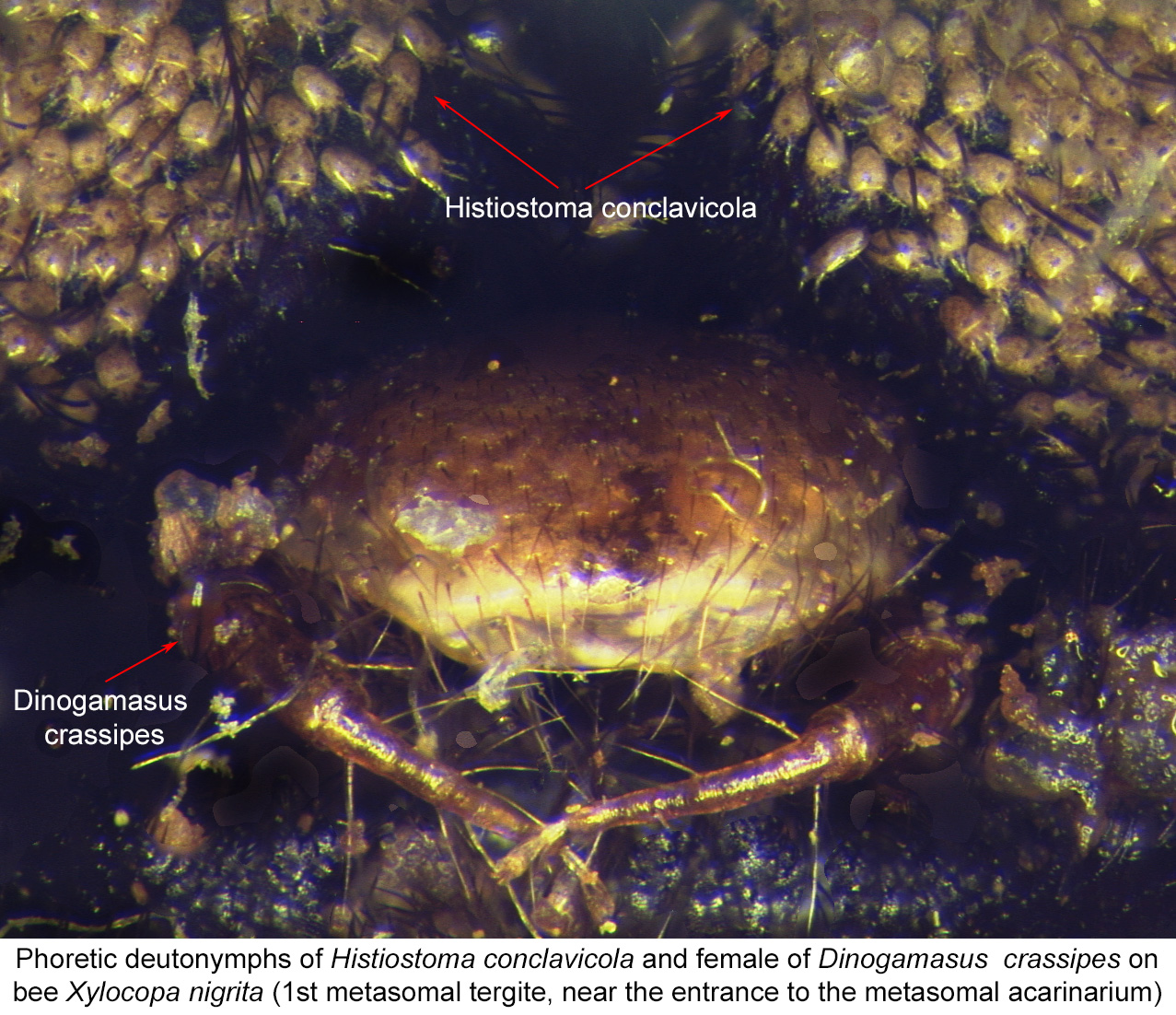
Ontogenetic stage between protonymph and tritonymph (or adult, if tritonymph is absent). See <a href="index.cfm?pageID=1720">Life stages page</a> for more details.:: Histiostoma conclavicola, Histiostoma heriades, and Histiostoma inquilinus. It is interesting that Histiostoma inquilinus may be found in the metasomal acarinariumacarinarium:
A specialized morphological structure that facilitates retention of mites on the body of an organism, typically a bee or wasp.
(pockets between overlapping tergites and sternites 1-3) of bees of the genus Xenoglossa (Figs. 25, 26). This may suggest a mutualistic association similar to that described for Anoetus, offering protection from microorganisms to developing bees and their food. H. conclavicola has been found in the unpaired metasomal acarinariumacarinarium:
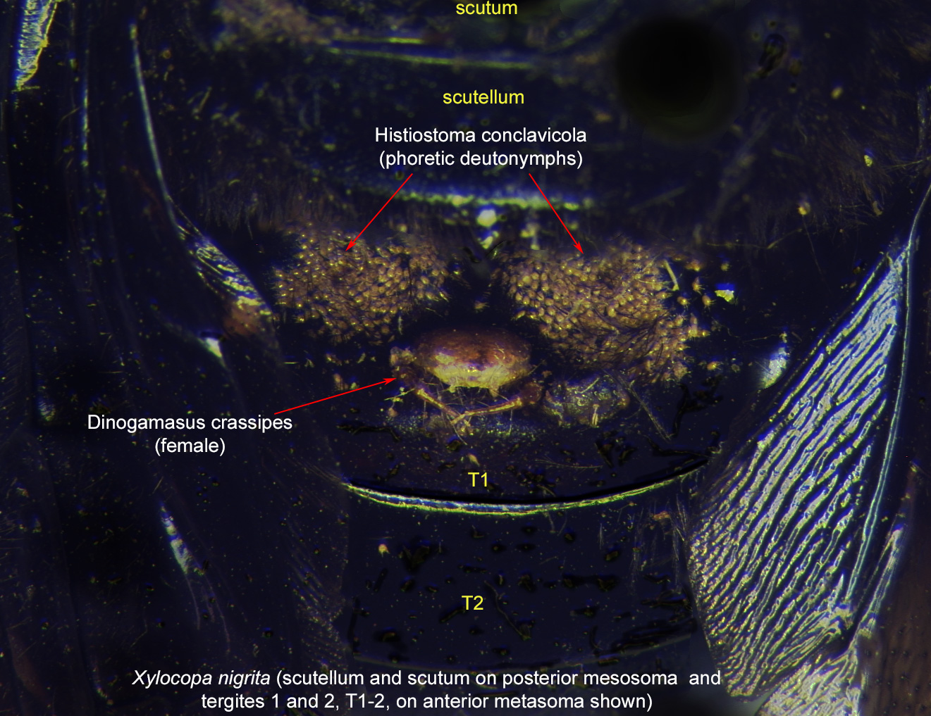
A specialized morphological structure that facilitates retention of mites on the body of an organism, typically a bee or wasp.
of the large carpenter bee Xylocopa in Africa (Figs. 27, 28). H. conclavicola occurs relatively more rarely than other mite species (e.g., Dinogamasus and Sennertia), so it is unlikely that the bee's acarinariaacarinarium:
A specialized morphological structure that facilitates retention of mites on the body of an organism, typically a bee or wasp.
have evolved in response to this mite.
Histiostoma feroniarum is a generalist, probably cosmopolitan species, known from a variety of habitats with moist, decomposing organic matter such as rotten vegetables, soil, compost, litter, and bird nests. This mite species, like many other histiostomatids, is a microorganism-feeder that uses its modified cheliceraechelicera:
Anterior, paired appendage of the body. Primary organ for food acquisition, adapted for chewing, piercing, tearing, sucking, or filtering.
to filter microorganisms from the substrate. Phoreticphoretic:
Pertaining to phoresy; using another organism (i.e., a host) for dispersal to new habitats. Phoresy can be distinguished from parasitism because feeding typically does not occur during phoresy.
deutonymphsdeutonymph:
Ontogenetic stage between protonymph and tritonymph (or adult, if tritonymph is absent). See <a href="index.cfm?pageID=1720">Life stages page</a> for more details. have been found on a great variety of hosts, ranging from Myriapoda to Coleoptera. This species occasionally enters beehives, probably with their insect carriers, which are attracted to various food sources available in beehives.
Histiostoma polypori is normally associated with the earwig Forficula auricularia, which builds a nest and has advanced parental care. The mites usually stay on the adult female earwig as phoreticphoretic:
Pertaining to phoresy; using another organism (i.e., a host) for dispersal to new habitats. Phoresy can be distinguished from parasitism because feeding typically does not occur during phoresy.
deutonymphsdeutonymph:
Ontogenetic stage between protonymph and tritonymph (or adult, if tritonymph is absent). See <a href="index.cfm?pageID=1720">Life stages page</a> for more details.. Forficula auricularia can enter beehives and bring with it both Histiostoma polypori (an earwig specialist) and Histiostoma feroniarum (a very broad generalist) (Chmielewski, 2010Chmielewski, 2010:
Chmielewski, W. 2010. Powiazania foretyczne roztoczy (Acarina) ze skorkami, Forficula auricularia L. (Insecta: Dermaptera) spotykanymi w srodowisku pasiecznym (=Phoretic relations of mites (Acarina) with earwigs, Forficula auricularia L. (Insecta: Dermaptera) occurring in apicultural environment). Wiadomosci Entomologiczne. 29: 115-118.). This can account for records of these mite species in beehives (Chmielewski, 1991aChmielewski, 1991a:
Chmielewski, W. 1991a. Roztocze (Acarida) pszczoly miodnej ( Apis mellifera L.) w Polsce [Mites (Acarida) of honeybee ( Apis mellifera L.) in Poland]. Wiadomosci Parazytologiczne 37: 91-94.; Sumangala and Haq, 2001Sumangala and Haq, 2001:
Sumangala, K. amp; M. A. Haq. 2001. Survey of the mite fauna associated with Apis spp. in Kerala, southern India. In Acarology: Proceedings of the 10th International Congress., eds. R. B. Halliday, D. E. Walter, H. C. Proctor, R. A. Norton amp; M. J. Colloff, 565-568. Melbourne: CSIRO Publishing.). Histiostoma polypori, being an earwig specialist, probably cannot form sustainable populations in beehives in the absence of its principal host.
|
Fig. 27. Phoretic deutonymphs of Histiostoma conclavicola and female of Dinogamasus crassipes on bee Xylocopa nigrita at first metasomal tergite, near the entrance to the metasomal acarinarium. On the bee, scutellum and scutum on the posterior mesosoma and tergites 1 and 2 (T1-2) on the anterior metasoma are labeled. Mites are usually found inside the acarinarium; photo by Barry OConnor, University of Michigan. |
Authors: P. Klimov, B. OConnor, R. Ochoa, G. Bauchan, A. Redford, J. Scher
Last updated October 2016
tool images at ITP Node
idtools.org