some species are kleptoparasitic or predatory, feeding on pollen or causing detah of eggs or larvae; some species suspected to be neutral or mutualistic where bees have acarinariaacarinarium:
A specialized morphological structure that facilitates retention of mites on the body of an organism, typically a bee or wasp.
to hold mites; details of biology unknown
Sennertia Oudemans, 1905
Superorder Acariformes » Order Sarcoptiformes » Suborder Oribatida » Infraorder Desmonomata » Hyporder Astigmata » Family Chaetodactylidae » Genus Sennertia
Pediculus cerambycinus Scopoli, 1763
Phoretic phoretic:
Pertaining to phoresy; using another organism (i.e., a host) for dispersal to new habitats. Phoresy can be distinguished from parasitism because feeding typically does not occur during phoresy.
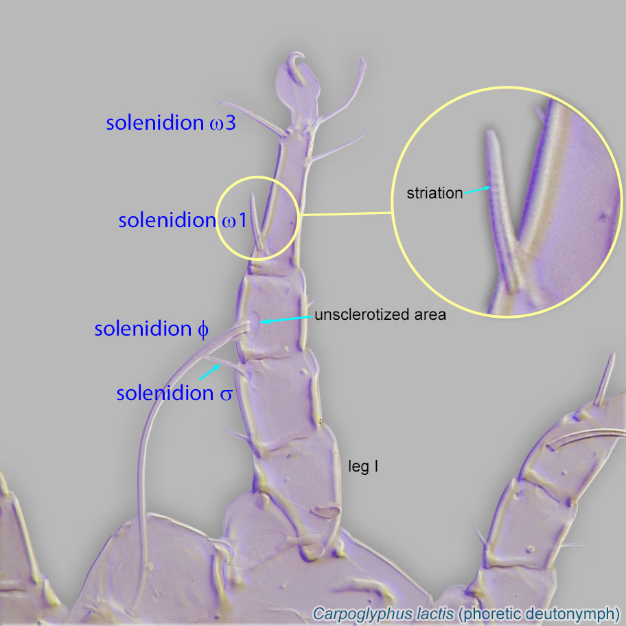
deutonymph: Gnathosomal solenidionsolenidion:
Thin-walled, terminally rounded or pointed filiform or peglike structure that is not birefringent in polarized light (unlike common setae in Acariformes). Often appears striated because of its internal structure. Found on the palpal tarsus on the gnathosoma and may also occur on the tarsus and tibia, less frequently on the genu, and occasionally on the femur of legs I-IV. In Acariformes, leg solenidia often arise from unsclerotized areas.
 present, setae on free palpi absent, and free palpi absent (Fig. 16). Only hysterosomal shieldhysterosomal shield:
Unpaired shield situated on the dorsal hysterosoma.
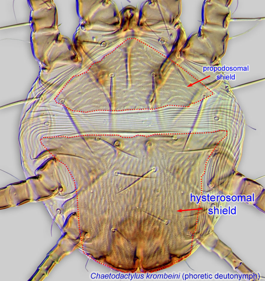 present on idiosomaidiosoma:
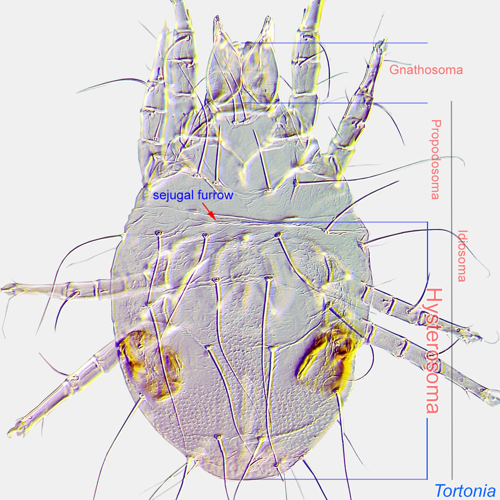
Body not including the gnathosoma.
 ; propodosomal shieldpropodosomal shield:
Unpaired shield situated on the dorsal propodosoma.
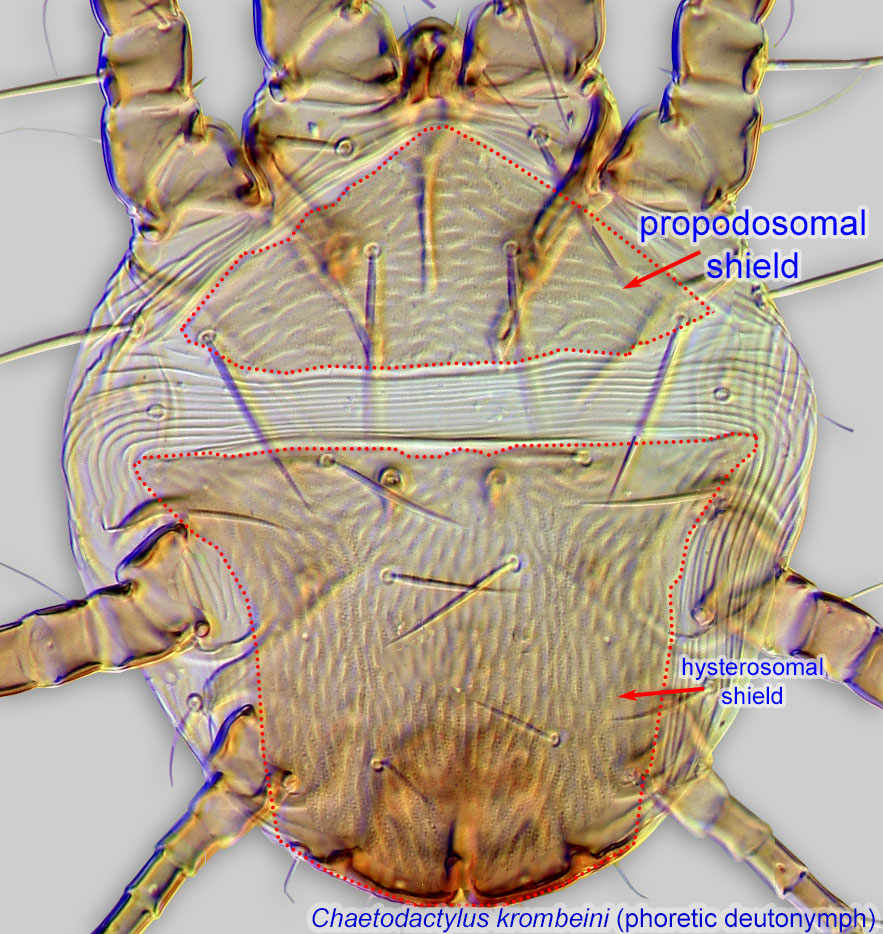 absent (Fig. 7). Cupules im situated at level of trochanterstrochanter:
Leg or palp segment (also known as podomere or palpomere) between femur and coxa. In Acariformes this is the most basal movable leg segment (or podomere) forming a joint with the body.
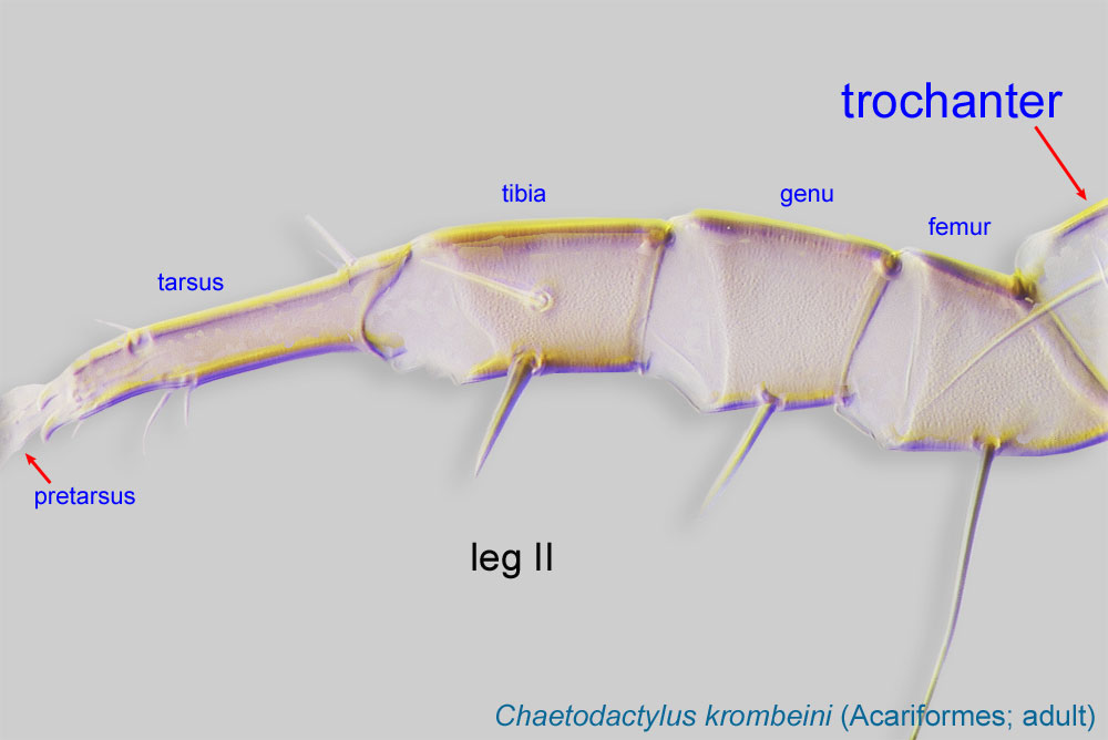 of legs III (Fig. 17).
Adults: Cupules im dorsal (correlates with character in deutonymphdeutonymph:
Ontogenetic stage between protonymph and tritonymph (or adult, if tritonymph is absent). See <a href="index.cfm?pageID=1720">Life stages page</a> for more details.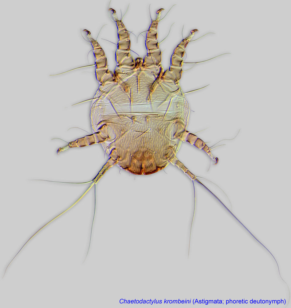 ) (Fig. 18).
Male: Progenital scleritesProgenital sclerite:
A paired or unpaired sclerite situated anterior to the oviporus (female) or genital apparatus (male). In some astigmatid females it is a single, enlarged sclerite, which is often called an epigynum in descriprive works.
lateral to genital capsulegenital capsule:
A caudal or ventrocaudal capsule in males of Pygmephoroidea, Pyemotoidea, and Tarsonemidae formed by consolidated plates H, PS, and Ag. In many mites with short, sclerotized aedeagus flanked by setigenous processes and surrounded by adhesive flange or disc.
(Fig. 19). Progenital scleritesProgenital sclerite:
A paired or unpaired sclerite situated anterior to the oviporus (female) or genital apparatus (male). In some astigmatid females it is a single, enlarged sclerite, which is often called an epigynum in descriprive works.
distinctly separated and not touching each other (Fig. 20). Dorsal supporting scleritesclerite:
A component section of an exoskeleton; a plate forming the skeleton of an arthropod.
not extending posterior to base of aedeagusaedeagus:
An external organ of a male arthropod that is specialized to deliver sperm during copulation.
(Fig. 20).
The key by A. Fain (Fain, 1981Fain, 1981:
Fain, A. 1981. A revision of the phoretic deutonymphs (hypopi) of the genus Sennertia Oudemans, 1905 (Acari, Astigmata, Chaetodactylidae). Systematic Parasitology.3: 145-183.) should be used with caution as it does not include recently described species. A key to New World Sennertia (phoretic deutonymphsdeutonymph:
Ontogenetic stage between protonymph and tritonymph (or adult, if tritonymph is absent). See <a href="index.cfm?pageID=1720">Life stages page</a> for more details., males, and females) is available from the bee-associated mites website and Klimov and OConnor, 2008Klimov and OConnor, 2008:
Klimov, P. B. amp; B. M. OConnor. 2008. Morphology, evolution, and host associations of bee-associated mites of the family Chaetodactylidae (Acari: Astigmata), with a monographic revision of North American taxa. Miscellaneous Publications Museum of Zoology University of Michigan.199: 1-243..
Worldwide, except for Antarctica. Most mites associated with Xylocopa form clear morphology-based New and Old World lineages. The Sennertia zhelochovtsevi species group is an exception. It is distributed in the Old World but is related to the New World lineage. In contrast, species of Sennertia associated with Ceratina do not form distinct, geographically defined lineages.
Most species ocur on small carpenter bees (Ceratina) and large carpenter bees (Xylocopa) of the family Apidae. A few species (Sennertia vaga-group) are associated with Centris (Paracentris) in the Neotropics.
permanentpermanent:
associated exclusively with bees or their close relative, wasps; cannot live without these hosts
disperse on adult bees from one nest to another, either externally (Figs. 9-13) or internally in the genital acarinariumacarinarium:The interactions of Sennertia with their hosts in nests remain largely unknown. There are conflicting accounts suggesting either negative or neutral effect of the mite presence. In the former case, the damage to developing bees was marginal and always substantially less than that of Chaetodactylus.
Feeding stages of the Sennertia vaga-group are unusual because they can disperse on adult bees of the genera Centris (Paracentris) and Xylocopa (Fig. 8). Reproduction and feeding can also occur during dispersal. Species of the Sennertia vaga group probably do not form phoreticphoretic:
Pertaining to phoresy; using another organism (i.e., a host) for dispersal to new habitats. Phoresy can be distinguished from parasitism because feeding typically does not occur during phoresy.
deutonymphsdeutonymph:
Ontogenetic stage between protonymph and tritonymph (or adult, if tritonymph is absent). See <a href="index.cfm?pageID=1720">Life stages page</a> for more details. and disperse exclusively as feeding stages. Observations on these mites inside nests are lacking.
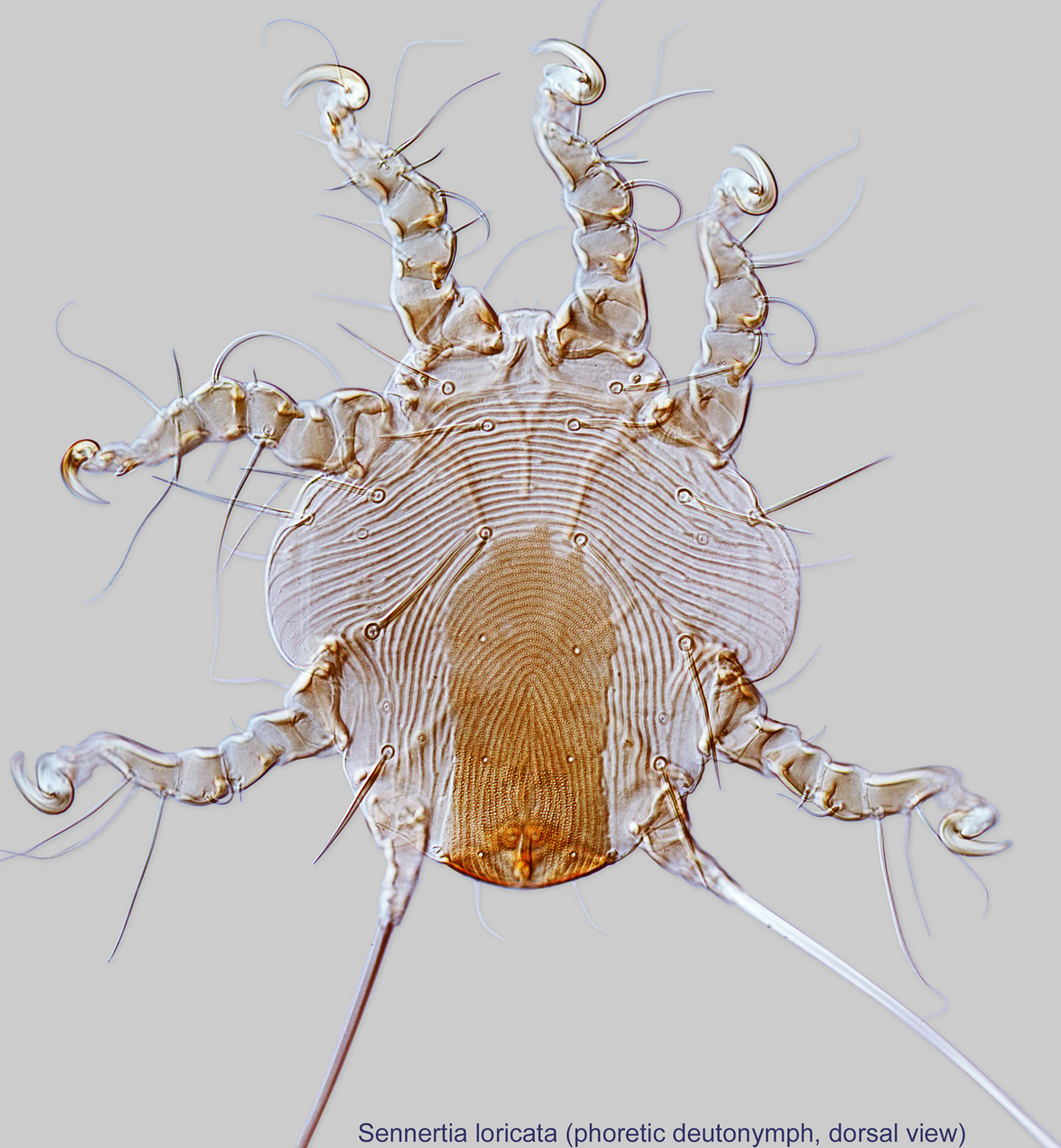
Phoretic phoretic:
Pertaining to phoresy; using another organism (i.e., a host) for dispersal to new habitats. Phoresy can be distinguished from parasitism because feeding typically does not occur during phoresy.
deutonymphsdeutonymph:
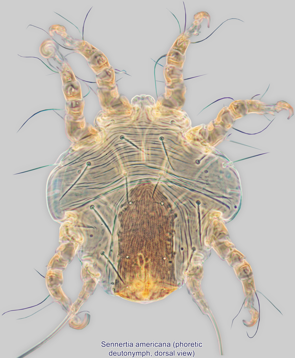
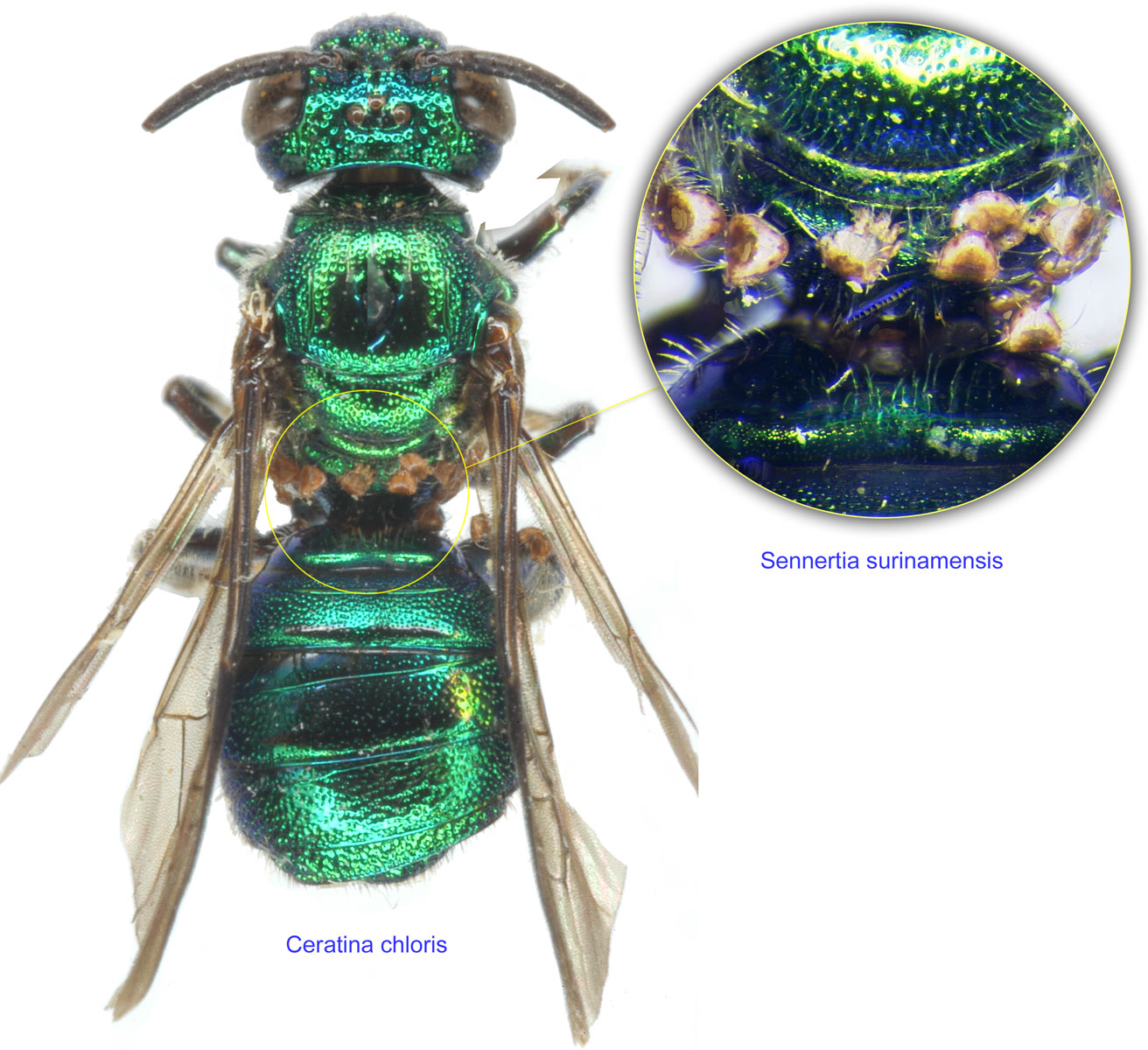
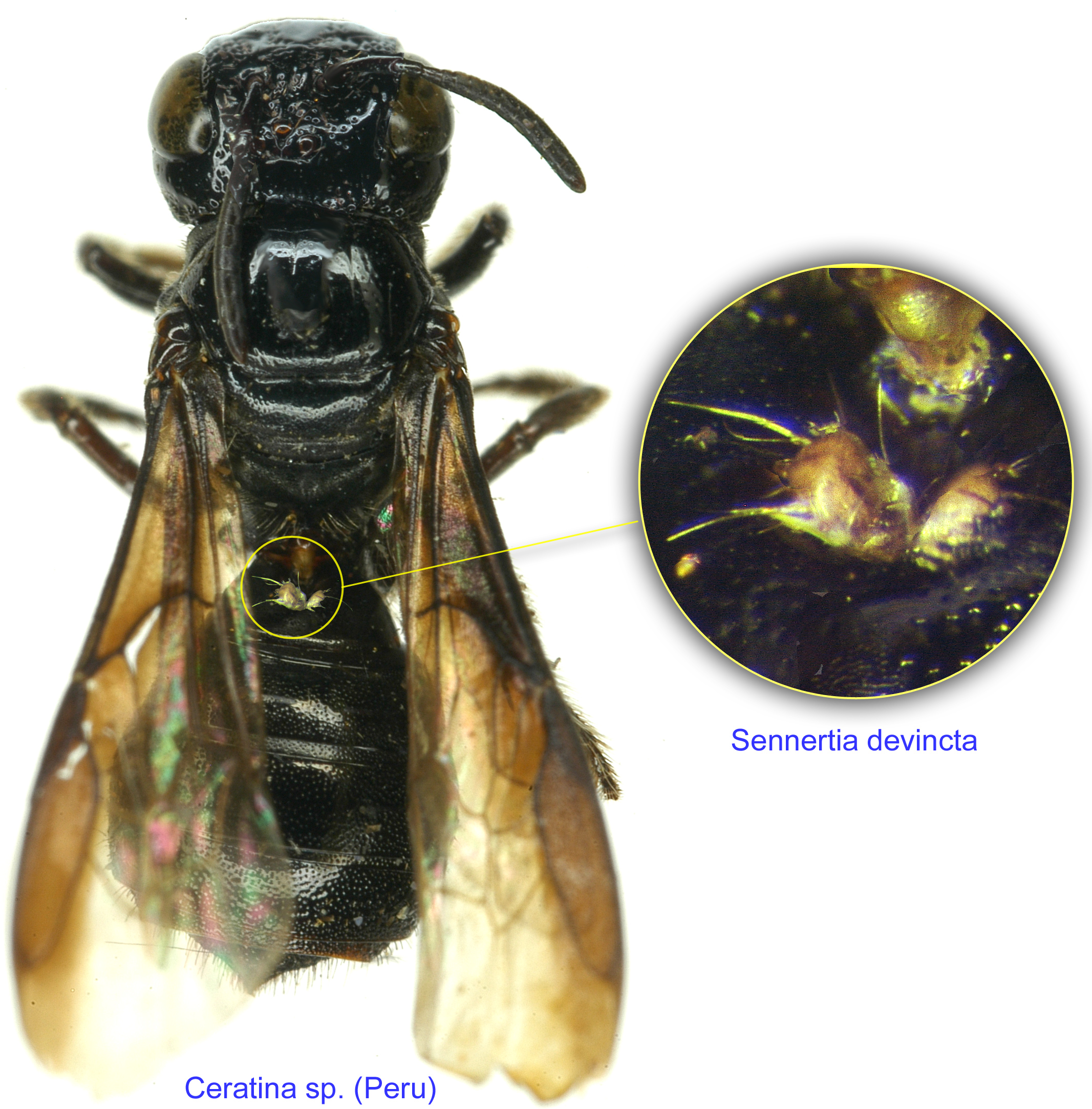
Ontogenetic stage between protonymph and tritonymph (or adult, if tritonymph is absent). See <a href="index.cfm?pageID=1720">Life stages page</a> for more details. (non-feeding stages, Figs. 1-7) disperse on adult bees from one nest to another (Figs. 9-13). Ceratina bees associated with Sennertia (S. sayutara and S. devincta) have acarinariaacarinarium:
A specialized morphological structure that facilitates retention of mites on the body of an organism, typically a bee or wasp.
, indirectly suggesting mutualistic relationship between the mites and the bees (Fig. 14). Some other bees have rudimentary acarinariaacarinarium:
A specialized morphological structure that facilitates retention of mites on the body of an organism, typically a bee or wasp.
that their mites take advantage of for phoresyphoresy:
Attaching to or boarding another organism (i.e., a host) for dispersal to new habitats. Can be distinguished from parasitism because feeding typically does not occur.
. Sennertia koptorthosomae and S. hipposideros, which are associated with Asian large carpenter bees (Xylocopa), have special pouches filled with fungal spores (sporothecae) on the hysterosomahysterosoma:
Division of body posterior to the sejugal furrow, bearing legs III and IV.
 (Klimov and OConnor, 2008Klimov and OConnor, 2008:
Klimov, P. B. amp; B. M. OConnor. 2008. Morphology, evolution, and host associations of bee-associated mites of the family Chaetodactylidae (Acari: Astigmata), with a monographic revision of North American taxa. Miscellaneous Publications Museum of Zoology University of Michigan.199: 1-243.) (Fig. 15), suggesting that they may transfer fungi from one bee nest to another. It is unknown whether these fungi can cause harm to the bee host.
Sennertia cerambycina (Fig. 2) is normally associated with Xylocopa, feeding on the provisioned pollen in the bee nest. Rarely, dead bee eggs and larvae can be found in the infested cells. However, it is unclear if the mite directly or indirectly causes death of eggs or larvae (Vicidomini, 1996Vicidomini, 1996:
Vicidomini, S. 1996. Biologia di Xylocopa (Xylocopa) violacea (L., 1758) (Hymenoptera: Apidae): interazione con Sennertia ( Sennertia ) cerambycina (Acari Chaetodactylidae). Bollettino di Zoologia Agraria e di Bachicoltura.28: 71-76.). Adult bees of Xylocopa violacea extensively infested with S. cerambycina show slow flight, lack of precision in flying trajectories, difficult and imprecise landing, heavy take-off, and reduced feeding rates (Vicidomini, 1999Vicidomini, 1999:
Vicidomini, S. 1999. Nuovi dati sulla distribuzione italiana e sugli ospiti degli acari Chaetodactylidae e nuova ipotesi sul tipo di interazione con gli Xylocopini (Hymenoptera: Apidae). Gli Uccelli d'Italia.24: 97-102.).
Duchemin (1886) reported the death of 30 honey bee (Apis mellifera) hives in France attributable to a mite identified originally as Sennertia cerambycina. The mite infests honey bees visiting sunflowers, eventually causing death of colonies. Similar cases (e.g., death of honey bee colonies with the introduction of sunflowers) were reported in Russia at the end of 19th to the beginning of the 20th century (reviewed in Grobov, 1978Grobov, 1978:
Grobov, O. F. 1978. Kleshchi medonosnoy pchely ( Apis mellifera L.): ikh znachenie i osnovnue printsypy bor'by s kleschchevymi porazheniyami [=Mites of the honeybee ( Apis mellifera L.): their significance and main principles of control of diseases caused by mites]. Doctor of Sciences (Habilitation) Thesis. 536. Moscow: All-Union Institute of Experimental Veterinary, All-Union Academy of Agricultural Sciences.). Experimental data suggesting that the mite is responsible for the death of bees are, however, absent. Furthermore, misidentification is possible here and it may be that another mite species, not belonging to the genus Sennertia, was involved in these cases.
|
Fig. 16. Gnathosomal morphology is a key character separating genera of the family Chaetodactylidae (Centriacarus, Roubikia, Achaetodactylus, Chaetodactylus, and Sennertia) based on phoretic deutonymphs. In particular, Sennertia can be distinguished by its gnathosomal solenidion present and free palpi and setae on free palpi absent; drawing of Achaetodactylus leleupi courtesy of Belgian GTI Focal Point 2009, http://www.taxonomy.be. |
|
|
Fig. 17. Cupules im of deutonymphs of Chaetodactylidae. The genus Sennertia is unique in having cupules im situated at the level of trochanters III, while in other genera these cupules are situated posterior to the level of trochanters III. Also, cupules im are dorsal in Sennertia and ventral in all other genera (but may appear dorsal on slide-mounted specimens of Chaetodactylus, due to flattening). |
|
Authors: P. Klimov, B. OConnor, R. Ochoa, G. Bauchan, A. Redford, J. Scher
Last updated October 2016
tool images at ITP Node
idtools.org