mutualists; feed on microorganisms on provisioned pollen and developing bees and reduce fungal infestation in nest cells via fungicides, increasing survival rates of developing bees.
Anoetus Dujardin, 1842
Superorder Acariformes » Order Sarcoptiformes » Suborder Oribatida » Infraorder Desmonomata » Hyporder Astigmata » Family Histiostomatidae » Genus Anoetus
Hypopus alicola Dujardin, 1849
In old literature, the name Anoetus was used to group species now assigned to genera Histiostoma and Anoetus.
Phoretic phoretic:
Pertaining to phoresy; using another organism (i.e., a host) for dispersal to new habitats. Phoresy can be distinguished from parasitism because feeding typically does not occur during phoresy.
deutonymph: Solenidionsolenidion:
Thin-walled, terminally rounded or pointed filiform or peglike structure that is not birefringent in polarized light (unlike common setae in Acariformes). Often appears striated because of its internal structure. Found on the palpal tarsus on the gnathosoma and may also occur on the tarsus and tibia, less frequently on the genu, and occasionally on the femur of legs I-IV. In Acariformes, leg solenidia often arise from unsclerotized areas.
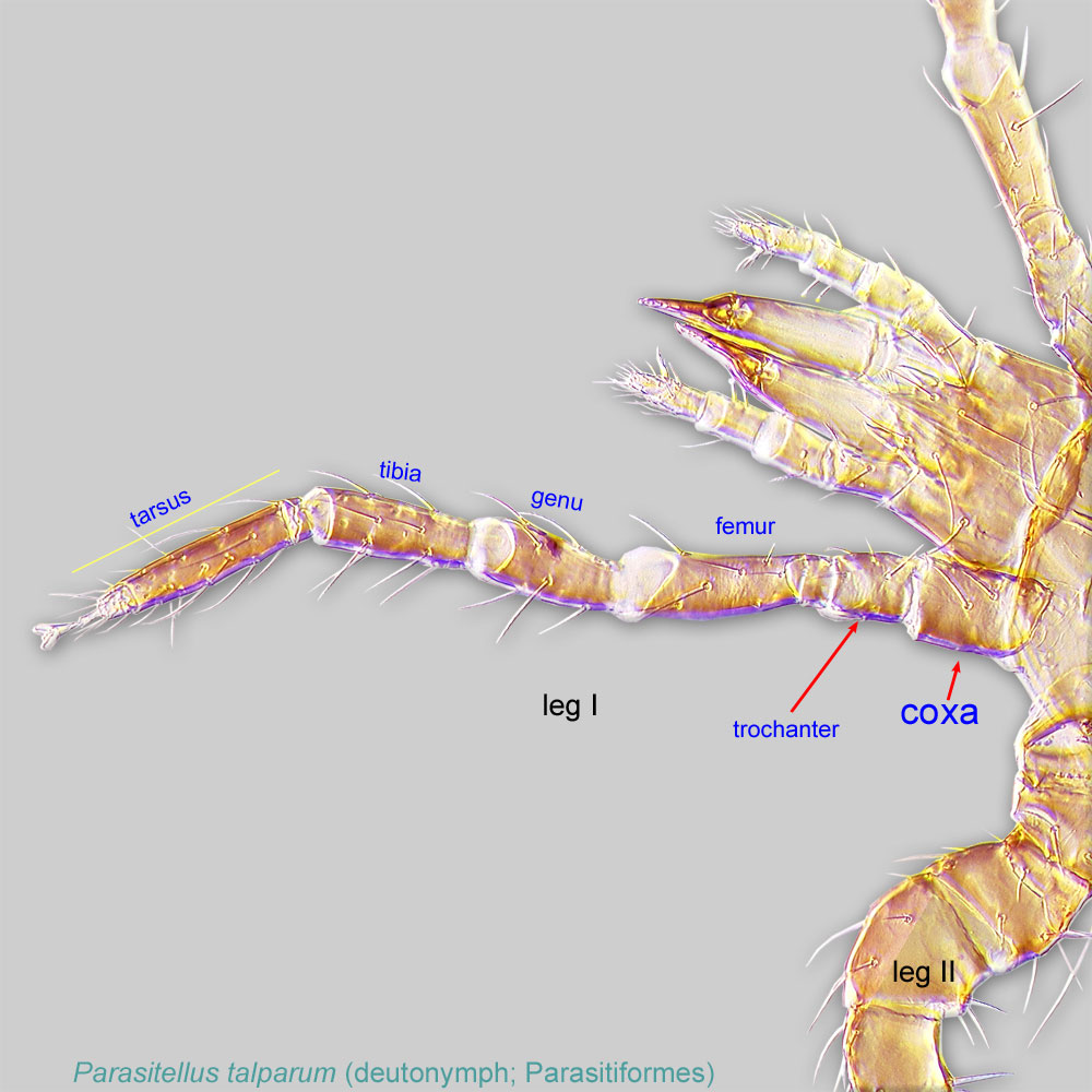
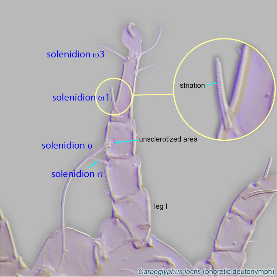 ω1 of leg I positioned directly on tibiatibia:
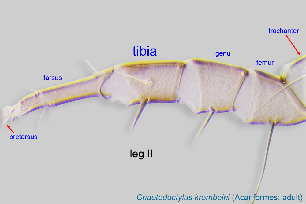
Leg or palp segment (also known as podomere or palpomere) between tarsus and genu.
 , associated with tibial solenidionsolenidion:
Thin-walled, terminally rounded or pointed filiform or peglike structure that is not birefringent in polarized light (unlike common setae in Acariformes). Often appears striated because of its internal structure. Found on the palpal tarsus on the gnathosoma and may also occur on the tarsus and tibia, less frequently on the genu, and occasionally on the femur of legs I-IV. In Acariformes, leg solenidia often arise from unsclerotized areas.
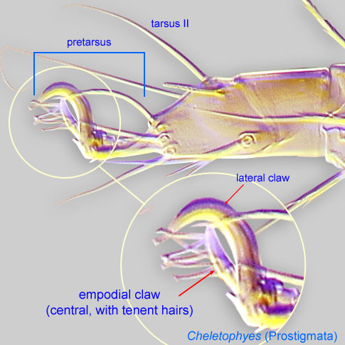
phi (φ) (Fig. 6) and claws III and IV small (as compared to membranous ambulacraambulacrum:
The claws and empodium of the apotele or pretarsus.
), thin, and linear (Fig. 8).
Phoretic phoretic:
Pertaining to phoresy; using another organism (i.e., a host) for dispersal to new habitats. Phoresy can be distinguished from parasitism because feeding typically does not occur during phoresy.
deutonymph: Empodial clawsEmpodial claw:
Claw-like, membranous, or pad-like structure of setal origin. Present only on the pretarsus in Acariformes. In Astigmata, it is the only claw on the pretarsus and often referred to simply as the claw. In the remaining Acariformes, may be accomanied by two lateral claws. Also known as empodium, pretarsal empodium, or central claw.
 I-IV simplesimple:
Of claws or setae; not modified or not bi- or trifurcate at tip.
(not bifurcated) (Figs. 7-8). TarsiTarsus:
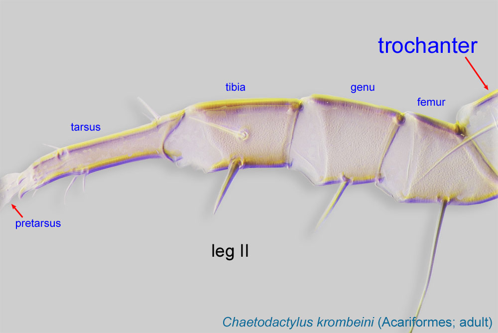
Terminal segment (also known as podomere or palpomere) of legs or palps. In Parasitoformes it can be subdivided into telotarsus and basitarsus.
 III and IV with a weak, flexible region in middle of segment (Fig. 8). PretarsiPretarsus:
Terminal leg or palpal segment distal to tarsus.
III and IV with empodial clawsempodial claw:
Claw-like, membranous, or pad-like structure of setal origin. Present only on the pretarsus in Acariformes. In Astigmata, it is the only claw on the pretarsus and often referred to simply as the claw. In the remaining Acariformes, may be accomanied by two lateral claws. Also known as empodium, pretarsal empodium, or central claw.
(Fig. 8). TrochantersTrochanter:
Leg or palp segment (also known as podomere or palpomere) between femur and coxa. In Acariformes this is the most basal movable leg segment (or podomere) forming a joint with the body.
 I-II without setae. Anterior prodorsumprodorsum:
Dorsal surface of propodosoma.
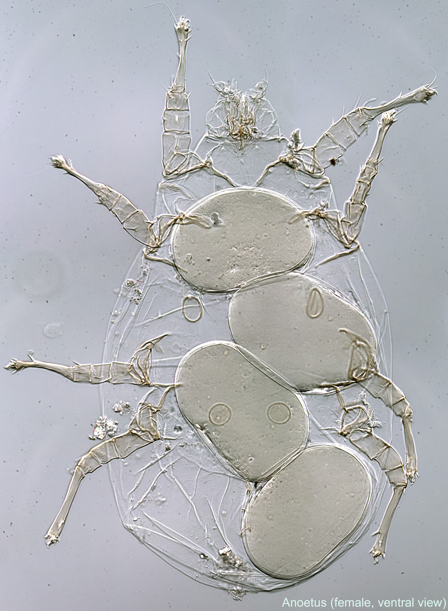
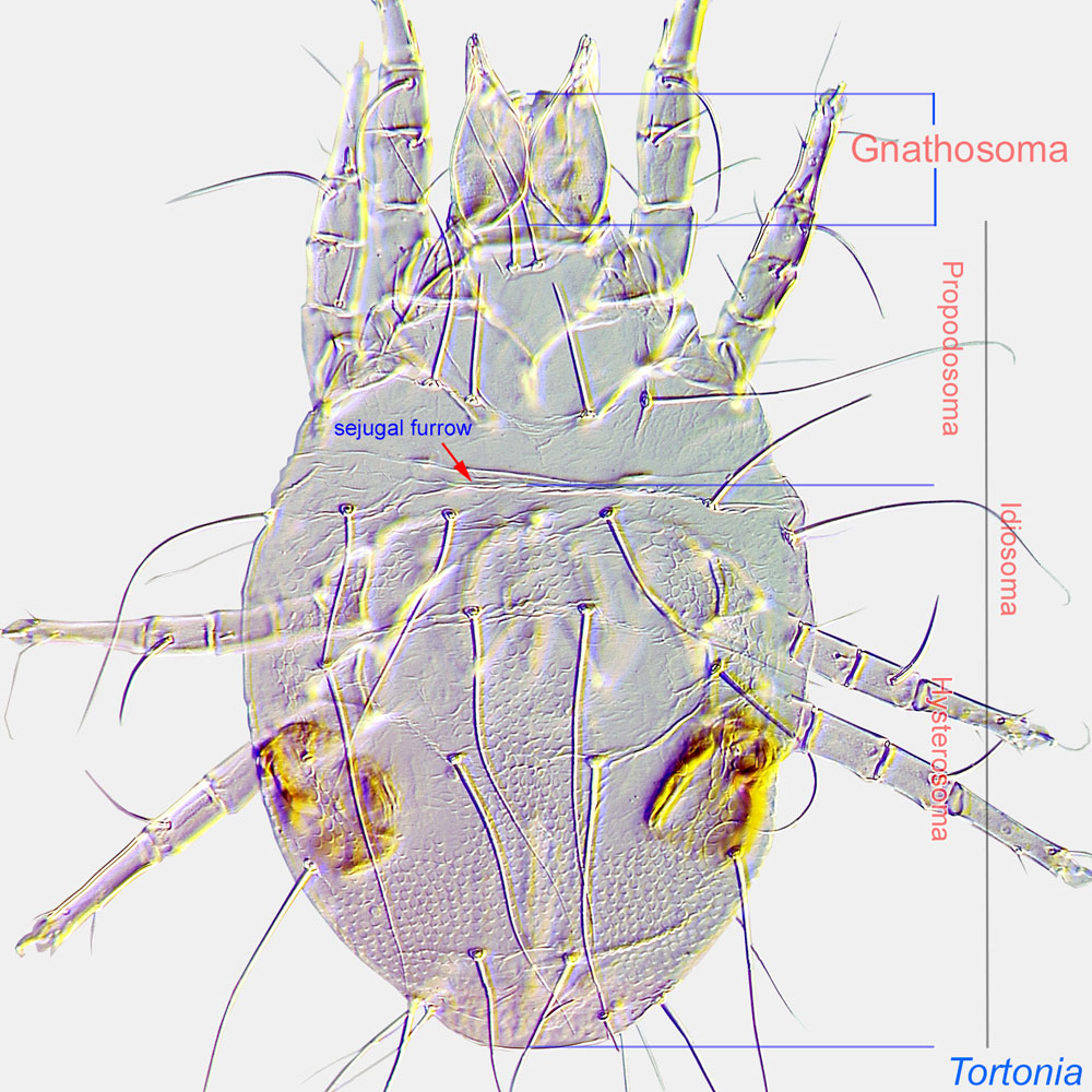
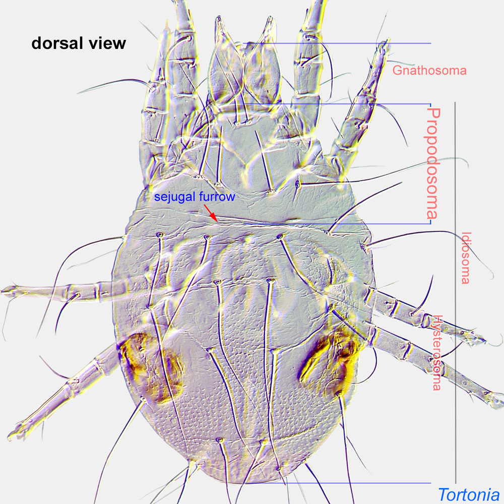 without a pair of brown pigmented areas (Fig. 1). Anterior edge of dorsal hysterosomahysterosoma:
Division of body posterior to the sejugal furrow, bearing legs III and IV.
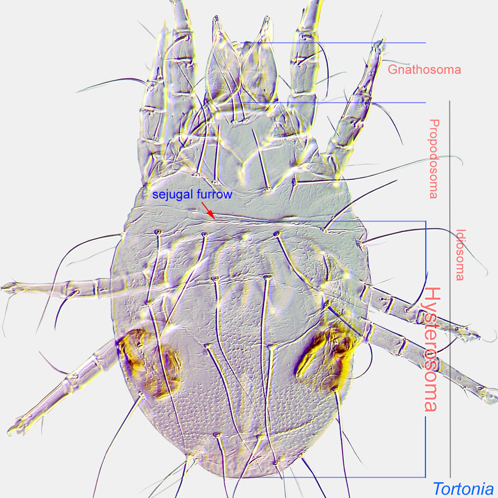 entire, smooth (not scalloped) (Fig. 1). Anterolateral region of hysterosomahysterosoma:
Division of body posterior to the sejugal furrow, bearing legs III and IV.
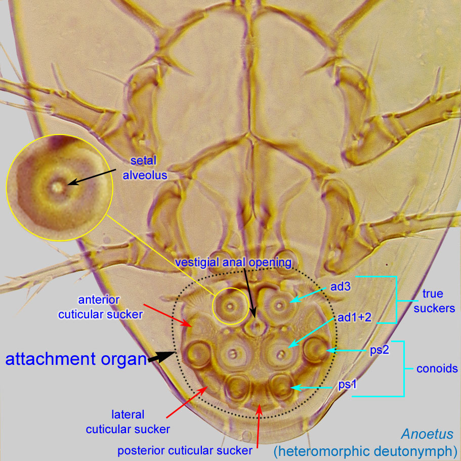
without lens-like organs (Fig. 1). Hysterosomal setae c1, d1 and e1 filiform (not lanceolate) (Fig. 1). Attachment organattachment organ:
Complex unpaired structure in phoretic deutonymphs of Astigmata situated on the posteroventral end of the body that serves for attachment to the host during phoresy. In deutonymphs phoretic on insects, attachment organ consists of a vestigial anal opening and two types of attachment elements: true suckers that create negative pressure and conoids that create adhesive forces. Not to be confused with pedicel.
 wider than long (Fig. 5). Coxal fields IV closed (Fig. 2). Setae of coxacoxa:
In Parasitiformes, most basal leg segment (or podomere) forming a joint with the body. Areas delimited by coxal apodemes are called coxal fields in Astigmata or coxisternal plates in Prostigmata.
 I and III (1a and 3a) either all filiform (Fig. 4) or conoidal. Gnathosomagnathosoma:
Division of body anterior to the propodosoma bearing two pairs of appendages (palps and chelicerae).
 subquadrate to trapezoidal, generally wider than long (Fig. 3).
Female: PretarsiPretarsus:
Terminal leg or palpal segment distal to tarsus.
with membranous ambulacrumambulacrum:
The claws and empodium of the apotele or pretarsus.
short, not bilobed. Posterior genital "rings" not associated with the anus (Fig. 12). Coxal setae I, III, IV present (may be small) (Fig. 12). PalpsPalp:
Second (after chelicera) paired appendage of the gnathosoma. Has a sensory function, but may be variously modified for other functions (e.g., raptorial, attachment to host, or filtering).
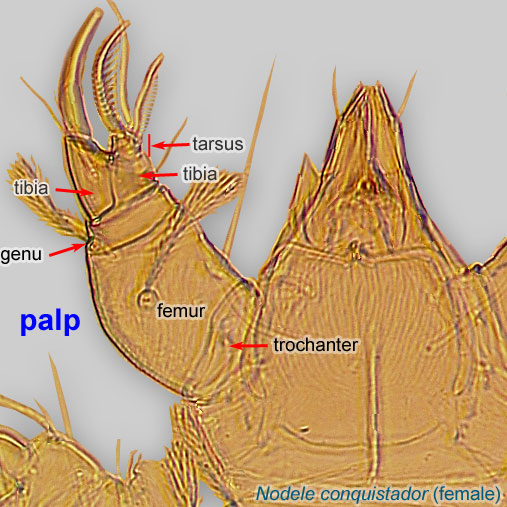 very small and bulbous (Fig. 13). Dorsal scleritesclerite:
A component section of an exoskeleton; a plate forming the skeleton of an arthropod.
very reduced or absent (Fig. 11).
Male: Paranal suckers absent. Aedeagusaedeagus:
An external organ of a male arthropod that is specialized to deliver sperm during copulation.
thin, usually short and often posteriorly directed. Without large spine-like setae at base of aedeagusaedeagus:
An external organ of a male arthropod that is specialized to deliver sperm during copulation.
. With only one pair of genital "rings." Mouthparts vestigial. Leg and body setation strongly reduced.
A dichotomous key to phoreticphoretic:
Pertaining to phoresy; using another organism (i.e., a host) for dispersal to new habitats. Phoresy can be distinguished from parasitism because feeding typically does not occur during phoresy.
deutonymphsdeutonymph:
Ontogenetic stage between protonymph and tritonymph (or adult, if tritonymph is absent). See <a href="index.cfm?pageID=1720">Life stages page</a> for more details.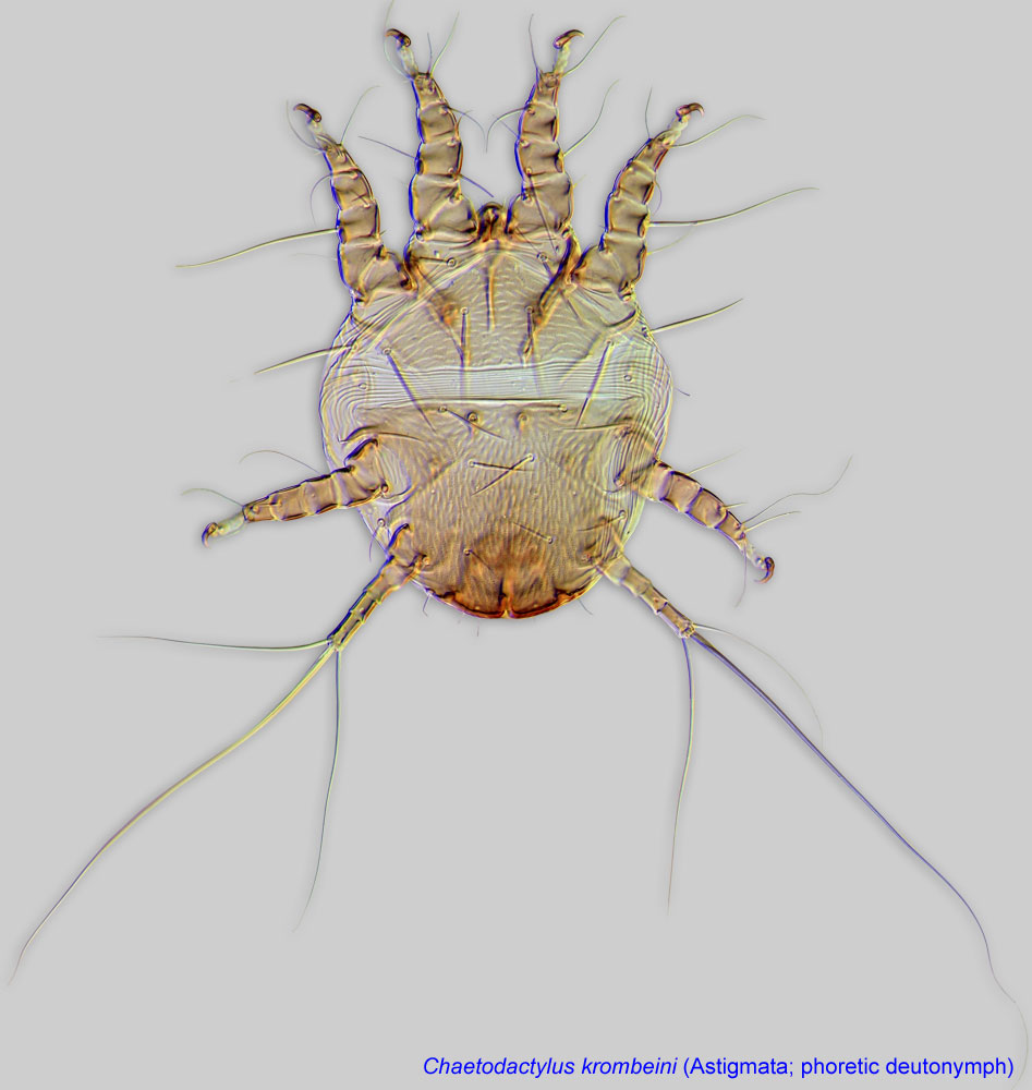 of Palaearctic species is available in Mahunka, 1974Mahunka, 1974:
Mahunka, S. 1974. Beiträge zur Kenntnis der an Hymenopteren lebenden Milben (Acari). 2. Folia Entomologica Hungarica. 27: 99-108.. For Nearctic species, descriptions of both phoreticphoretic:
Pertaining to phoresy; using another organism (i.e., a host) for dispersal to new habitats. Phoresy can be distinguished from parasitism because feeding typically does not occur during phoresy.
stages and females are available in Woodring, 1973Woodring, 1973:
Woodring, J. P. 1973. Four new anoetid mites associated with halictid bees. Journal of the Kansas Entomological Society. 46: 310-327.. However, most species remain undescribed.
Phoretic phoretic:
Pertaining to phoresy; using another organism (i.e., a host) for dispersal to new habitats. Phoresy can be distinguished from parasitism because feeding typically does not occur during phoresy.
deutonymphsdeutonymph:
Ontogenetic stage between protonymph and tritonymph (or adult, if tritonymph is absent). See <a href="index.cfm?pageID=1720">Life stages page</a> for more details. of Anoetus are very similar to those of Histiostoma. In Anoetus, claws III - IV are very small (as compared to width of membranous ambulacraambulacrum:
The claws and empodium of the apotele or pretarsus.
) and thin, linear; gnathosomagnathosoma:
Division of body anterior to the propodosoma bearing two pairs of appendages (palps and chelicerae).
subquadrate. In Histiostoma claws III-IV are large (as compared to width of membranous ambulacraambulacrum:
The claws and empodium of the apotele or pretarsus.
), claw III is claw-like, claw IV either claw-like or linear; gnathosomagnathosoma:
Division of body anterior to the propodosoma bearing two pairs of appendages (palps and chelicerae).
subquadrate or elongated.
Nearctic, Palaearctic (including N. Africa), Neotropical, and Australian regions.
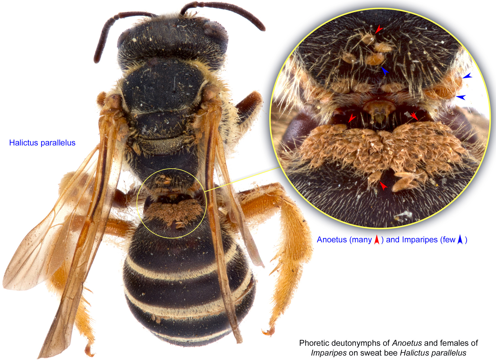
Halictid bees of the genera Lasioglossum, Lasioglossum (Dialictus), Halictus, Augochlora, and Megalopta. In old literature the honey bee, Apis mellifera, was cited as a host of Anoetus alicola; these records need to be verified.
permanentpermanent:
associated exclusively with bees or their close relative, wasps; cannot live without these hosts
disperse on adult bees, often forming large clusters on propodeum or first metasomal tergite T1 of female bees, which function as a rudimentary acarinariumacarinarium:Biological observations are available for Anoetus halictonida, associated with Halictus rubicundus, and an undescribed species of Anoetus associated with Lasioglossum lineatulum (Eickwort, 1979Eickwort, 1979:
Eickwort, G. C. 1979. Mites associated with sweat bees (Halictidae). in (ed.) Recent Advances in Acarology, Vol. 1. ed. J. G. Rodriguez, 575-581. New York: Academic Press.; Eickwort, 1994Eickwort, 1994:
Eickwort, G. C. 1994. Evolution and life-history patterns of mites associated with bees. In Mites: Ecological and Evolutionary Analyses of Life-History Patterns, ed. M. A. Houck, 218-251. New York: Chapman amp; Hall.):
Phoretic phoretic:
Pertaining to phoresy; using another organism (i.e., a host) for dispersal to new habitats. Phoresy can be distinguished from parasitism because feeding typically does not occur during phoresy.
deutonymphsdeutonymph:
Ontogenetic stage between protonymph and tritonymph (or adult, if tritonymph is absent). See <a href="index.cfm?pageID=1720">Life stages page</a> for more details. attach to the host's wings. On female bees they also often attach in shingle-like rows to metasomal tergum I or II, while on males they may occur on the venter of the head, thorax, and sometimes the metasoma. Phoreticphoretic:
Pertaining to phoresy; using another organism (i.e., a host) for dispersal to new habitats. Phoresy can be distinguished from parasitism because feeding typically does not occur during phoresy.
deutonymphsdeutonymph:
Ontogenetic stage between protonymph and tritonymph (or adult, if tritonymph is absent). See <a href="index.cfm?pageID=1720">Life stages page</a> for more details. move from their phoreticphoretic:
Pertaining to phoresy; using another organism (i.e., a host) for dispersal to new habitats. Phoresy can be distinguished from parasitism because feeding typically does not occur during phoresy.
host onto the provision mass in a new cell, and transform into tritonymphstritonymph:
Ontogenetic stage between the deutonymph and adult. Sometimes this stage is absent and deutonymph transforms directly to the adult stage. See <a href="index.cfm?pageID=1720">Life stages page</a> for more details.
that soon molt into adults.
The adult females stay on the provision mass and swell greatly. The males remain very small and crawl onto the dorsum of the female when the host larva is about half grown. It is possible that these males develop rapidly from the first unfertilized eggs laid by the females, perhaps skipping all nymphal instars, and then mate with females of the parental generation. The inseminated females then lay fertilized eggs, all of which develop into females.
Those female eggs are laid on the bee nest cell wall and bee larvae, and mite larvae and protonymphsprotonymph:
Ontogenetic stage between larva and deutonymph. See <a href="index.cfm?pageID=1720">Life stages page</a> for more details.
feed on the surfaces of the bee larvae and pupae, apparently consuming microorganisms; they do not harm the bees upon which they feed. Mite protonymphsprotonymph:
Ontogenetic stage between larva and deutonymph. See <a href="index.cfm?pageID=1720">Life stages page</a> for more details.
begin to appear when the bee larva pupates, when they crawl on the pupa, especially ventrally. Molting to phoreticphoretic:
Pertaining to phoresy; using another organism (i.e., a host) for dispersal to new habitats. Phoresy can be distinguished from parasitism because feeding typically does not occur during phoresy.
deutonymphsdeutonymph:
Ontogenetic stage between protonymph and tritonymph (or adult, if tritonymph is absent). See <a href="index.cfm?pageID=1720">Life stages page</a> for more details. begins about half way through the pupal stadium. Phoreticphoretic:
Pertaining to phoresy; using another organism (i.e., a host) for dispersal to new habitats. Phoresy can be distinguished from parasitism because feeding typically does not occur during phoresy.
deutonymphsdeutonymph:
Ontogenetic stage between protonymph and tritonymph (or adult, if tritonymph is absent). See <a href="index.cfm?pageID=1720">Life stages page</a> for more details. preferentially cluster on the pupa's dorsal propodeal surface and about the wing bases. They transfer to the adult bee when it emerges. DeutonymphsDeutonymph:
Ontogenetic stage between protonymph and tritonymph (or adult, if tritonymph is absent). See <a href="index.cfm?pageID=1720">Life stages page</a> for more details. remain on their female hosts while the hosts hibernate, and they detach from host bees as new cells are constructed in the spring.
In addition, field observations and laboratory experiments on Anoetus associated with halictid bees of the genus Megalopta showed that mites reduce fungal infestation of the nest cells, thus decreasing bee mortality (Biani et al., 2009Biani et al., 2009:
Biani, N. B., U. G. Mueller amp; W. T. Wcislo. 2009. Cleaner Mites: Sanitary Mutualism in the Miniature Ecosystem of Neotropical Bee Nests. American Naturalist. 173: 841-847.).
Authors: P. Klimov, B. OConnor, R. Ochoa, G. Bauchan, A. Redford, J. Scher
Last updated October 2016
tool images at ITP Node
idtools.org