


This tool is a response to significant pollinator losses that pose a threat to agricultural crops and native plants, as summarized in a Presidential Memorandum of June, 2014, and expanded upon in the Pollinator Research Action Plan. The loss of bee pollinators is a result of many causes, including habitat loss, exposure to pesticides, and introduced diseases and parasites. Bee Mite ID: Bee-Associated Mite Genera of the World strives to help reduce pollinator losses through accurate identification of bee-asssociated mites that may be harmful to their hosts.
Bee Mite ID can be used to:
Bee Mite ID contains an interactive key, fact sheets, an image gallery, and abundant supporting information. The interactive key allows you to choose characters to obtain a list of mite genera possibly matching your specimen. Consult fact sheets to find images and information for a particular mite genus, including its harmfulness rating, diagnostic characters, and information about its bee hosts and the biology of their association. Use the filterable image gallery of over 850 mite images to compare images from multiple taxa.
A number of the components of this tool were specifically designed to help non-experts. Mite identification generally relies on microscopic characters, so slide-mounted specimens are usually required. In order to use this tool's key, fact sheets, and image gallery effectively, consult the preparation and photography page to learn how to slide-mount mites. There are also seven quick reference guides showing mites that disperse on the seven bee genera most often used for pollination. Many of these mites can be distinguished by shape without a compound microscope; see the quick reference page for tips on using these guides. The glossary provides definitions of mite terminology and is illustrated. It is supplemented by a mite morphology page that provides an overview, specifics on setae, solenidiasolenidion:
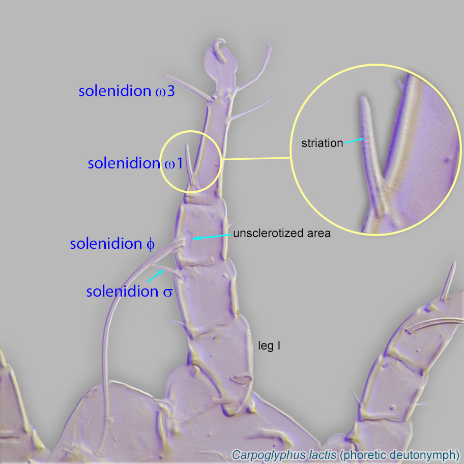
Thin-walled, terminally rounded or pointed filiform or peglike structure that is not birefringent in polarized light (unlike common setae in Acariformes). Often appears striated because of its internal structure. Found on the palpal tarsus on the gnathosoma and may also occur on the tarsus and tibia, less frequently on the genu, and occasionally on the femur of legs I-IV. In Acariformes, leg solenidia often arise from unsclerotized areas.
 , and suckers, and ways to determine sex. See the life stages page to learn even more about mites, and the bee morphology page, which points out bee body features referred to throughout the tool.
If you know the bee's genus name, you can use it to reduce the set of taxa remaining in the interactive key, in a fact sheet search to find fact sheets mentioning the bee as a mite host, or to take advantage of a quick reference guide if your bee is one of the seven.
Tips on using the fact sheets
Authors: P. Klimov, B. OConnor, R. Ochoa, G. Bauchan, A. Redford, J. Scher
Last updated October 2016
tool images at ITP Node
idtools.org
