probably mutualistic because bee hosts developed acarinariaacarinarium:
A specialized morphological structure that facilitates retention of mites on the body of an organism, typically a bee or wasp.
to transfer mites, though evidence somewhat contradictory
Dinogamasus Kramer, 1898
Superorder Parasitiformes » Order Mesostigmata » Suborder Monogynaspida » Hyporder Dermanyssiae » Family Laelapidae » Genus Dinogamasus
Dinogamasus crassipes Kramer, 1898
Greenia Oudemans, 1901; Greeniella Banks, 1904; Dolaea Oudemans, 1912
Adult: Sternal seta st3 situated off sternal shieldsternal shield:
A shield in the anterior intercoxal region of parasitiform mites that bears one or more pairs of sternal setae.
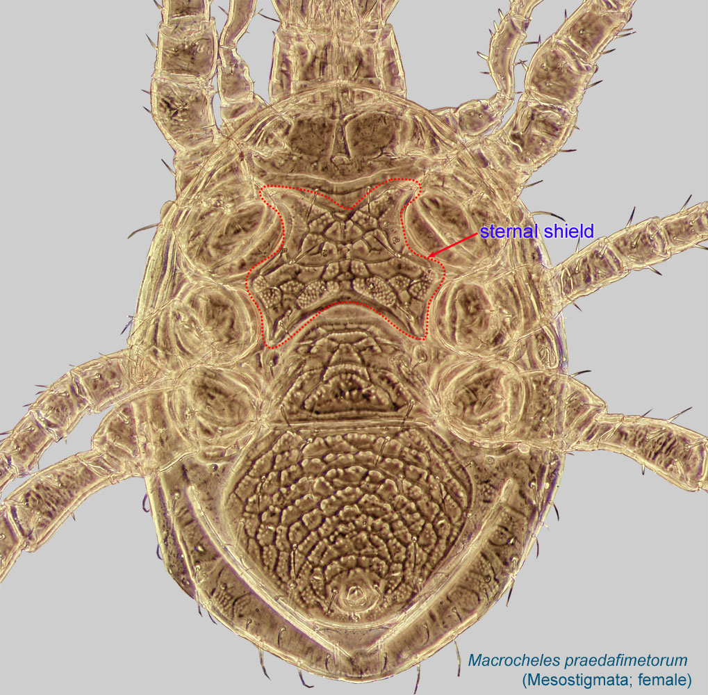 (Figs. 2, 3). Sternal and epigynal shieldsepigynal shield:
A shield protecting the female genital opening. Well-developed in Mesostigmata. Also known as epigynial shield.
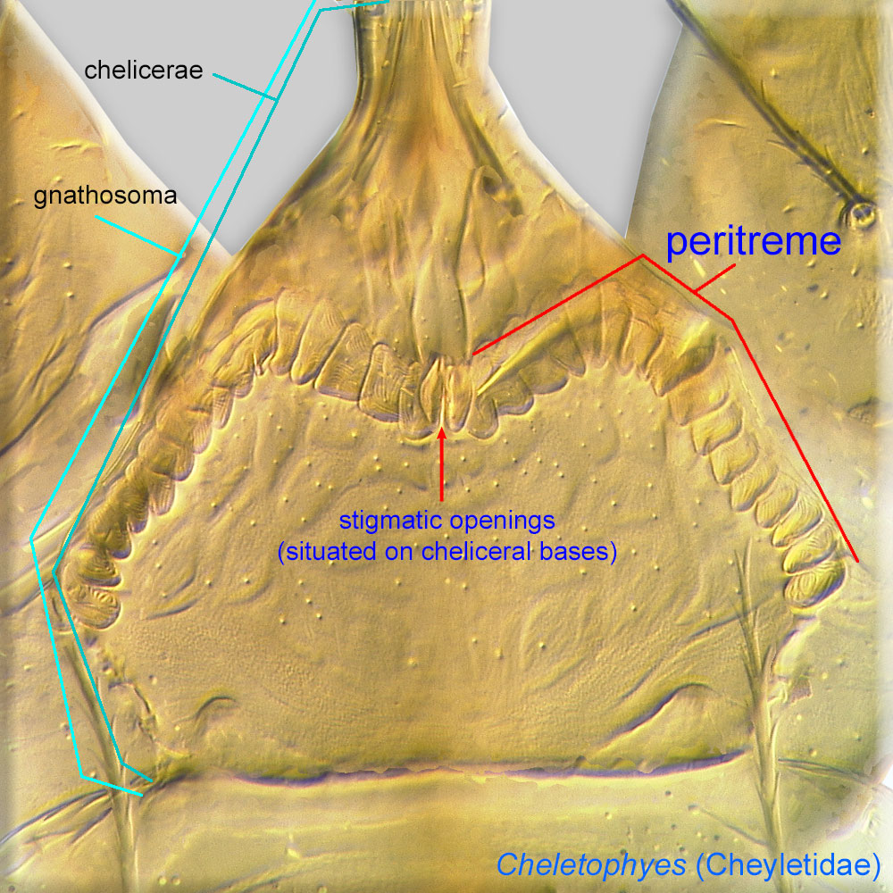
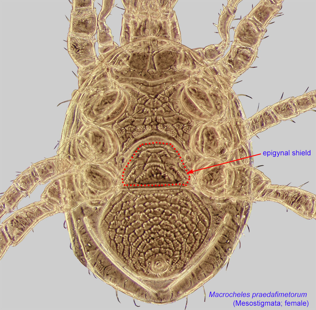 distinctly separated (Figs. 2, 3). PeritremesPeritreme:
Paired, tubular, elaborated extensions of a tracheal system associated with stigmatic openings. Can be chambered, arch-like, and situated on the bases of chelicerae as in Cheyletidae (Prostigmata) or, in Mesostigmata, linear and situated on the lateral sides of the body.
 reduced or absent, not elongated (Figs. 4, 5). Dorsum hypertrichoushypertrichous:
With many irregularly arranged setae.
, especially at body margins (Fig. 1).
A dichotomous key is available in Lundqvist, 1998Lundqvist, 1998:
Lundqvist, L. 1998. Taxonomic revision of the genus Dinogamasus (Acari: Mesostigmata: Laelapidae). Entomologica Scandinavica Supplement. 54:1-109..
Old World tropics
large carpenter bees (Xylocopa)
permanentpermanent:
associated exclusively with bees or their close relative, wasps; cannot live without these hosts
The biology of Dinogamasus in the bee nest is not entirely understood. Based on observations of Dinogamasus braunsi and D. villosior (Skaife, 1952Skaife, 1952:
Skaife, S. H. 1952. The yellow-banded carpenter bee Mesotrichia caffra Linn., and its symbiotic mite, Dinogamasus braunsi Vitzthum. The Journal of the Entomological Society of Southern Africa.15: 63-76.; Madel, 1975Madel, 1975:
Madel, G. 1975. Vergesellschaftung der Milbenart Dinogamasus villosior mit der ostafrikanischen Holzbiene Xylocopa flavorufa (Acarina: Laelaptidae / Hymenoptera: Xylocopidae). Entomologica Germanica. 1: 144-150.), the mites apparently feed on exudates produced by the developing bee larvae and pupae, or on fungi and other microorganisms associated with them. Mites could not survive on pollen in laboratory conditions, and they cannot pierce the soft cuticule of bee larvae with their cheliceraechelicera:
Anterior, paired appendage of the body. Primary organ for food acquisition, adapted for chewing, piercing, tearing, sucking, or filtering.
 .
.
After eclosion, the adult bee breaks the cell partition and spends its first days in the nest. During this time, adult females of Dinogamasus enter the metasomal acarinariumacarinarium:
A specialized morphological structure that facilitates retention of mites on the body of an organism, typically a bee or wasp.
of adult female bees. The metasomal acarinariaacarinarium:
A specialized morphological structure that facilitates retention of mites on the body of an organism, typically a bee or wasp.
probably serve for protected dispersal for newly emerged adult females during the period of bee dispersal from the parent nest. On average, Xylocopa flavorufa carries 21 mites of D. villosior (maximum 35) in the metasomal acarinariumacarinarium:
A specialized morphological structure that facilitates retention of mites on the body of an organism, typically a bee or wasp.
, and X. caffra carries 10-12 (maximum 18). Since only female bees build nests, dispersal on males will result in mites dying without establishing colonies in new nests.
Experimental evidence is contradictory. It has been found that mites do not harm the pupa, but bees do not benefit from the mite presence either (Skaife, 1952Skaife, 1952:
Skaife, S. H. 1952. The yellow-banded carpenter bee Mesotrichia caffra Linn., and its symbiotic mite, Dinogamasus braunsi Vitzthum. The Journal of the Entomological Society of Southern Africa.15: 63-76.). Alternatively, it has been demonstrated that bee pupae lose weight proportionally to the number of mites (Watmough, 1974Watmough, 1974:
Watmough, R. H. 1974. Biology and behaviour of carpenter bees in southern Africa. Journal of the Entomological Society of Southern Africa. 37:26-281.), indicating that these mites can be harmful to their hosts. On the other hand, the presence of acarinariaacarinarium:
A specialized morphological structure that facilitates retention of mites on the body of an organism, typically a bee or wasp.
is indicative of mutualistic associations. Given the contradictory evidence, more research is needed to elucidate the nature of mite-bee interactions in this system.
|
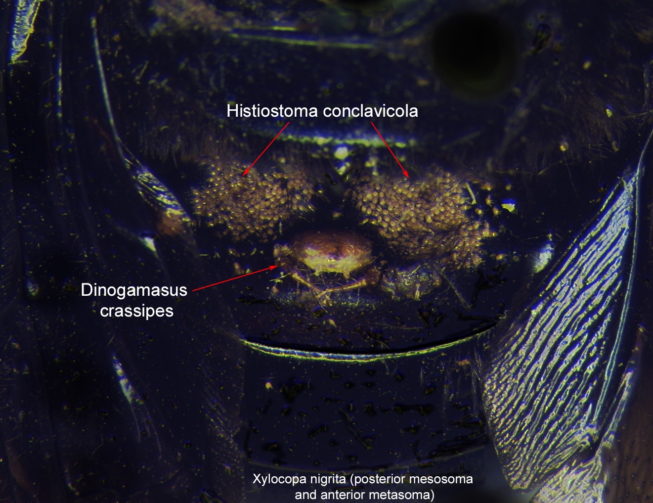
Fig. 7. Posterior mesosoma and anterior metasoma of the bee Xylocopa nigrita. First metasomal tergite of the bee has two large clusters of phoretic deutonymphs of Histiostoma conclavicola and a single female of Dinogamasus crassipes. The anterior body of D. crassipes is partially inserted in the metasomal acarinarium and is hidden, while the posterior body is visible from the outside; photo by Barry OConnor, University of Michigan. |
|
|
Fig. 9. First metasomal tergite of the large carpenter bee, Xylocopa nigrita. There are two large clusters of phoretic deutonymphs of Histiostoma conclavicola and a single female of Dinogamasus crassipes. The anterior body of D. crassipes is partially inserted in the metasomal acarinarium and is hidden, while the posterior body and legs are visible from the outside; photo by Barry OConnor, University of Michigan. |
Authors: P. Klimov, B. OConnor, R. Ochoa, G. Bauchan, A. Redford, J. Scher
Last updated October 2016
tool images at ITP Node
idtools.org