
About flat mites
Collecting the specimens is the easy part of working with mites. The hard part is getting to know your mite. A flat mite looks very different in real life under a stereoscope compared to after it has been squashed flat and mounted on a slide. Very often the natural 3-D shape of the mite is lost and the flat monoplane distorted image that results is what we are left with to produce identification tools.
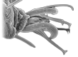
This distorted flat image of a mounted mite can vary due to many factors, both biotic and abiotic. With Brevipalpus for example, if your specimen is a young female that has recently moulted, or a female that has not yet fed, the mounting media will soften and clear the specimen significantly more so than it would an older darker female (Fig. 1). In the process, many of the key characters on an over-cleared mite will be “ironed out” or washed away, resulting in an inability to identify the mite or more commonly a misidentification. This problem can be avoided by mounting several specimens with different colour patterns, if available, and not clearing the specimens too long prior to mounting. If multiple specimens are not available, and you have only young or pale coloured specimens, then cut the clearing time in half prior to mounting.
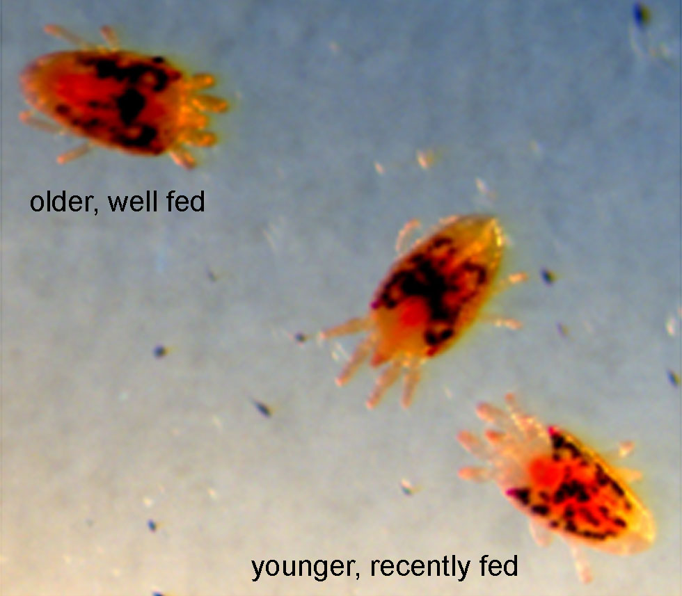
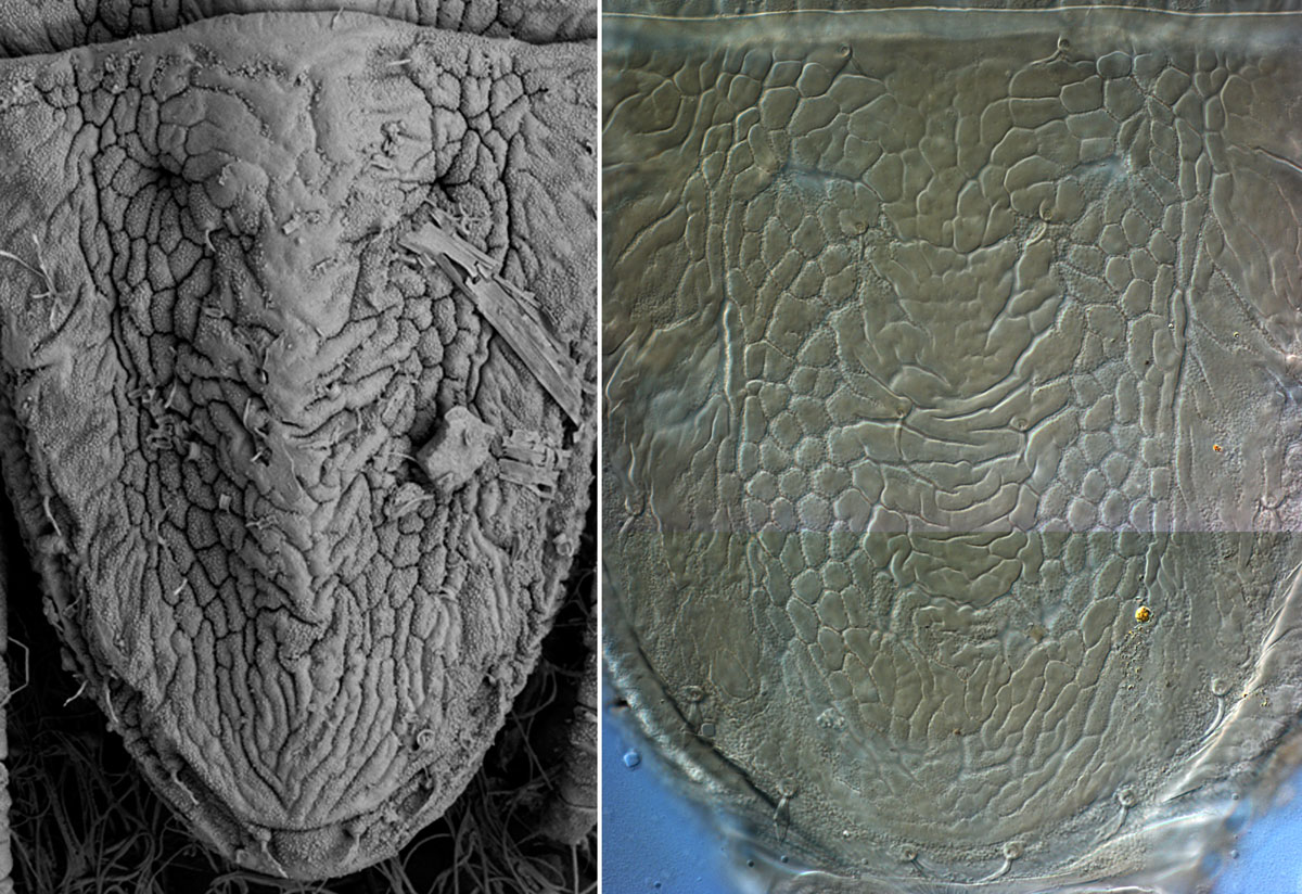
Strange as it may seem, flat mites are not actually flat. Brevipalpus mites usually have two distinct longitudinal furrows on either side of the dorsum that run the full length of their body (Fig. 2a). This pattern can alternatively be interpreted as a broad ridge or keel that runs down the centre of the body. This 3-D dorsal sculpturing is clearly lost during mounting (Fig. 2b); though if the specimen is not squashed flat the furrows will remain.
-
Figure 1: Brevipalpus females -
Figure 2a (left): furrows visible in LTSEM image, Figure 2b (right) 3-D sculpturing lost during slide mounting -
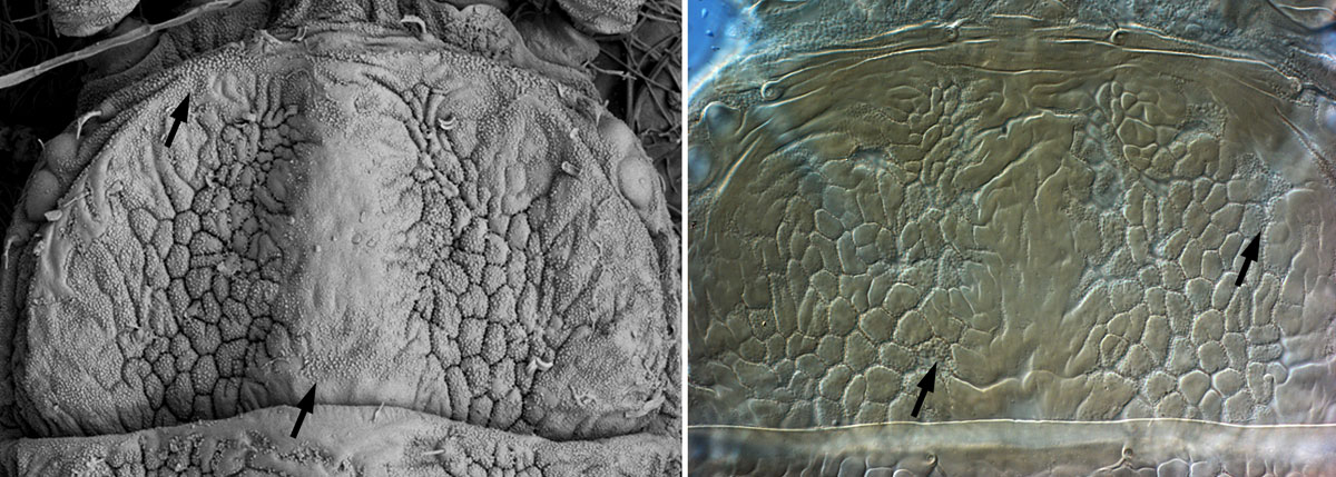
Figure 3a (left): reticulate sculpturing visible in LTSEM image, Figure 3b (right) 3-D sculpturing altered during slide mounting
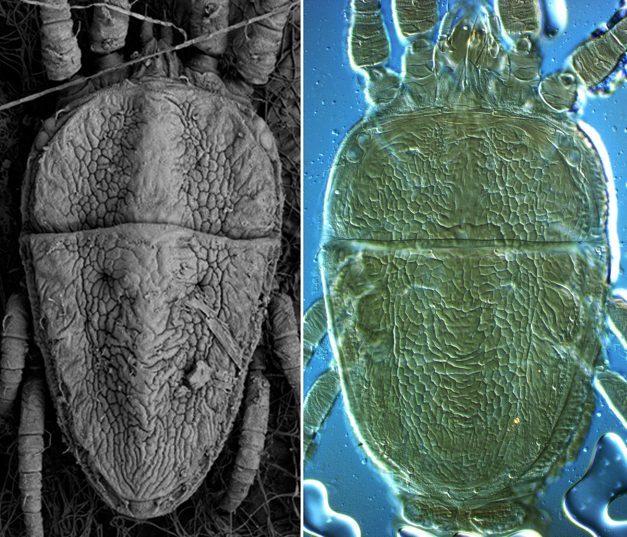
There are several significant consequences of the flattening process: 1) the entire body becomes broader and posterior margin of the body becomes very rounded (Figs 2b, 4b), in contrast to the more pointed body in real life (Figs 2a, 4a); 2) the deep 3-D reticulate sculpturing and deep folds evident on the mite cuticle become much more shallow and nearly 2-D when mounted, and this alters how we interpret the patterns (Fig. 3a,b); 3) each cell in the reticulate pattern becomes broader when flattened, altering the overall appearance of the reticulation; 4) lateral setae appear to be inserted in a more dorsal position; 5) dorsal pores lose their natural shape, and are often less dramatic in appearance (Fig. 4a,b).
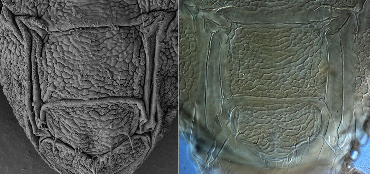
The pattern on the ventral and genital shields of a mounted specimen remains very close to that found in real life, although the deeply folded cuticle surrounding the shields becomes unfolded and flattened in a mounted specimen (Fig. 5a,b).
-
Figure 4a (left): real life appearance in LTSEM image, Figure 4b (right) body shape altered during slide mounting -
Figure 5a (left): real life appearance in LTSEM image, Figure 5b (right) body shape altered during slide mounting -
Figure 6: cuticular microplates
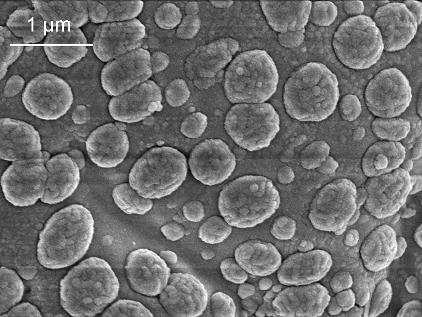
Brevipalpus immatures are soft and have not yet developed the strengthened plates present on the adult. Adult Brevipalpus, on the other hand, tend to be strengthened with plates ventrally and a thickened sculptured dorsum. When we look more closely at the cuticle of Brevipalpus, we see that it is further strengthened by a full-body covering of tiny microplates (Fig. 3a,b – indicated by arrows; Fig. 6). Preliminary research on Brevipalpus and other flat mite genera (Ochoa, Beard & Bauchan in prep.) indicates that the construction of individual cuticular microplates and their resulting shapes are species specific (Fig. 6) and that these patterns have potential as additional characters for species separation in the future.
