

About flat mites
DIC vs. phase contrast
It is a good idea to approach phase contrast and DIC (differential interference contrast) as complementary techniques when you are examining tissues and cells. The two techniques can be employed together to investigate the morphology of a specimen. In many cases, each technique will reveal specific details about a particular specimen that is not apparent from observing images captured by the other technique. However, when dealing with mites, we are interested mainly in the morphology and shape of surface structures, and DIC offers the best interpretation for these.
The greatest fundamental distinction between DIC and phase contrast is the optical basis upon which images are formed. Specimens examined with these methods produce images that are quite different in appearance and character. The use of both images can be greatly advantageous for interpretation of structures; however, depending on what you are examining, the use of only one or the other can result in significantly different interpretations.
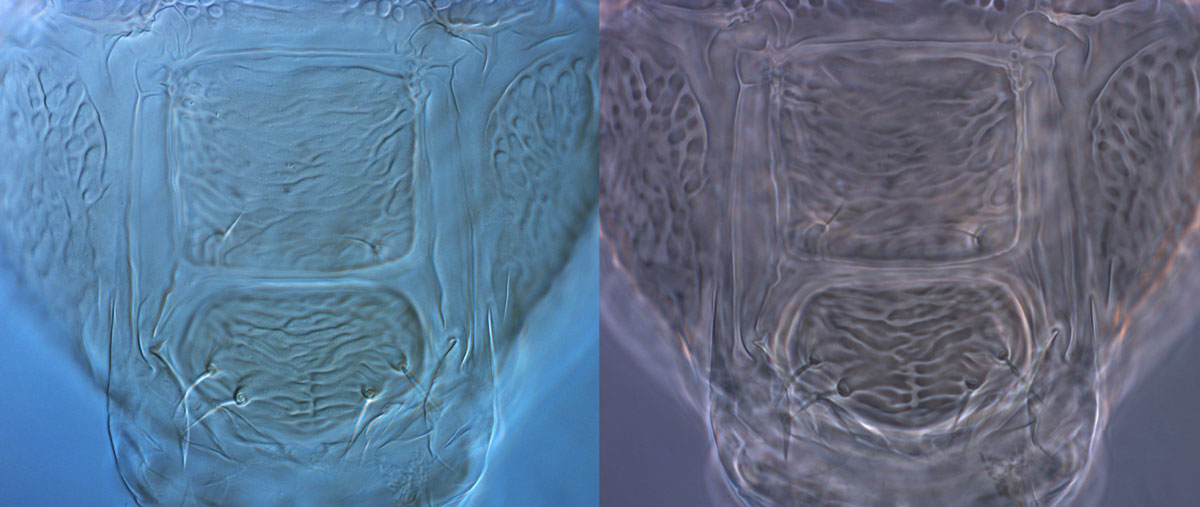
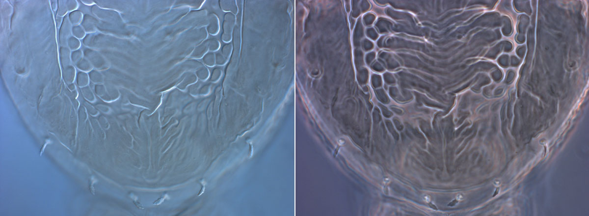
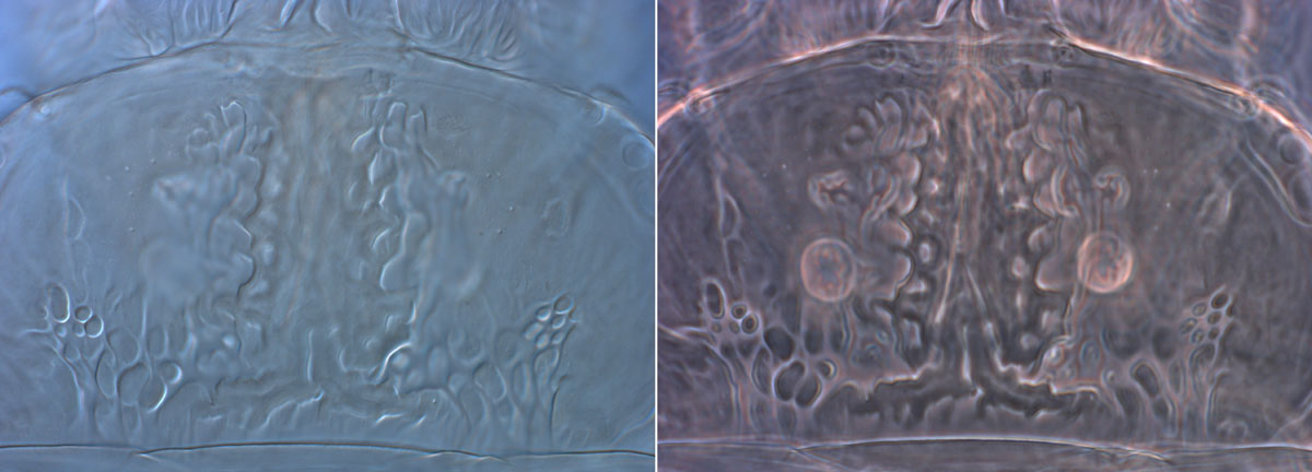
Phase contrast microscopy produces image intensity values that vary as a function of specimen optical path length magnitude. Very dense or thicker regions, such as setae, have large path lengths and hence appear darker than the background. Thinner specimen features, such as membranes, have relatively low thickness values, and so appear much lighter than the background (Figs 1, 2). In addition, internal structures are often visible when you are focused on the external surface of specimen. While this can be advantageous in certain circumstances, it is not so when examining mites, and it often contributes to confusion and results in misinterpretation (Fig. 3).
-
Figure 1: DIC view on left, phase contrast view on right -
Figure 2: DIC view on left, phase contrast view on right -
Figure 3: DIC view on left, phase contrast view on right; internal structures visible in phase contrast -
-
Brevipalpus phoenicis palps: Figure 4a (left) is DIC, Figure 4b (right) is phase contrast
-
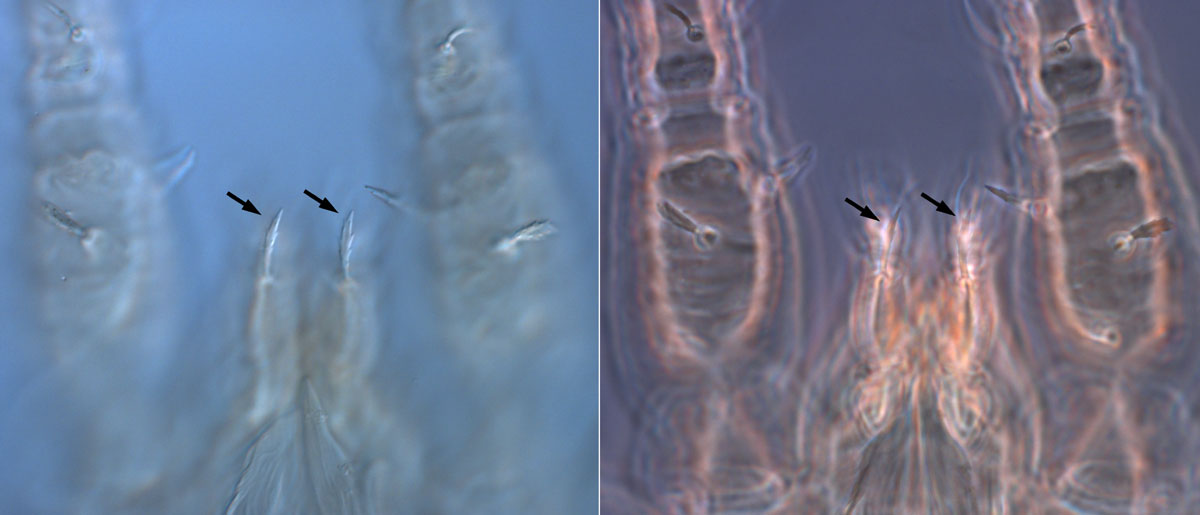
The situation is quite different for DIC, where gradients of optical path length are primarily responsible for contrast. Steeper gradients in path length produce excellent contrast. For example, the edges of structures are darker, and this creates the pseudo three-dimensional relief shading that is characteristic of DIC (Fig. 2). Regions with shallow optical path gradients, e.g. flat surfaces, produce insignificant contrast and often appear in the image at the same intensity level as the background. DIC does not produce the “halo” effect of Phase Contrast, keeping the image crisp and outlines clear. When there are several structures together the halo can effectively block structures from view. For example, the palps of Brevipalpus phoenicis type 1 are presented in Figure 4a using DIC and Figure 4b using phase contrast. The halo produced by the layers of cuticle in the region has made it impossible to see the key character of the seta indicated by the arrows.
For the study of mites - why is DIC better than phase?
The pseudo 3-D effect and crisper outlines produced by DIC allow a significantly clearer separation of dorsal and ventral surfaces, and external from internal structures. This in turns leads to a more reliable interpretation of surface patterns present on the cuticle, greater delineation of setae, and a clearer view of features.
For more information on the technical differences between DIC and phase contrast, see https://www.microscopyu.com/tutorials/comparison-of-phase-contrast-and-dic-microscopy and http://zeiss-campus.magnet.fsu.edu/articles/basics/contrast.html.
