
Gallery
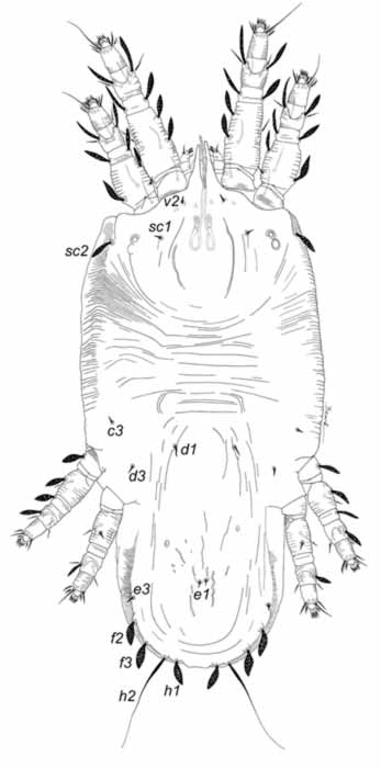
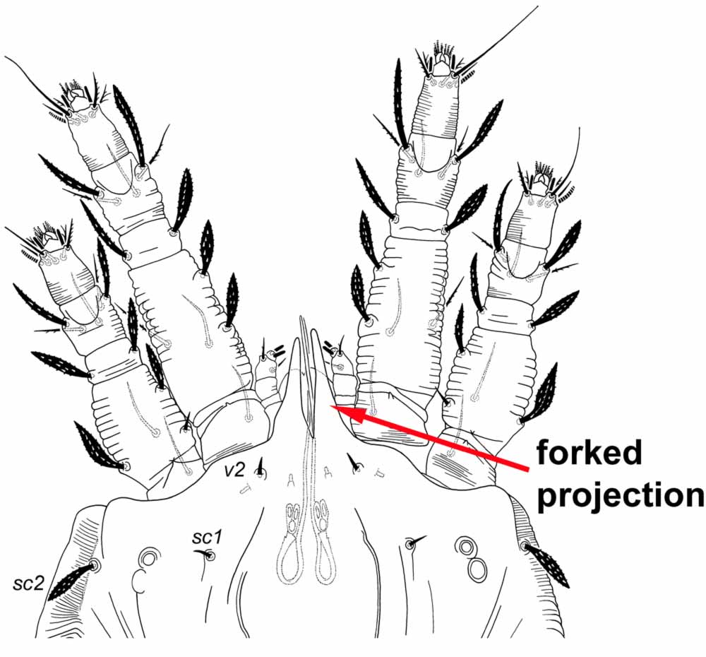
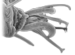
Fig. 2. Acaricis plana female anterior margin of the prodorsum indicating forked projection.
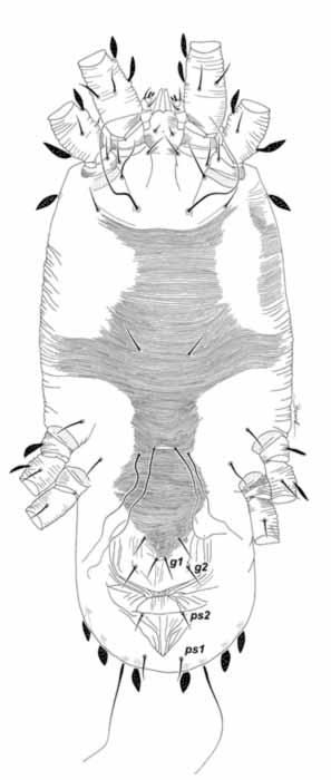
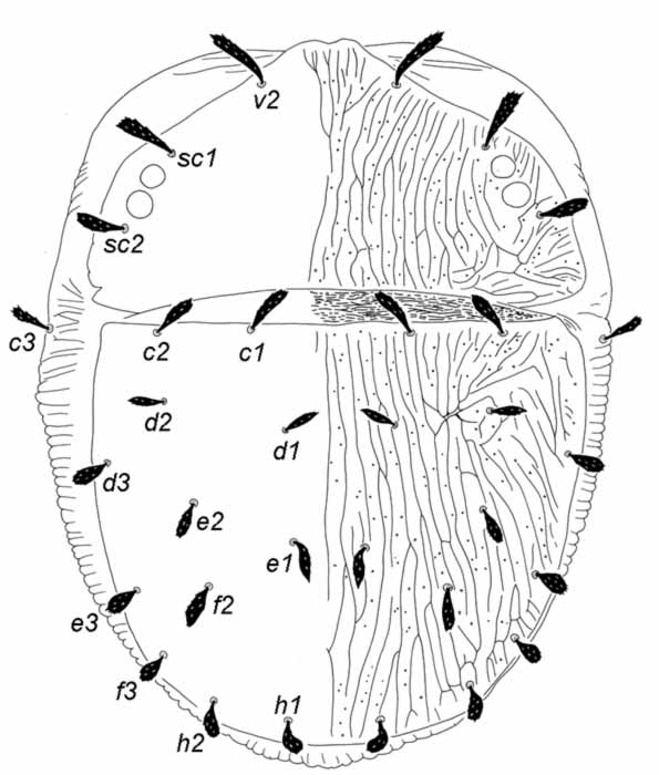
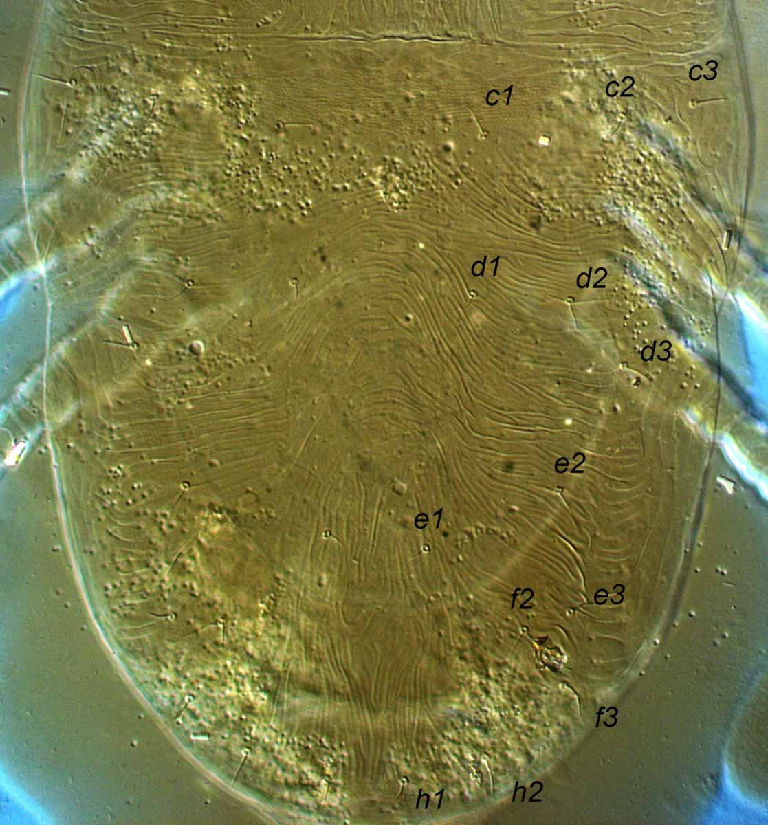
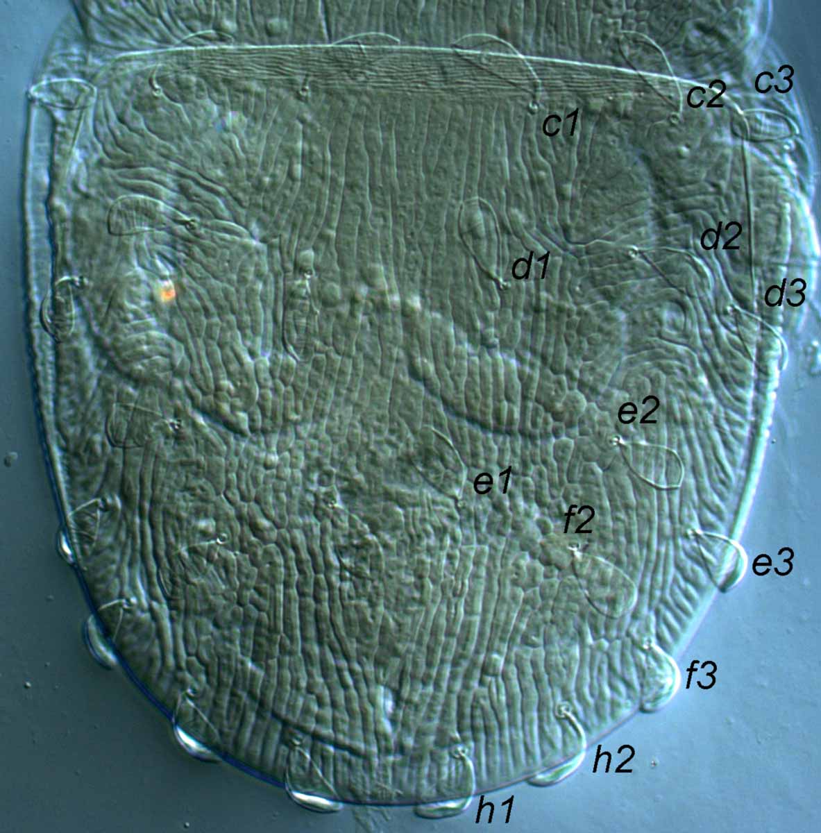
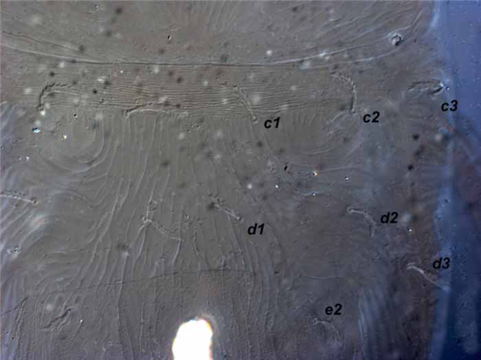
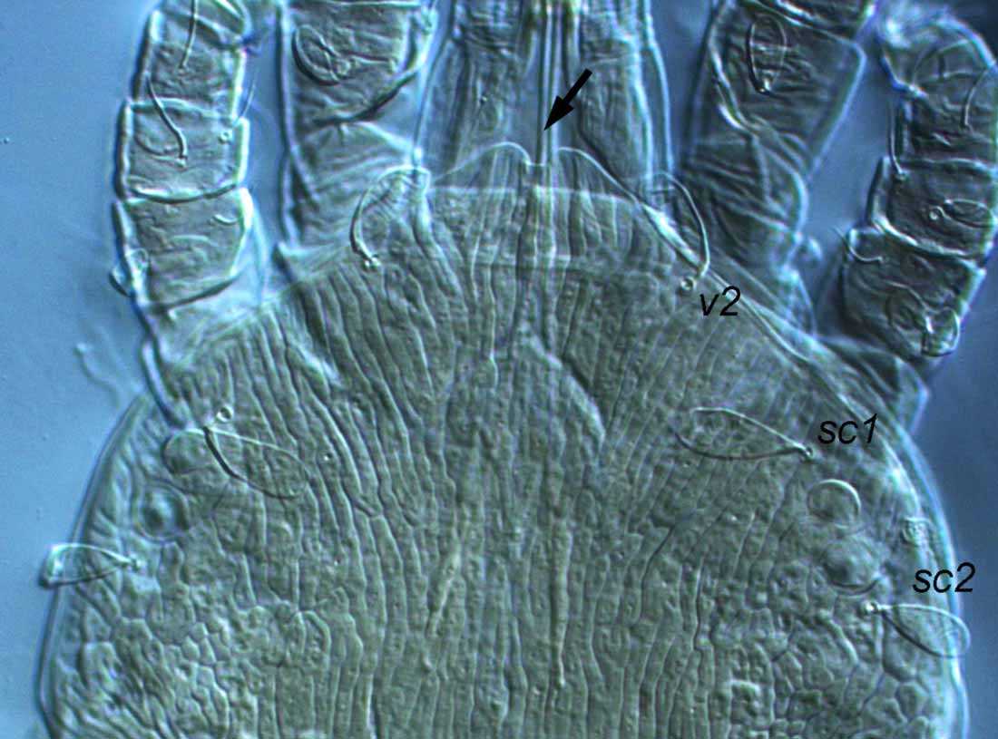
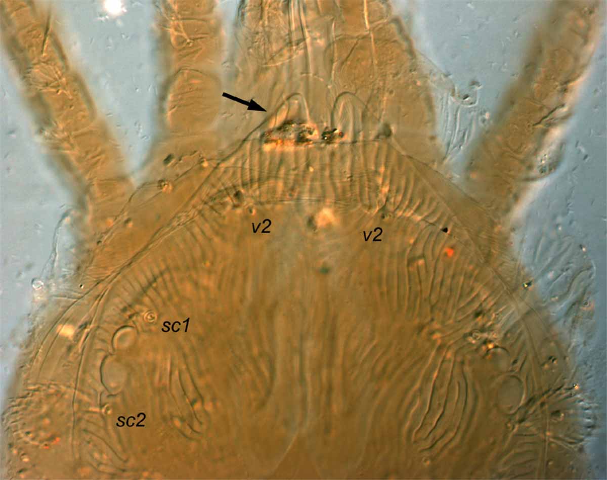
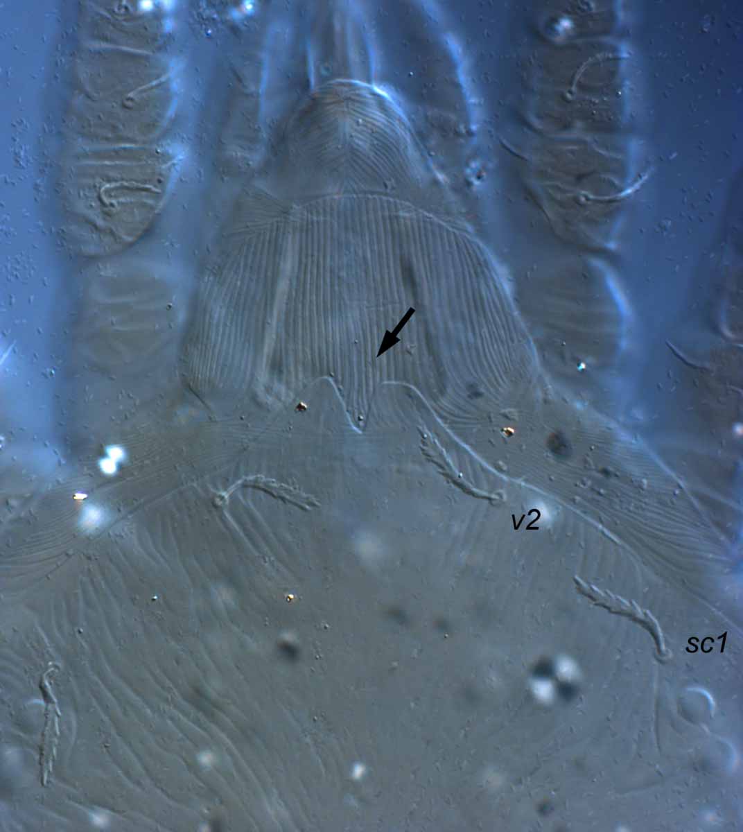
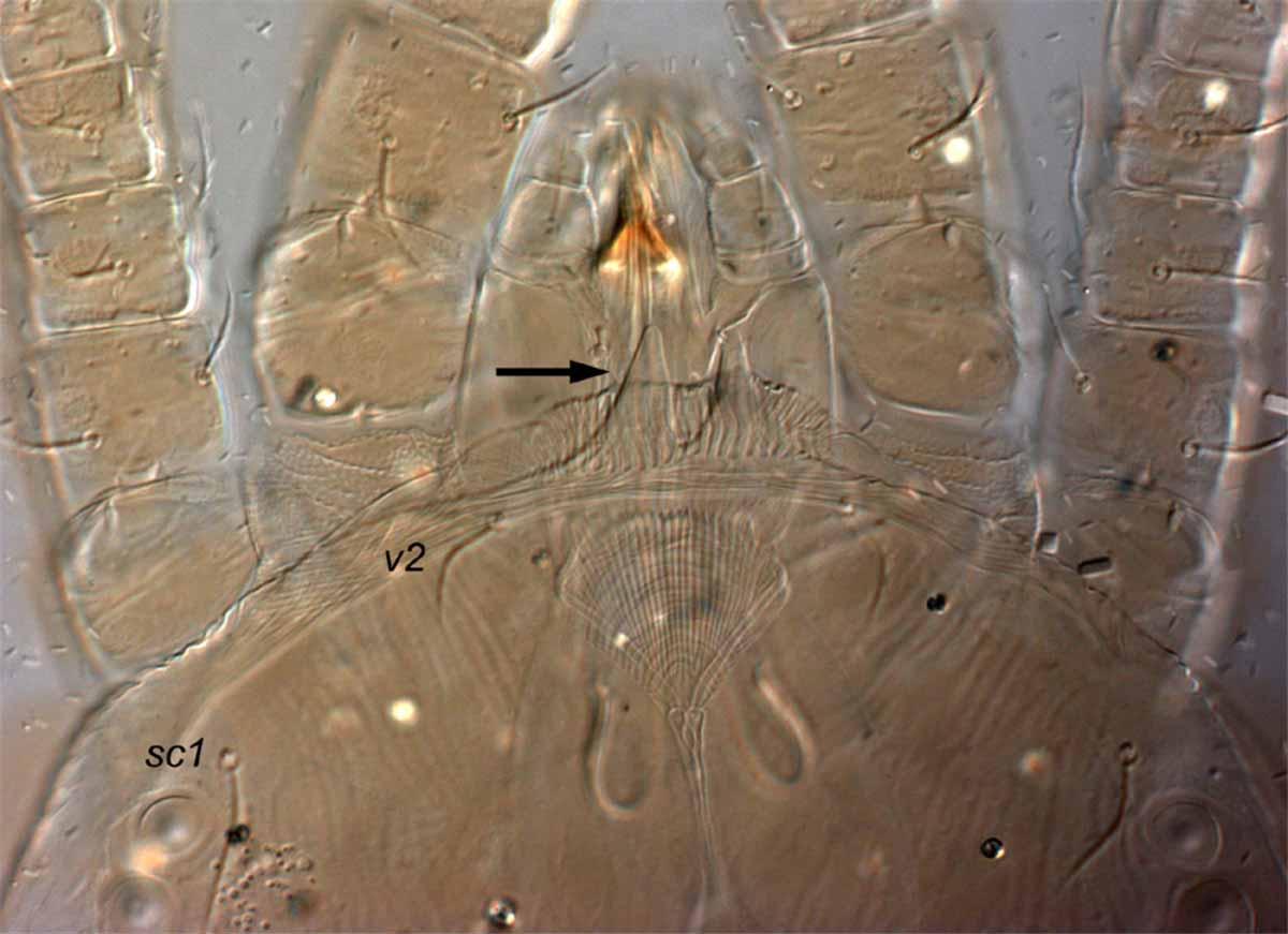
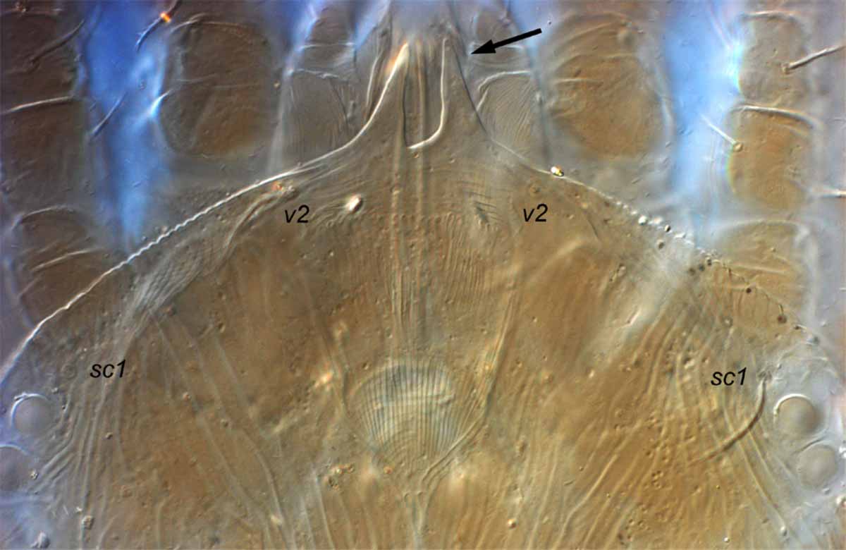
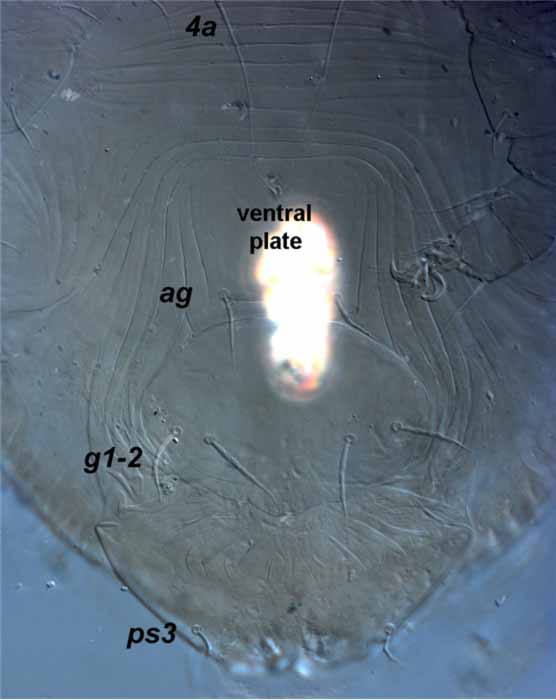 Fig. 11. Aegyptobia alpinensis female genital region - note the ventral plate indicated by change in striae on cuticle (type specimen).
Fig. 11. Aegyptobia alpinensis female genital region - note the ventral plate indicated by change in striae on cuticle (type specimen).
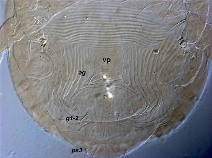
Fig. 12. Aegyptobia antenosoma female genital region (vp = ventral plate) (type specimen).
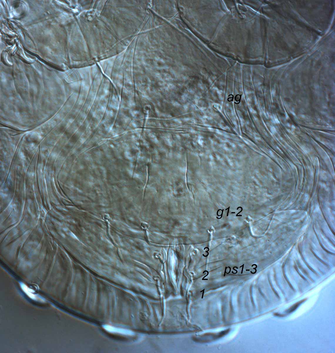
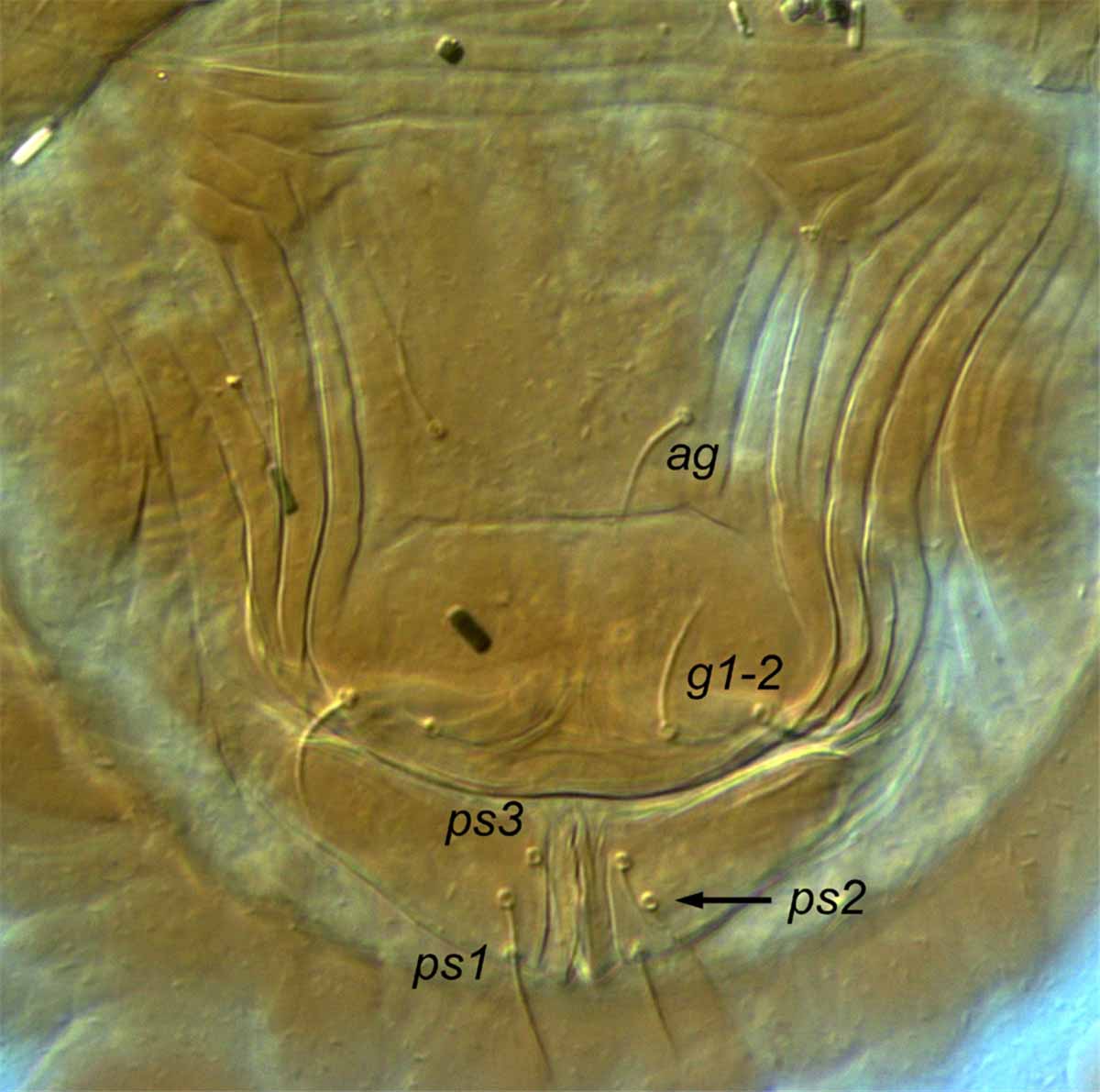
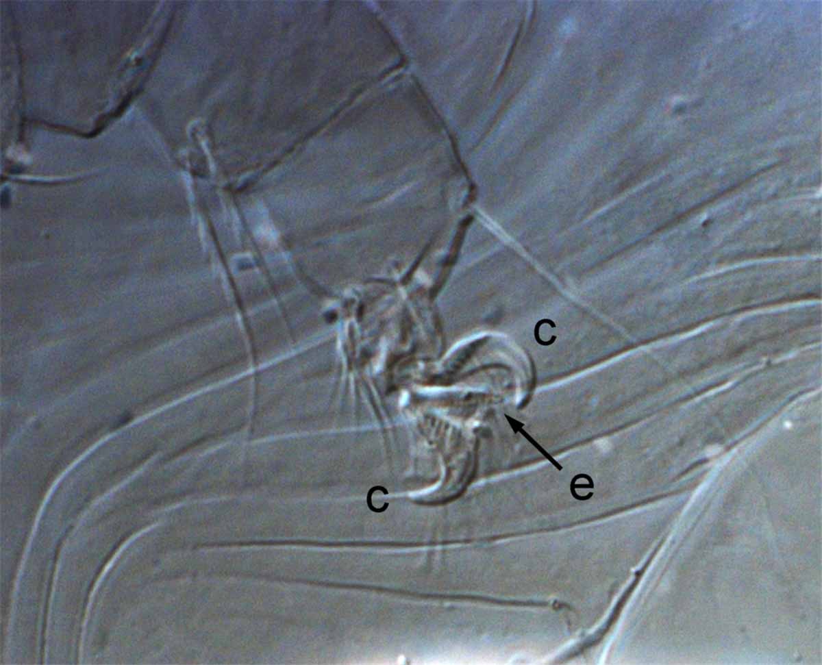
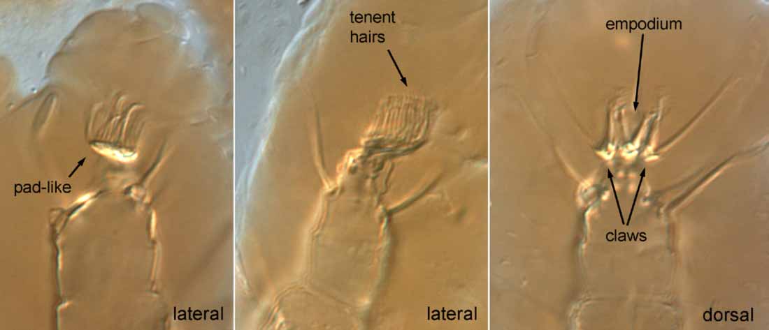
Fig. 16. Aegyptobia nomus female - claws are pad-like and empodium is pad-like (type specimen).
Fig. 17. Aegyptobia alpinensis female leg III indicating femur III (fe) with dorsal seta d present (arrow) (type specimen).
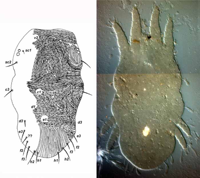
Fig. 1. Afronychus amnicus female dorsum (?? indicates possible presence of e2) (paratype).
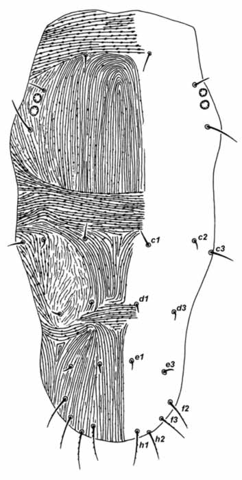
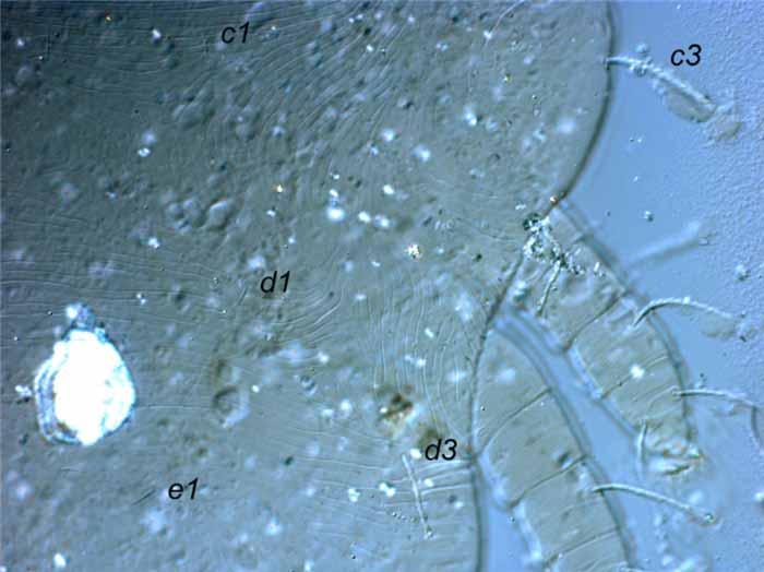
Fig. 3. Afronychus amnicus female anterior dorsal opisthosoma, indicating c2 absent (holotype).
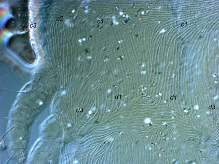
Fig. 4. Afronychus amnicus female anterior dorsal opisthosoma, indicating c2 present (paratype).
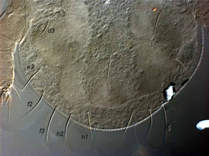
Fig. 5. Afronychus cliffortiae female posterior dorsum indicating e1 and e2 absent (paratype).
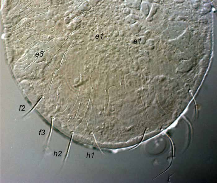
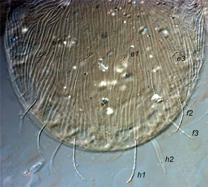
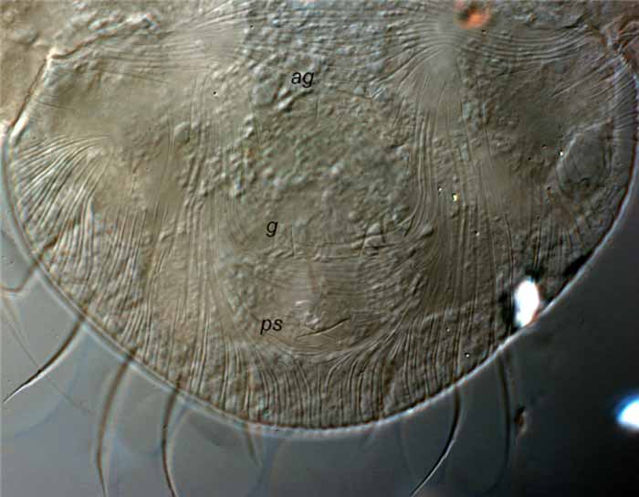 Fig. 8. Afronychus cliffortiae female posterior venter, indicating 1 pair of ag , g and ps setae ( ag = aggenital, g = genital, ps = psuedanal) (paratype).
Fig. 8. Afronychus cliffortiae female posterior venter, indicating 1 pair of ag, g and ps setae (ag = aggenital, g = genital, ps = psuedanal) (paratype).
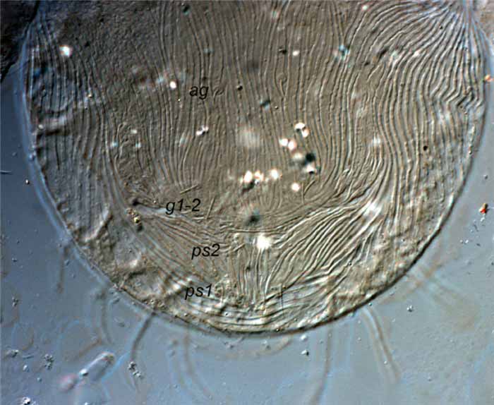 Fig. 9. Afronychus cliffortiae female posterior venter, indicating 2 pairs of g and ps setae ( ag = aggenital, g = genital, ps = psuedanal) (paratype).
Fig. 9. Afronychus cliffortiae female posterior venter, indicating 2 pairs of g and ps setae (ag = aggenital, g = genital, ps = psuedanal) (paratype).
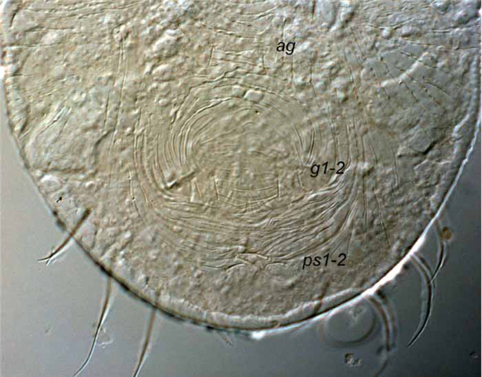 Fig. 10. Afronychus diastellae female posterior venter, indicating 2 pairs of g and ps setae ( ag = aggenital, g = genital, ps = psuedanal) (holotype).
Fig. 10. Afronychus diastellae female posterior venter, indicating 2 pairs of g and ps setae (ag = aggenital, g = genital, ps = psuedanal) (holotype).
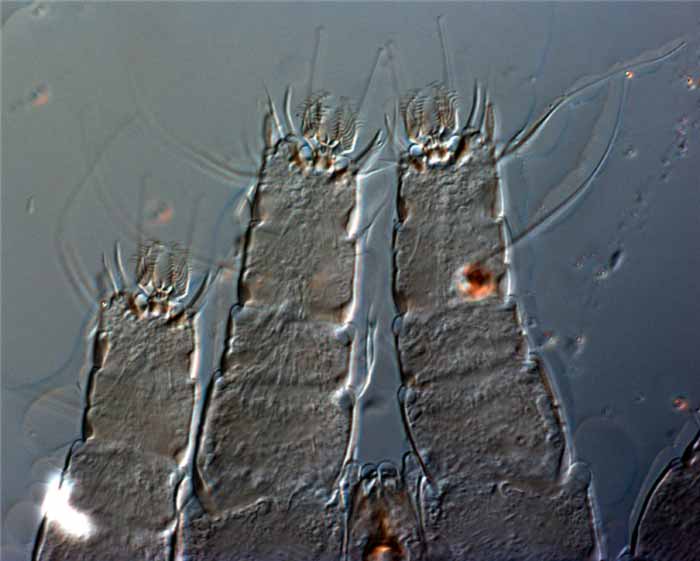
Fig. 11. Afronychus cliffortiae female legs I-II, highlighting short segments (paratype).
