
Dolichotetranychus floridanus
|
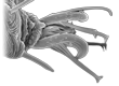
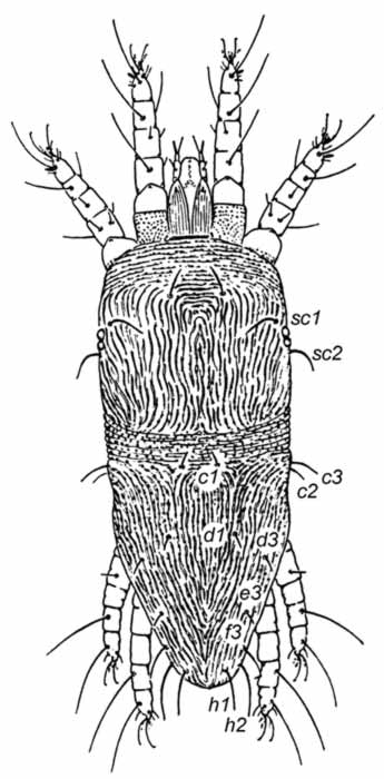
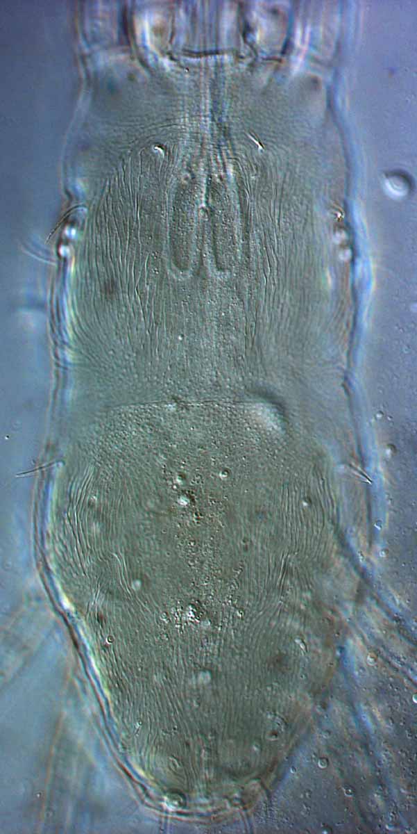
Fig. 1. Dolichotetranychus floridanus female prodorsum.
|
|
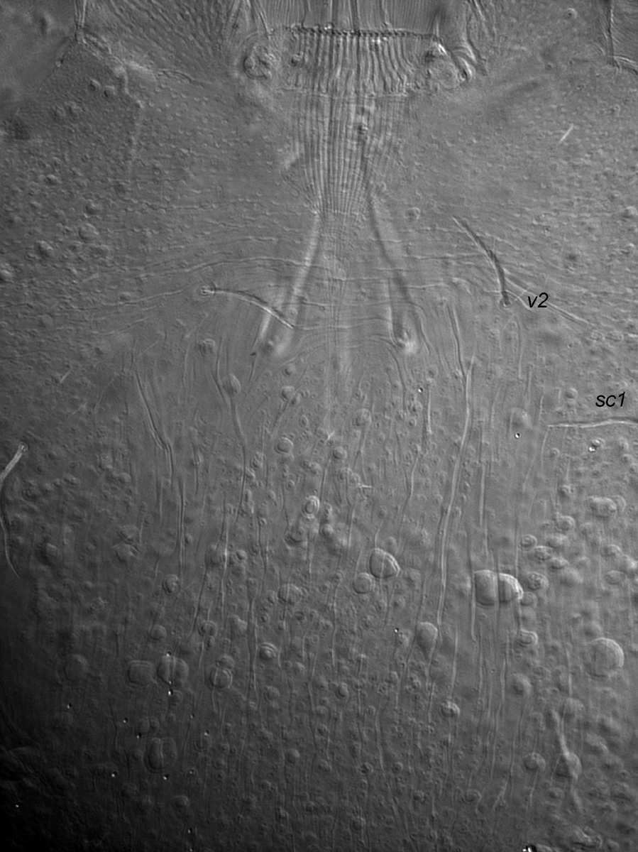
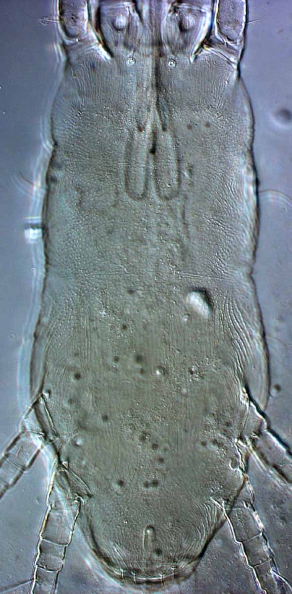
Fig. 2. Dolichotetranychus floridanus male prodorsum.
|
|
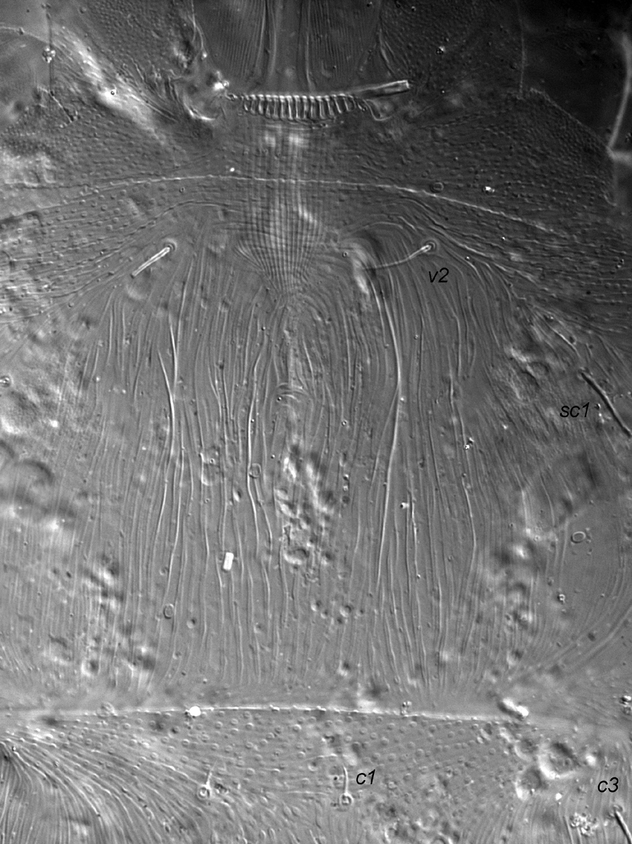
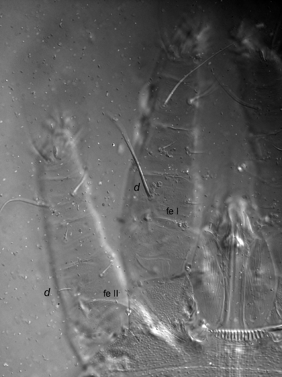
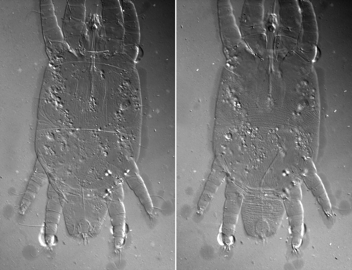
Fig. 3. Dolichotetranychus floridanus female, dorsal view of legs I-II indicating dorsal setae on femur I-II (fe I-II).
|
|
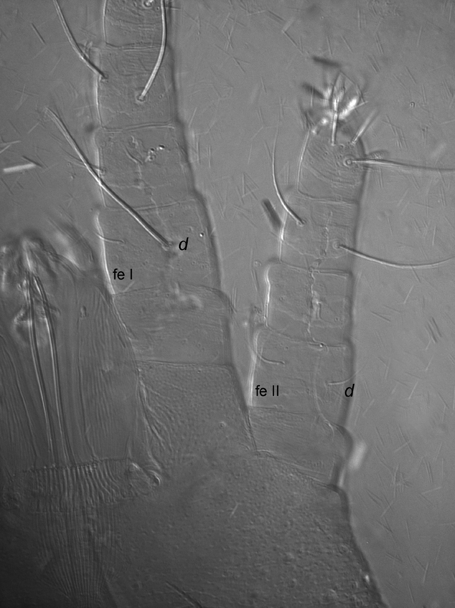
Fig. 4. Dolichotetranychus floridanus male, dorsal view of legs I-II indicating dorsal setae on femur I-II (fe I-II).
|
|
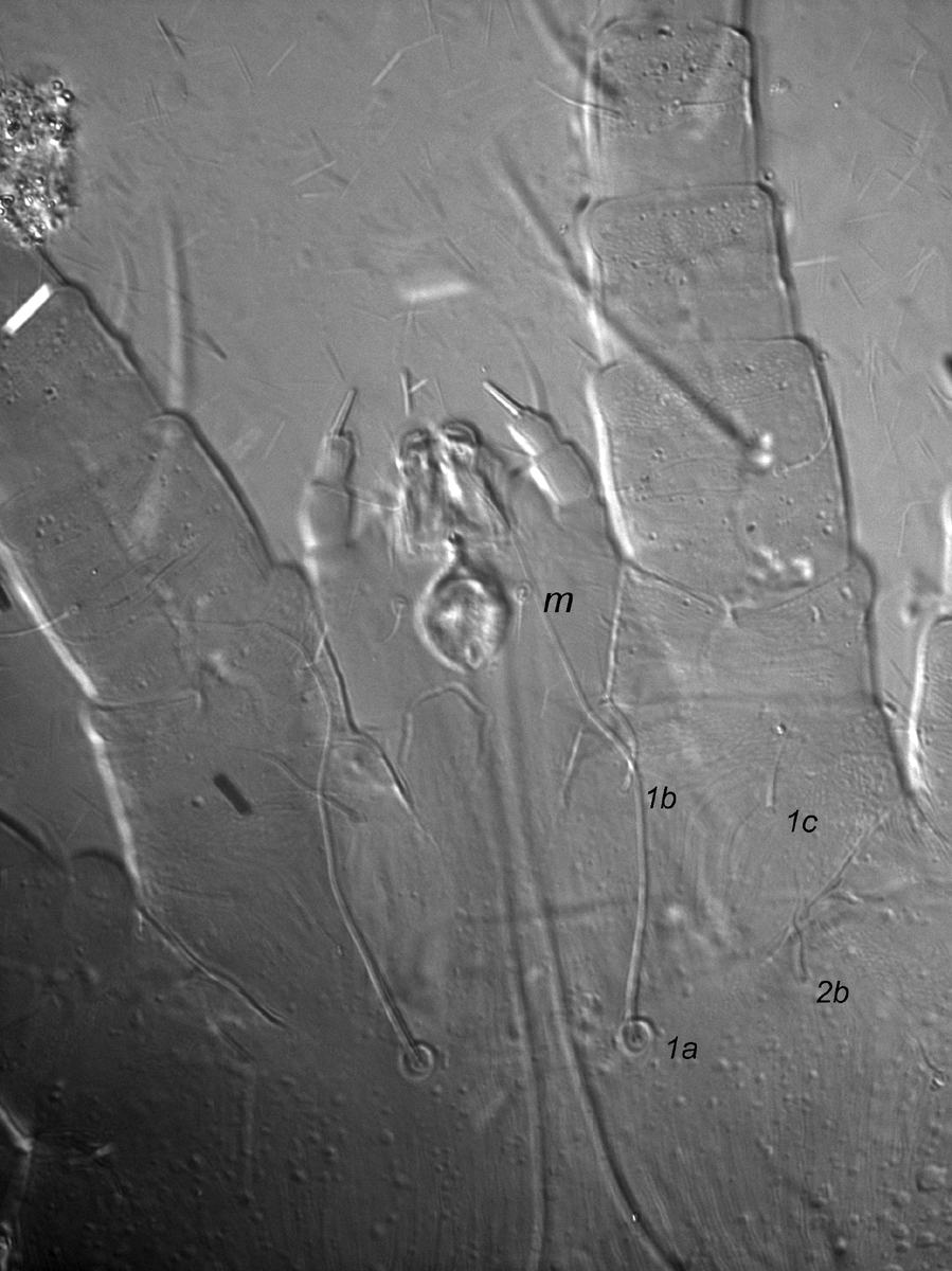
Fig. 5. Dolichotetranychus floridanus female, ventral gnathosoma indicating seta m, and 2 setae on coxa II (2b, 2c).
|
|
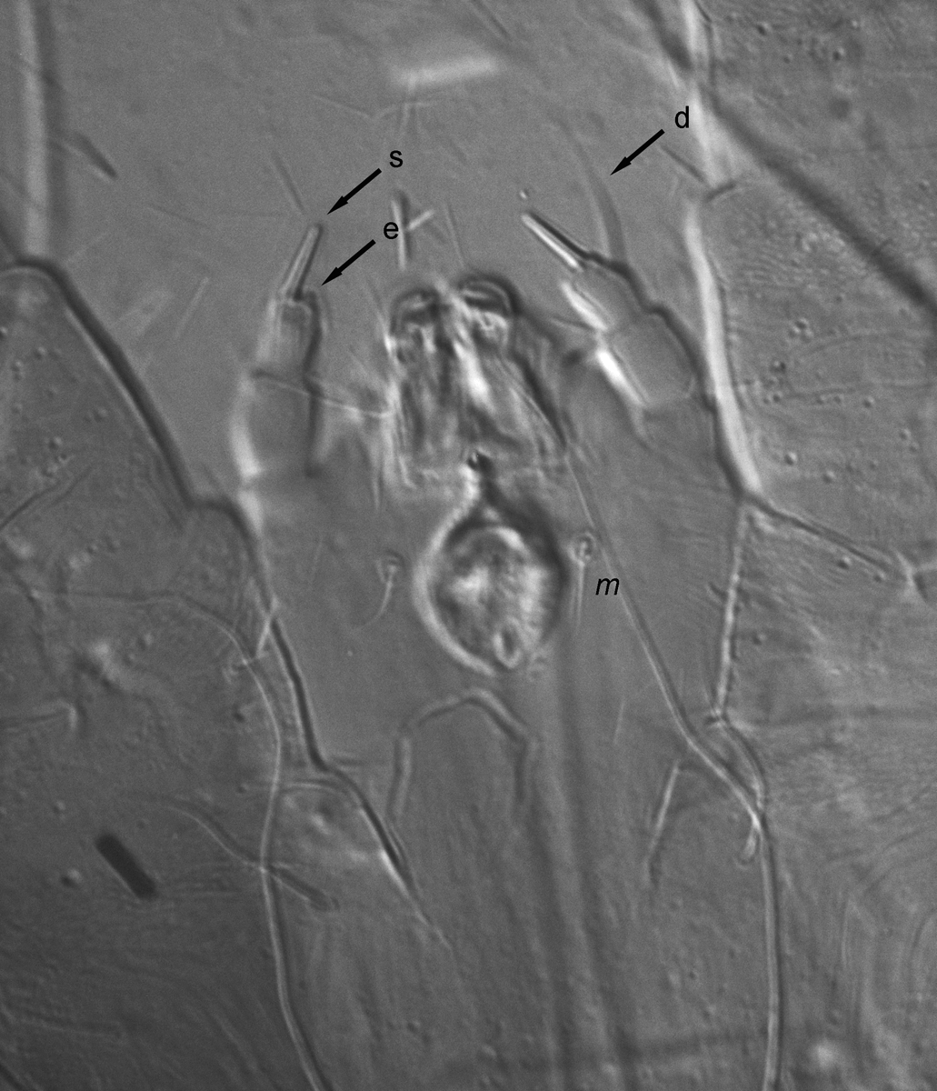
Fig. 6. Dolichotetranychus floridanus female, ventral gnathosoma indicating solenidion (s), eupathidium (e) and dorsal seta (d, out of focus) on palp tarsus, and ventral infracapitular seta m.
|
|
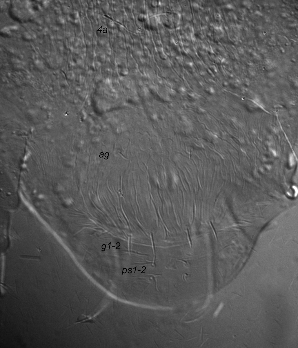
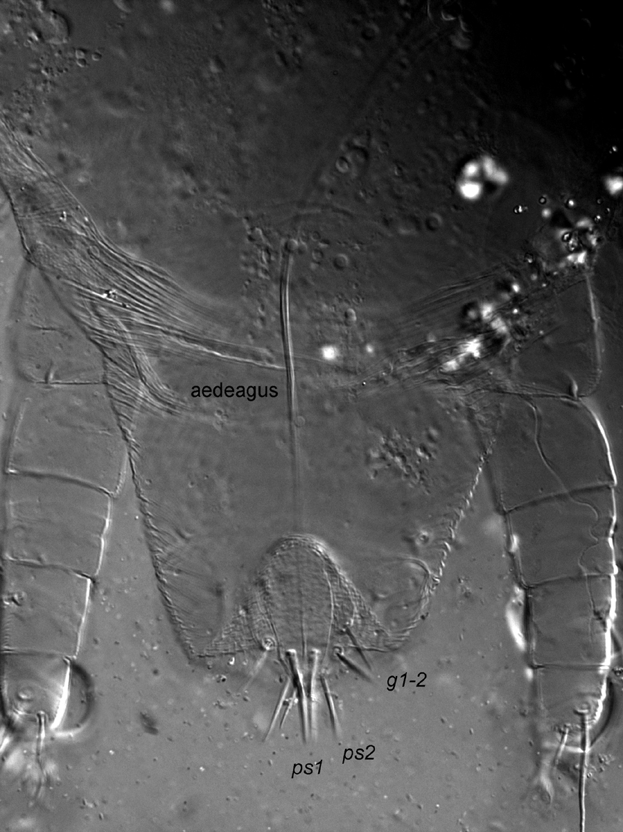
Fig. 7. Dolichotetranychus floridanus female posterior venter.
|
|
Fig. 8. Dolichotetranychus floridanus male posterior venter.
|
|
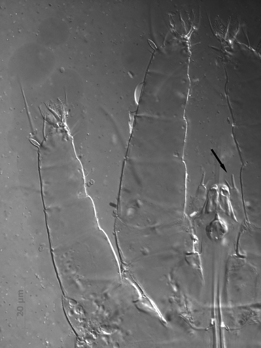
Fig. 9. Dolichotetranychus floridanus male palp, with arrow indicating the distal solenidion and eupathidium.
|
|
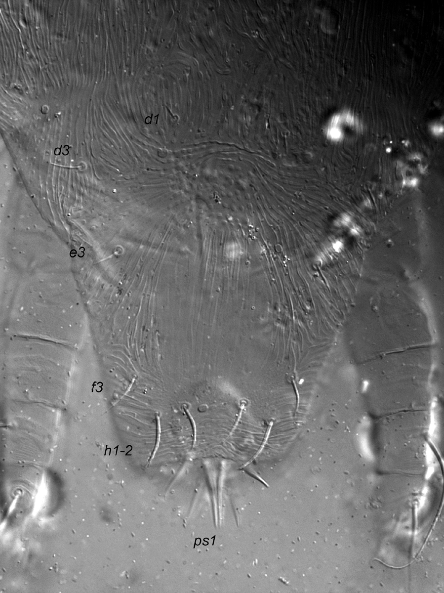
Fig. 10. Dolichotetranychus floridanus male, posterior dorsum, indicating ps1 setae.
|
|
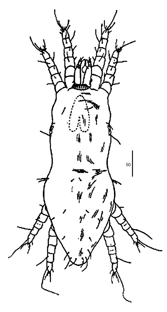
Fig. 11. Dolichotetranychus floridanus female, after Ehara 1966.
|
|
Fig. 12. Dolichotetranychus floridanus female, after Baker & Pritchard 1956.
|
|
Fig. 13. Dolichotetranychus floridanus female dorsum.
|
|
Fig. 14. Dolichotetranychus floridanus female venter.
|
|
Fig. 15. Dolichotetranychus floridanus male dorsum and venter.
|
Authority
(Banks)
Key characters
Female characters (Figs. 11-14) (with male where indicated (Fig. 15)):
- striae on prodorsum mostly smooth (f & m), sometimes with few tubercles centrally (Figs. 1, 2)
- femur I with dorsal seta long, reaching tarus (f & m) (Figs. 3, 4)
- femur II with dorsal seta short, about the length of the segment (f & m) (Figs. 3, 4)
- ventral infracapitular seta, m, present (Figs. 5, 6)
- coxa II with two setae, 2b & 2c (f & m) (Fig. 5)
- striae on genital flap without granular texture (Fig. 7)
- 2 pairs of genital setae (g1-2), shorter than distance between them (Fig. 7)
- 2 pairs ps setae (ps1-2) (f & m) (Fig) (Figs. 7, 8)
- female palp tarsus with 1 solenidion and 1 tiny eupathidium distally, and 1 dorsal seta (Fig. 6)
- male palp tarsus with 1 solenidion and 1 eupathidium distally, and 1 dorsal seta (Fig. 9)
- male with moderately long ps1 (Figs. 10, 15)
Recorded hosts
PineappleRecorded distribution
Australia, Brazil, Costa Rica, Cuba, Honduras, Japan, Java, Mexico, Panama, The Philippines, Puerto Rico, USARemarks
Adults, nymphs and eggs of this species are bright orange in colour when alive. The larvae are pale and almost translucent.
The tiny eupathidium on the palp tarsus has not been recorded before for this species. It is very difficult to see (Fig. 6).
There is some variation in the length of prodorsal setae v2 evident in the literature: prodorsal setae v2 about 1/3 as long as the distance v2-v2 (Ehara 2004); and prodorsal setae v2 just over 1/2 as long as the distance v2-v2 (El-Enany & Soliman 1987).
This mite causes damage at all stages of pineapple production, causing damage to both the leaves and fruit. Mite feeding causes the epidermal tissue to dry and crack, allowing fungae and bacteria to enter the plants and cause the tissue to rot. The dry lesions lead to scarring and tissue malformation (Jeppson et al. 1975; Sanches & Zem 1978; Poli 1991).
Within the plant, the mites are present most commonly on the lower surfaces of the leaves, near the mid ribs and veins. They can occur in very high numbers at the base of the leaves near the leaf stem, and between the bases of the tightly overlapping leaves (Poli 1991). The mites also have the ability to develop heavy infestations on detached crowns that are stored for a period of time for later planting (Poli 1991). In addition, significant populations of mites can be found in the soil at the base of the infested pineapples or on the pineapple stem itself. It is believed that the mites migrate to the soil to avoid extremems in climate (Poli 1991).
Plants of all ages are infested and young plants or freshly planted crowns can be seriously damaged or killed. If the plants are not killed, fruit yield is reduced.
This species is particularly prevalent in drier areas has the ability to build massive populations that require contol (Poli 1991).
Feeding mites have a characteristic posture, standing on their heads with the posterior raised in the air (Poli 1991).
The fungus Hirsutella thompsoni has been successfully used to control this mite (e.g. Ochoa et al. 1994).
References
*Banks (1900); Ehara (1966); Jeppson et al. (1975); Khanjani et al. (2011); Poli (1991); Pritchard & Baker (1958); Sanches & Zem (1978); Sanches & Flechtmann (1982); Sayed (1938)
* = original description
