This is a key to mesostigmatic mites likely to be found in quarantine intercepts compiled by Dr. David Evans Walter of Colorado State University and the University of Alberta (dew@ualberta.ca).
About one-quarter of all mite species belong to the monogynaspid Mesostigmata, including many economically important pests (e.g., varroa mite of bees, fowl mites, rat mites) and even more economically useful biocontrol agents (especially in the families Phytoseiidae, Laelapidae and Macrochelidae). Because of their pervasiveness, mesostigmatans are commonly found in quarantine inspections, but primarily because few are plant parasites, there are few taxonomic specialists to help in their identification. This key is designed to support the identification of members of the largest suborder of Mesostigmata, the Monogynaspida (See the key Major mite taxa to identify Mesostigmata to suborder). Monogynaspids are the mesostigmatans most likely to be found in a quarantine intercept and the Monogynaspida contains all of the economically important species.
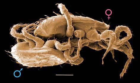
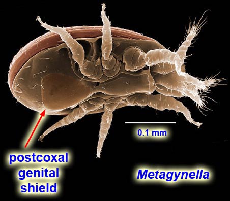
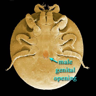
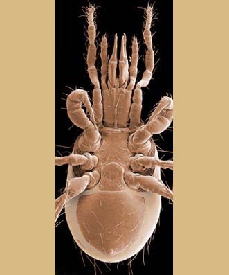
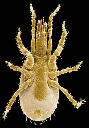
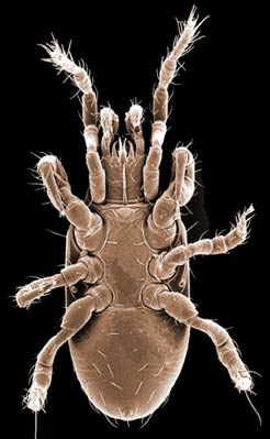
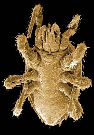
In most cases, identification of Mesostigmata to family or lower can be accomplished only if the specimen is an adult female. The easiest way to determine the stage and sex of your specimen is to look at the intercoxal region. Adult females have a genital opening that is almost invariably in the intercoxal region (species of Metagynella (Fig. 1) are exceptions) and covered by a sclerotized shield which may be truncate posteriorly or continue onto the ventral region. Adult males and immature stages of both sexes have a continuous intercoxal shield that may be truncate or continue onto the ventral region or even incorporate the entire ventral region. Adult males have a subcircular opening in the intercoxal shield (sometimes called a sternogenital or sternitogenital shield) somewhere between the posterior margins of coxae IV (Fig. 2) and the base of the tritosternum (Fig. 3). Immature stages (larva, protonymph, deutonymph) of Mesostigmata have no genital opening.
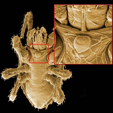
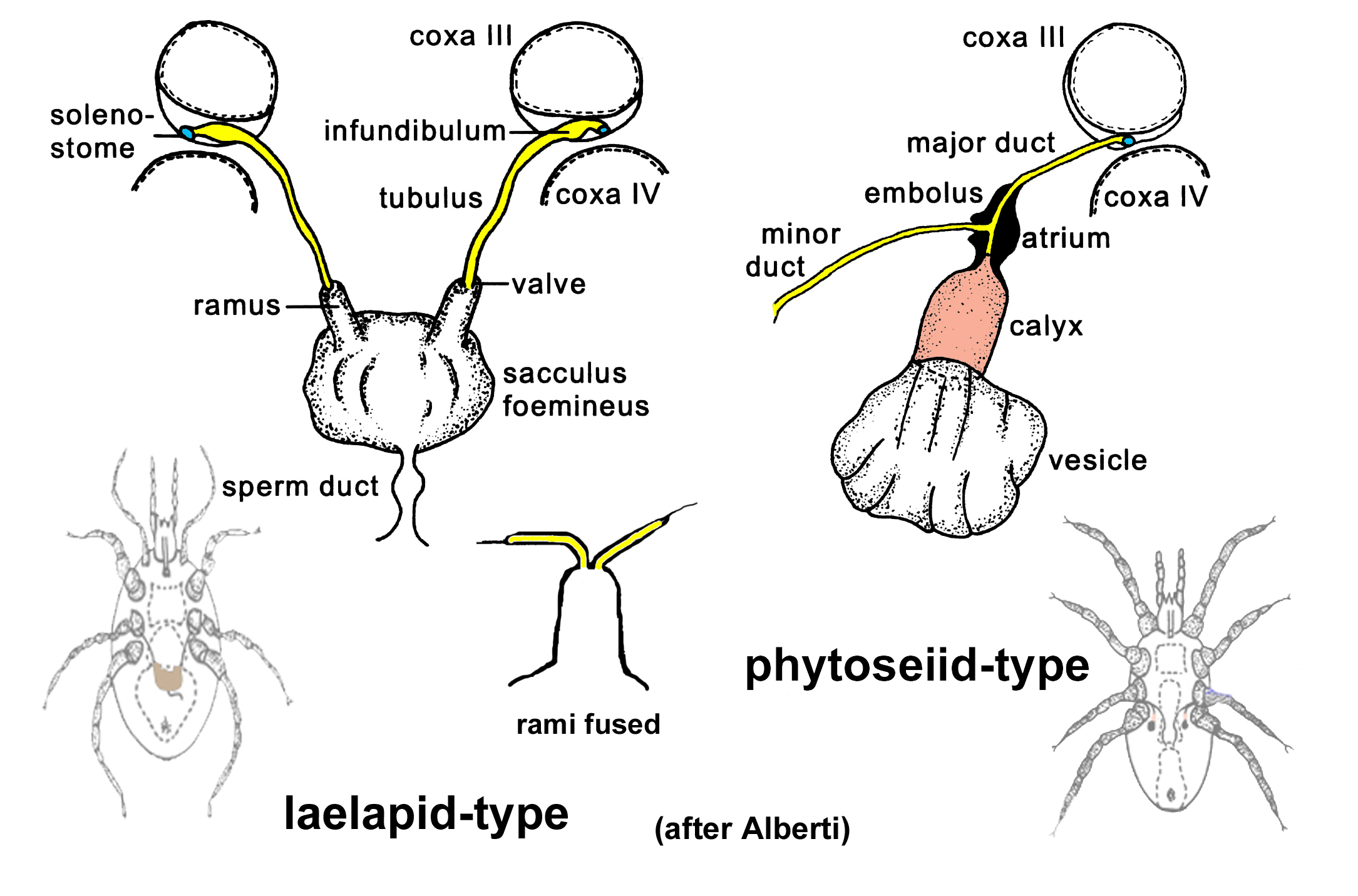
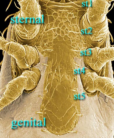
Female mesostigmatans are easily recognized by their genital opening, usually found between coxa IV and covered by one or more genital shields (Fig. 4). Although called "genital" (or epigynial, epigynal), the shield protects only the ovipore in mesostigmatans with a secondary sperm transfer system (Dermanyssiae; Fig. 5). Females lay eggs relatively large in comparison to their body size, so the genital opening and its shield are relatively large, and may be extended posteriorly to incorporate ventral or ventrianal elements.
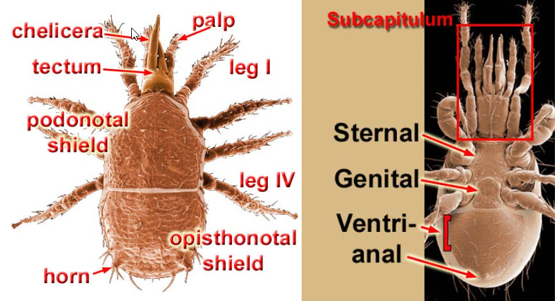
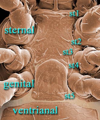
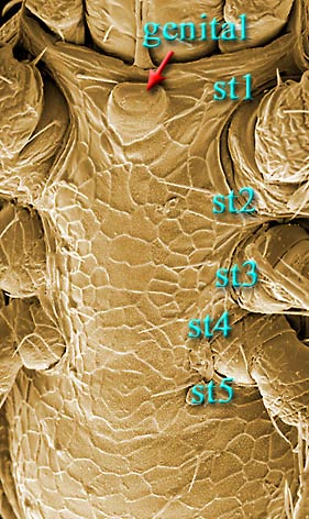
The genital shield often bears the 5th pair of sternal setae (st5), but may be nude and variously formed. Adult females also usually have one or more shields anterior to the genital shield(s) called sternal shields, and these usually sport one or more of sternal setae st1–4. Other shields cover the ventral and anal regions. The female ologamasid mite in Figures 6 and 7 has a large ventrianal shield, while the female laelapid mite in Figures 8 and 9 has a small anal shield.
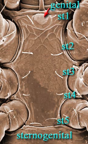
Males, however, produce relatively small sperm packets (spermatophores) and have much smaller genital openings. Although these openings also may lie between coxae IV, they are more typically towards the anterior end of the intercoxal region and often at the base of the tritosternum (see details in Figs. 10–13). In the male ologamasid mite (Figs. 10, 11), the genital opening is in a sternogenital (also sternitogenital) shield that bears 5 pairs of setae (st1–5). In the male laelapid mite (Figs. 12, 13), however, the sternogenital shield is fused to the ventrianal shield and other elements to form a ventral shield or plate. Males also often, but not always, have modifications to their chelicerae for transferring sperm, e.g., spermatodactyls or spermatotremes.
Click on an anatomical part below for a detailed illustration
Author: David E. Walter
Content last updated Sept. 2006, unless indicated otherwise
Lucid key player updated Sept. 2025
idtools.org