

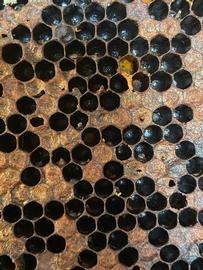
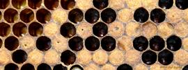
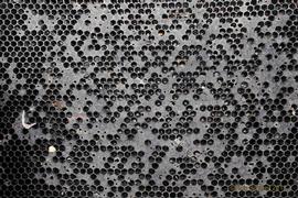
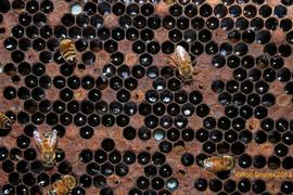
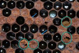
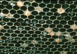
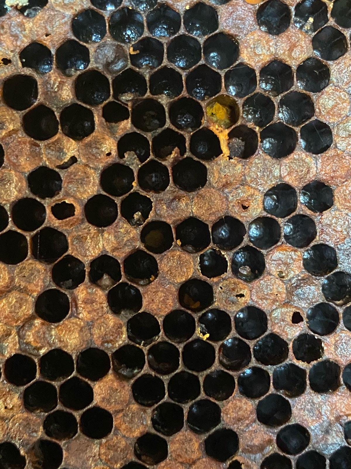
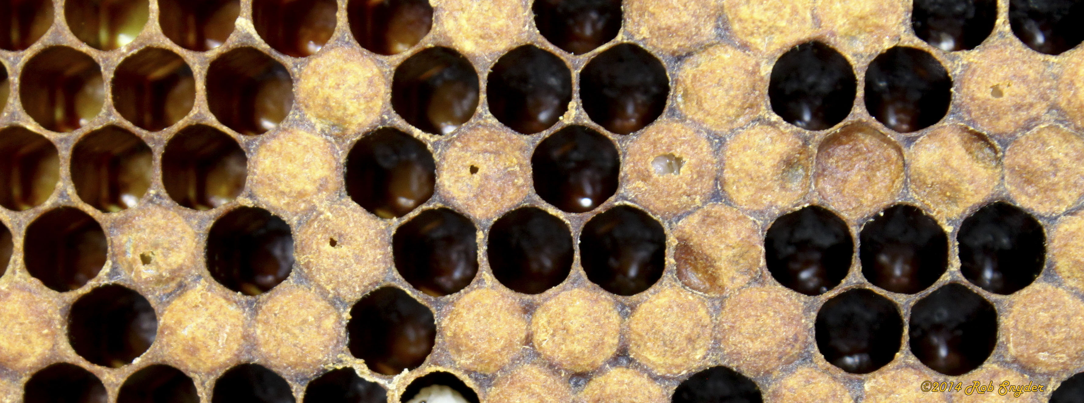
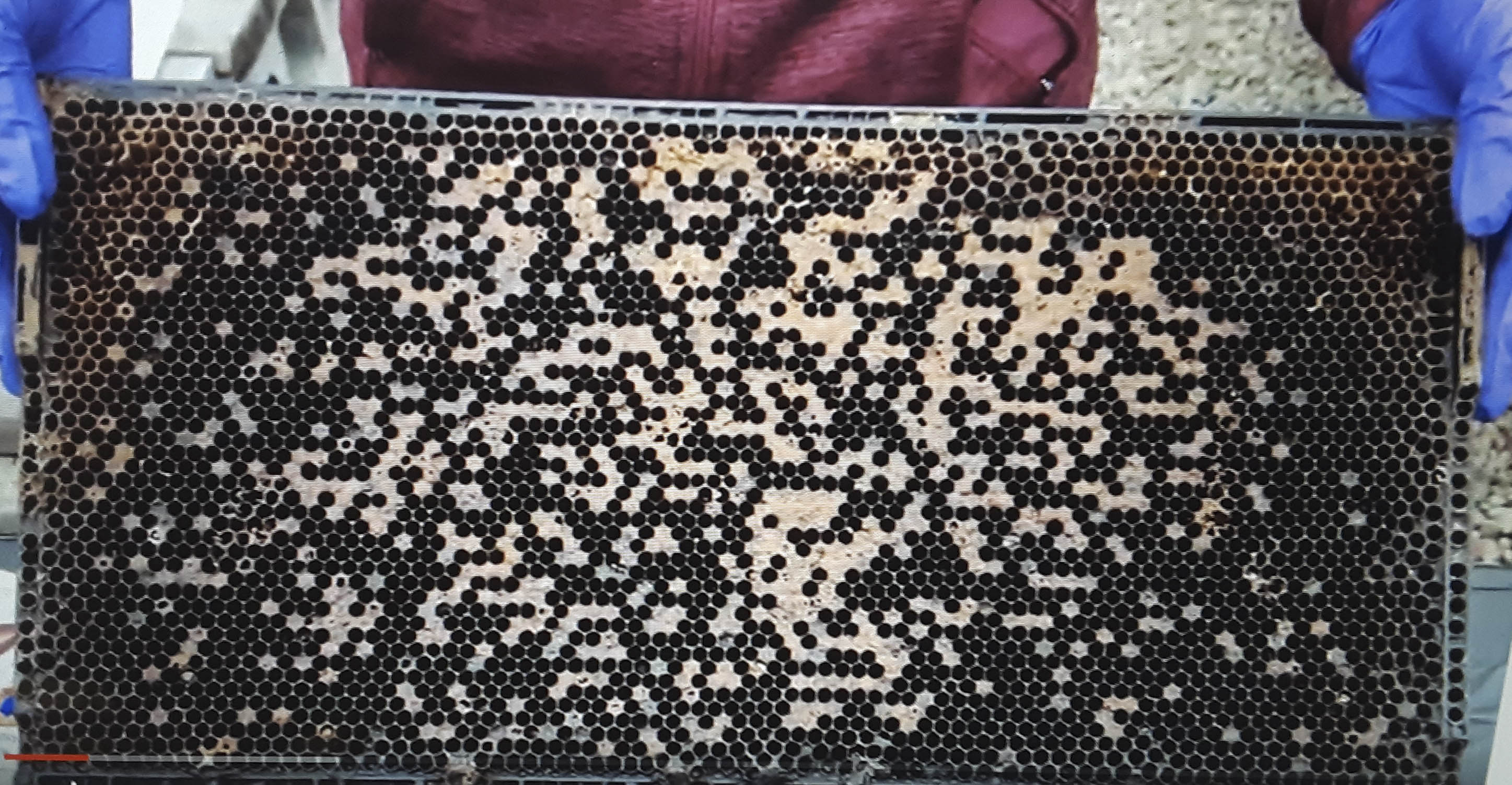
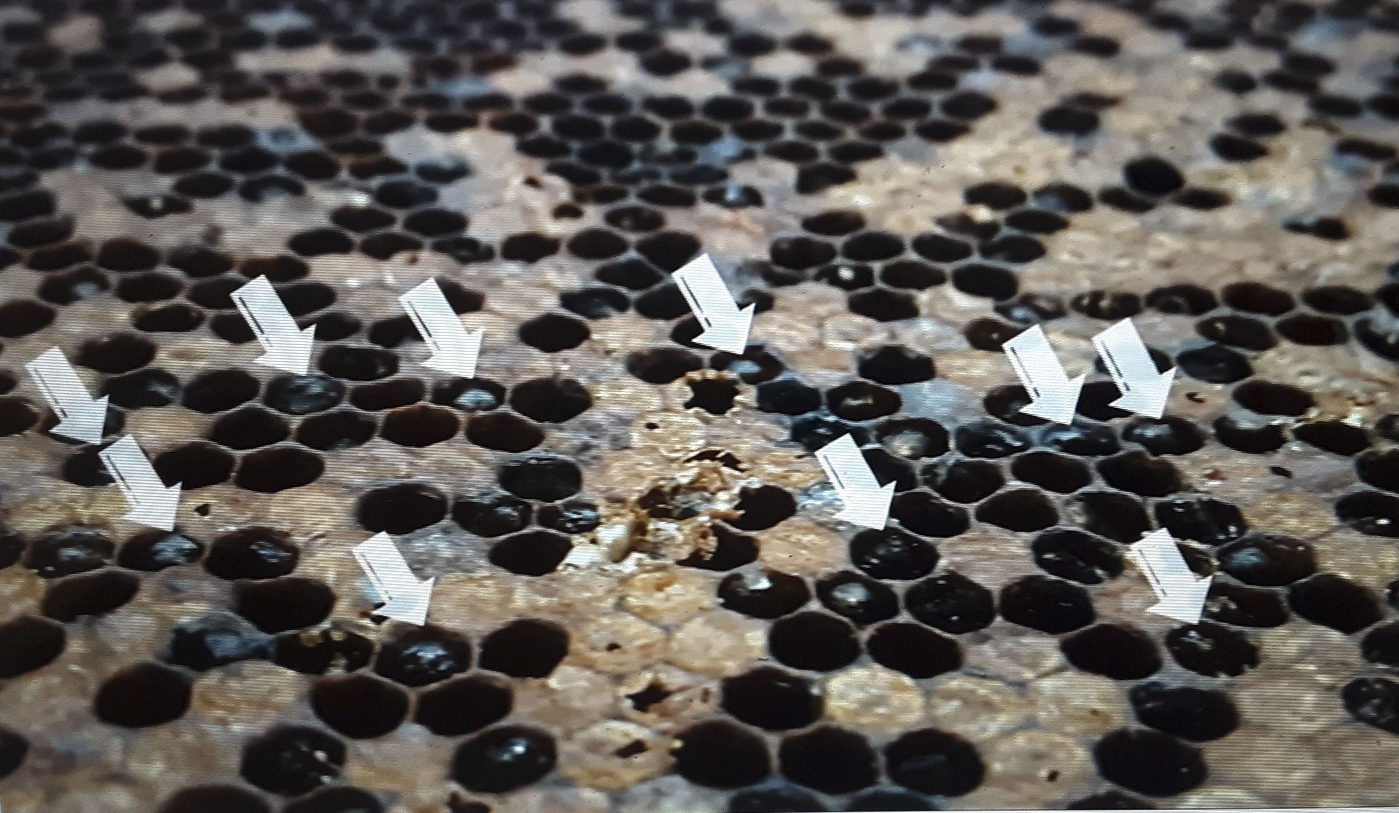
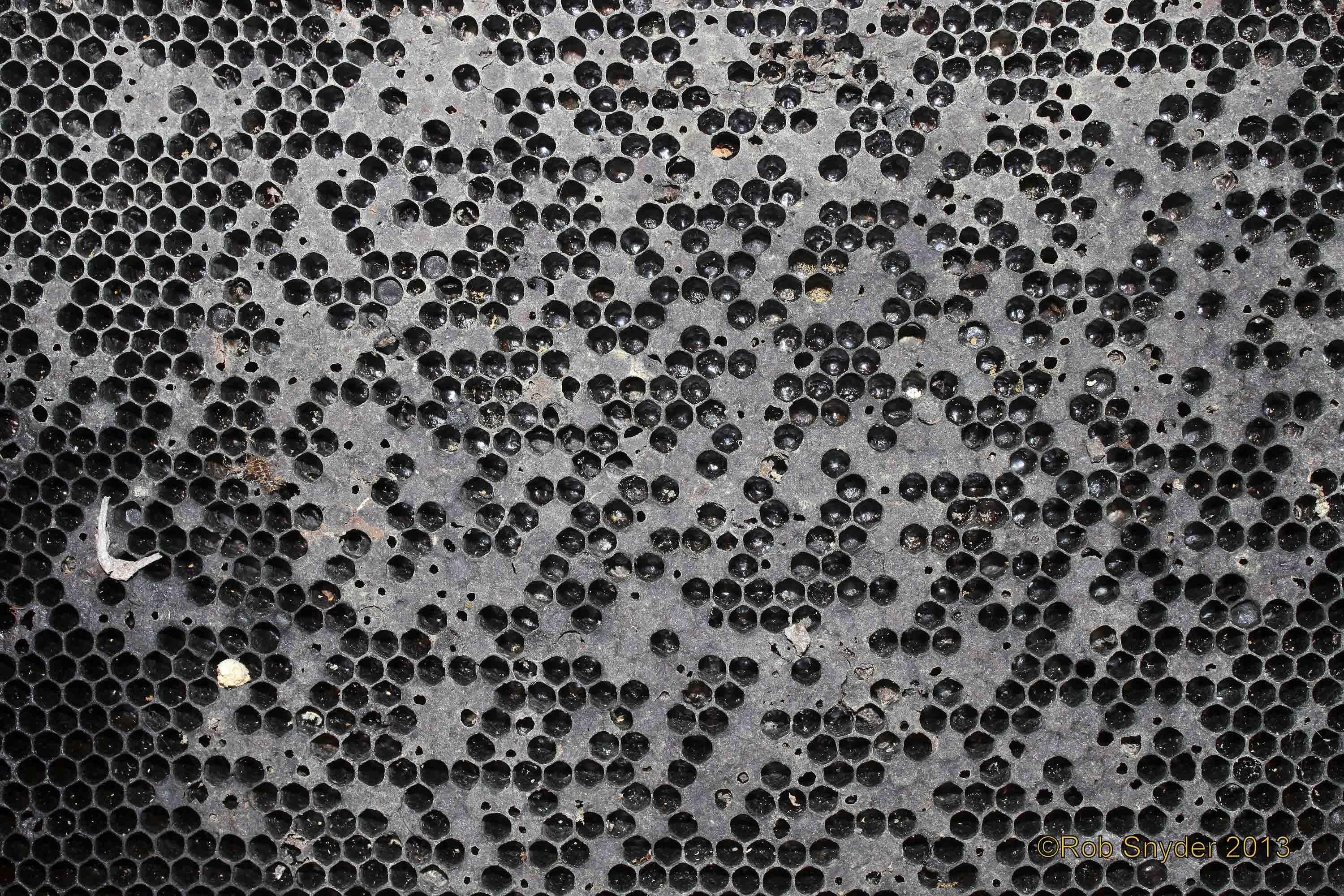
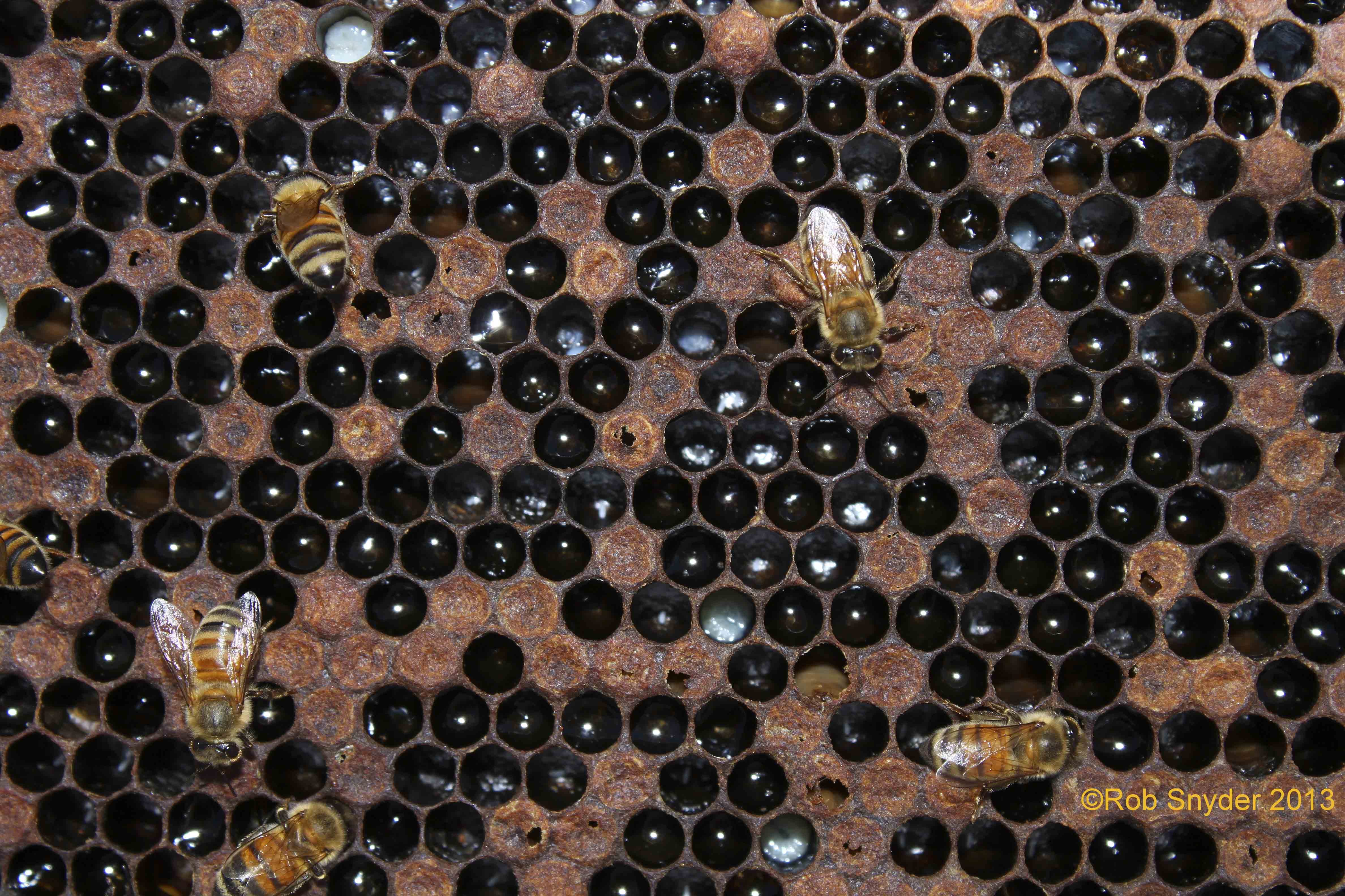
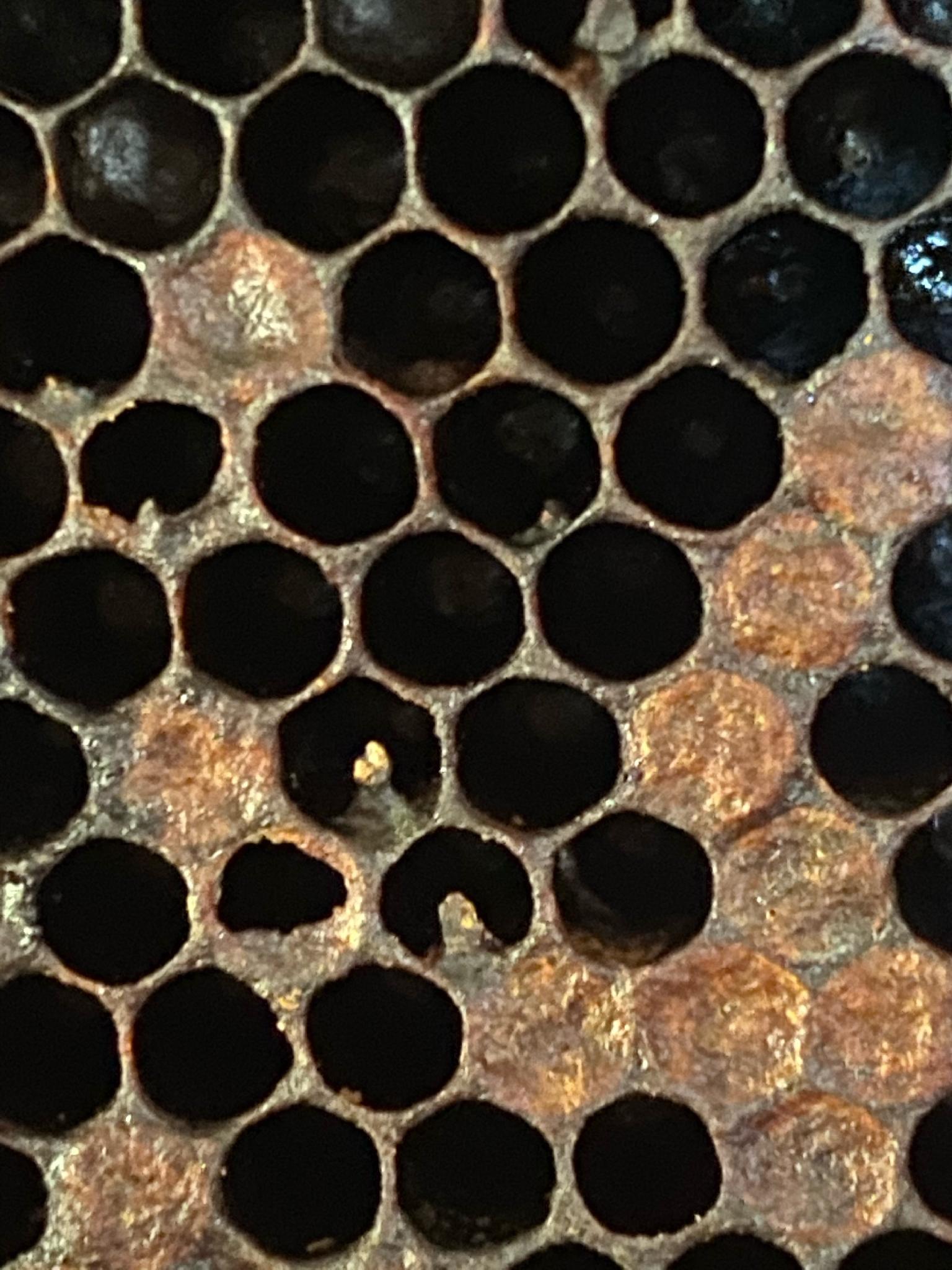
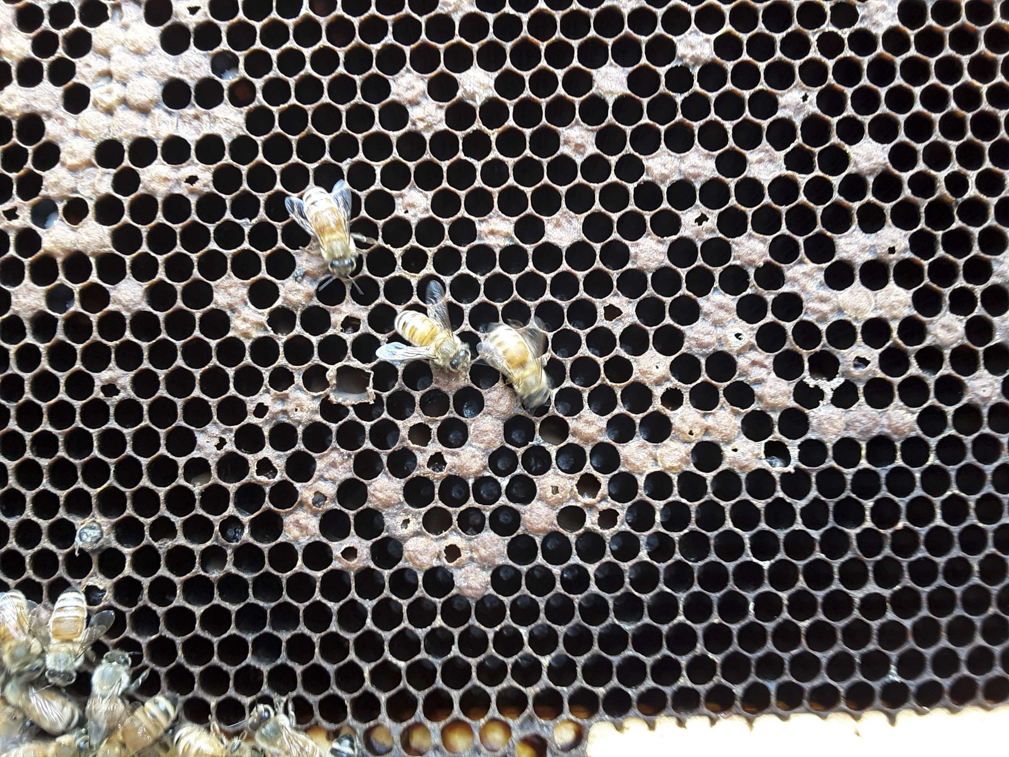
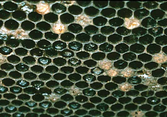
Brood cappings sunken, greasy-looking; darker cappings may have tiny perforations. Developing brood may have died underneath these cappings (uncap to see this). Larvae initially turn brown or caramel-colored, then dark brown to black. As they die, larvae and prepupae elongate in their brood cells on the lower cell margin.
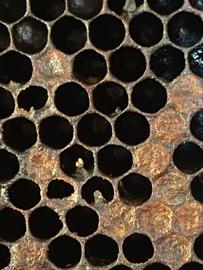
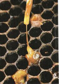
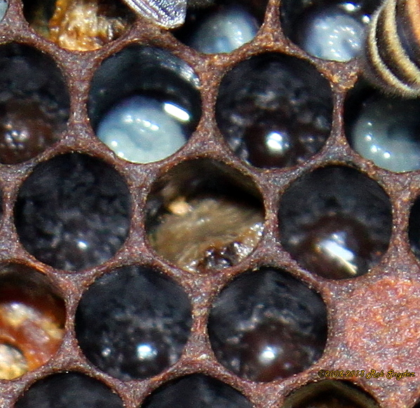
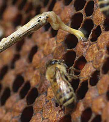
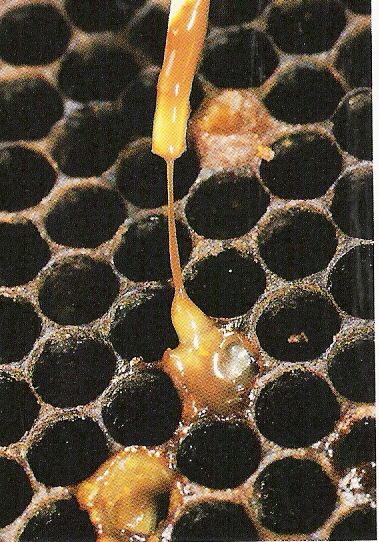
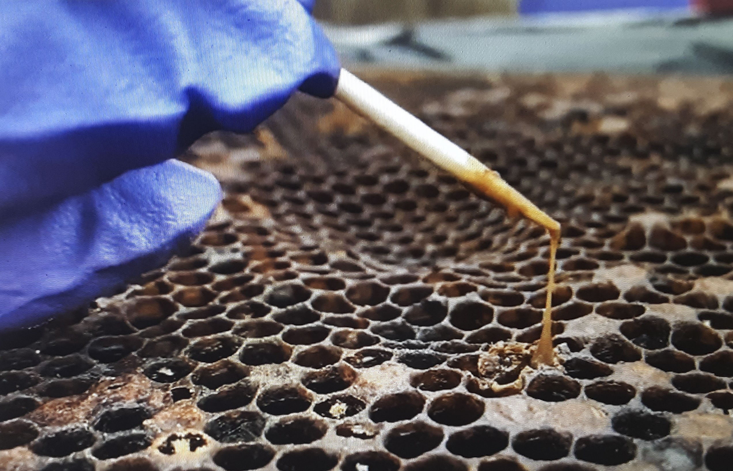
Dying or recently dead brood will “rope-out” (as if pulling taffy). Dead, desiccated brood (scale stage) adhere tightly to the bottom of the cell and are not removable without damaging the comb. Occasionally a false “pupal-tongue” may be observed in desiccating brood stretched across the cell opening.
A distinctive smell is associated with dying brood, described as: glue-pot, 1,000 dirty socks, or a filthy gym locker; it is not merely a sour, dead odor.
American foulbrood (AFB) is the most widespread and destructive of the brood diseases. It is caused by the spore-forming bacterium Paenibacillus larvae. It infects young larvae up to 36 hours old. If it lacks suitable-aged honey bee larvae, the pathogen encapsulates into a spore stage that is highly resistant to desiccation, heat, and chemical disinfectants. Spores can remain viable for more than seventy years in combs and honey. Adult bees spread AFB but are not impacted by it.
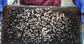
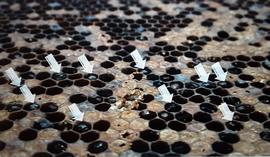
Signals of AFB occur in capped brood cells. Cappedcapping:
the covering that bees add over comb cells containing fully ripened honey or to cap brood that has reached the pupal stage; bee bread cells are not capped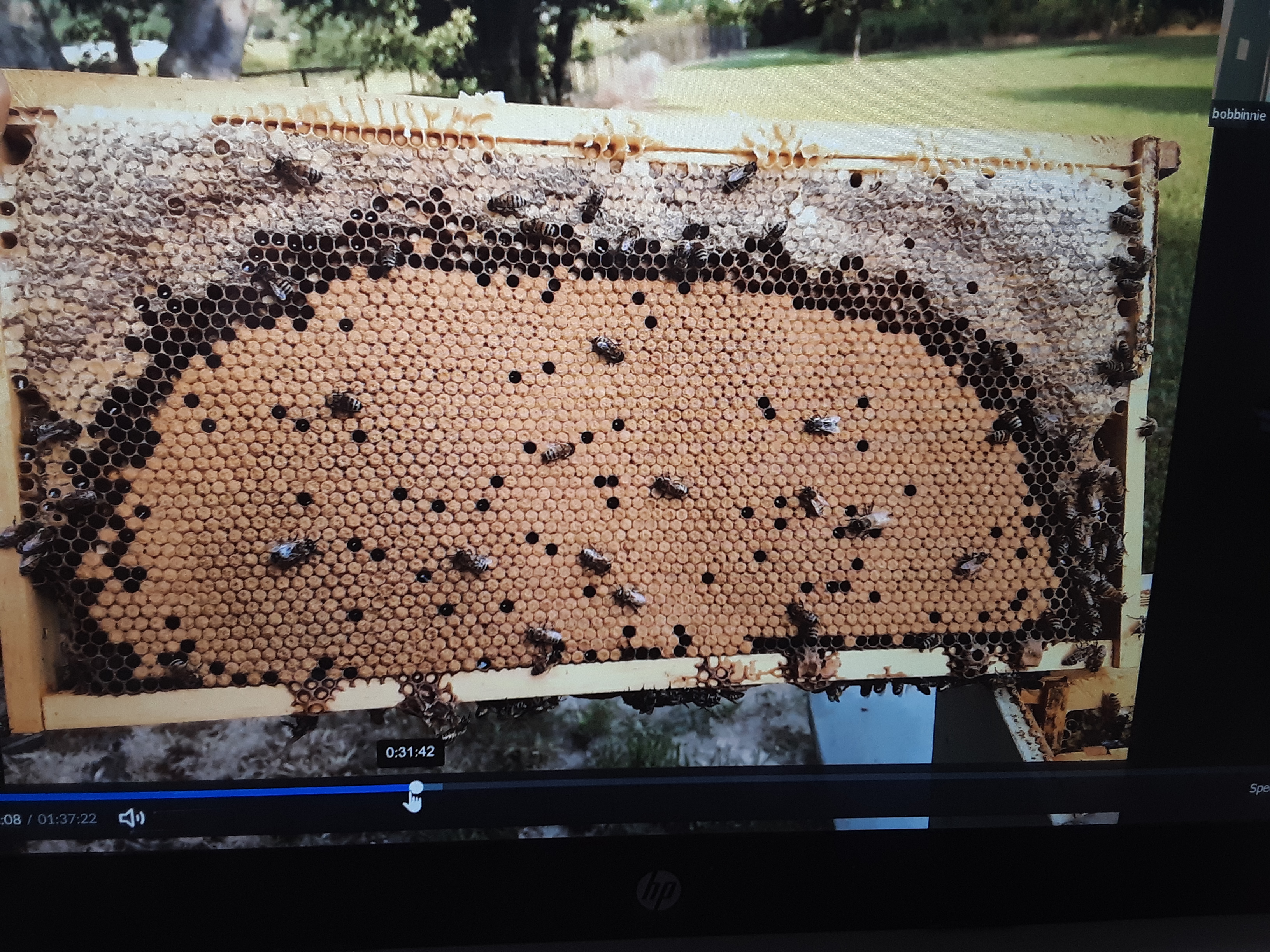 worker cells may be sunken, discolored, and have a tiny perforation in the cappingcapping:
the covering that bees add over comb cells containing fully ripened honey or to cap brood that has reached the pupal stage; bee bread cells are not capped. If cappingcapping:
the covering that bees add over comb cells containing fully ripened honey or to cap brood that has reached the pupal stage; bee bread cells are not capped is removed or partially removed, the decaying larva will be colored café-au-lait to caramel to brownish, finally turning black. Prior to complete dehydration, the remains, when poked with a stick or forceps, will rope out, as if pulling taffy. Dehydrated larvae sink to the lower cell wall, where they form a scale that adheres tightly; scales are not removable without destroying the cell itself.
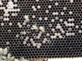
Initially, one or a few cells may break down with the disease. This might be discovered in a spring disease inspection. Bees attempting to clean out the dead remains may spread the bacteria (or the spores) to other developing larvae, leading to a downward spiral, with the disease finally resulting in death of the colony. When discovered as a deadout or heavily infected colony, the disease may have spread to other colonies via robbing or drifting of worker bees.
State and provincial apiaryapiary:
a place where beehives and beekeeping equipment are located; also called a bee yard. An out-apiary is a site away from the owner’s residence. laws generally prohibit maintaining colonies and combs with AFB and specify method of treatment when it is detected. Apiaryapiary:
a place where beehives and beekeeping equipment are located; also called a bee yard. An out-apiary is a site away from the owner’s residence. inspectors in some states and provinces actively examine colonies for AFB and only certify or permit interstate shipments of colonies that are AFB-free.
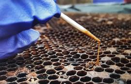
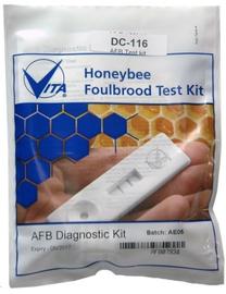
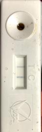
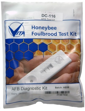
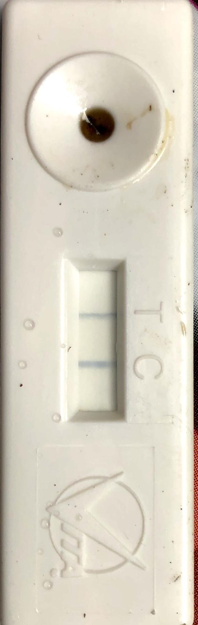
Since AFB is such a serious disease, it is best to obtain confirmation before attempting any mitigation. Get a local expert who is familiar with diseases to examine your colony or the frame you find with suspicious-looking cells, or send a sample of infected brood to the USDA Bee Disease Lab in Beltsville, MD. Some state agencies or universities will also do a disease diagnosis. You can purchase an AFB test kit (sold by Vita Bee Health) to test decaying brood cells; they have a high accuracy rate.
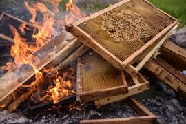
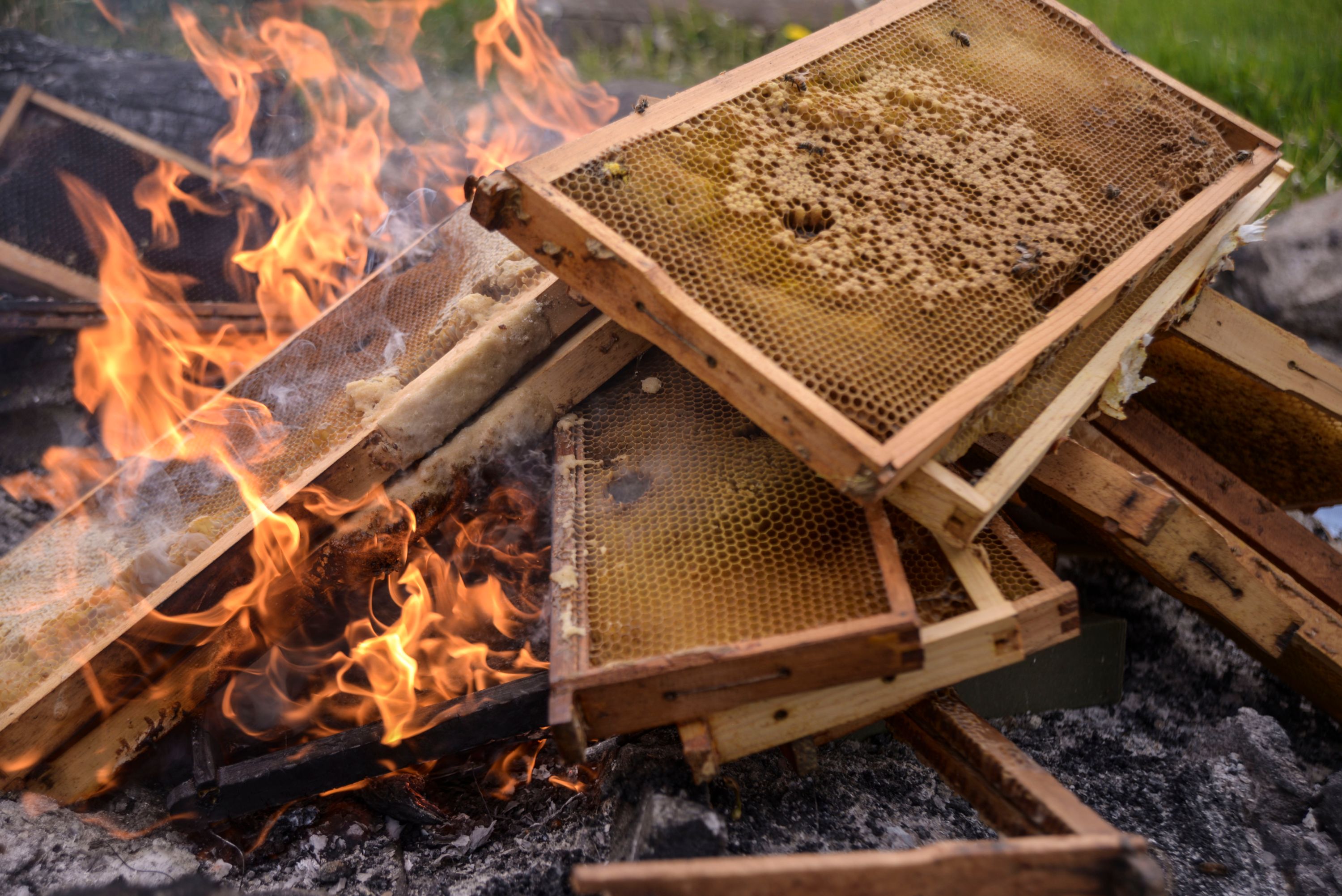
Once AFB has been confirmed, the colony (adult bees) normally needs to be euthanized, and bees and combs of brood and honey should be burned or sent to a landfill to be buried. Soapy water poured into the hive will kill the adults. In very early detection instances there are some other possible mitigation measures, but only practiced beekeepers should attempt such action. Antibiotics obtained through a veterinarian who confirms the disease continue to be used, but such treatment is not practical for the smaller scale beekeeper and/or individuals without animal husbandry experience.
European foulbrood, parasitic mite syndrome
Foulbrood. University of Florida/IFAS Honey Bee Research and Extension Lab. Accessed 2023. https://entnemdept.ufl.edu/honey-bee/beekeeper-resources/pest-and-disease-resources/foulbrood/
Shimanuki H and Knox D. 2000. Diagnosis of Honey Bee Diseases. US Department of Agriculture Agriculture Handbook 690. https://www.ars.usda.gov/is/np/honeybeediseases/honeybeediseases.pdf
MAAREC. 2005. Bee diseases & their control. Mid-Atlantic Apicultural Research & Extension Consortium Publication 4.9. https://canr.udel.edu/maarec/wp-content/uploads/sites/18/2010/03/Diseases_of_Honey_Bees_PM.pdf
Lόpez-Uribe M and Underwood R. 2019. Honey Bee Diseases: American Foulbrood. PennState Extension. Accessed 2023. https://extension.psu.edu/honey-bee-diseases-american-foulbrood
Milbrath M. 2018. Diagnosing and Treating American Foulbrood in Honey Bee Colonies. Michigan State University. Accessed 2023. https://pollinators.msu.edu/sites/_pollinators/assets/File/AmericanFoulbrood_Milbrath_2018.pdf
Caron DM. 2021. Dogs can smell disease. Bee Culture. Accessed 2023. https://www.beeculture.com/dogs-can-smell-honey-bee-disease/
Inglin K. 2020. Euthanizing a colony. The Beekeeper’s Corner. Accessed 2023. https://www.bkcorner.org/euthanizing-a-colony/
Breece, Carolyn. “Spotting American Foulbrood in your bee colony”. YouTube, uploaded by Oregon State Extension & Engagement, 21 July 2021. https://www.youtube.com/watch?app=desktop&v=6X9HKPFTl84&feature=emb_title


_and_Tukka._B._Ostendorf.jpg)
_and_Tukka._B._Ostendorf.jpg)