

more likely to be kleptoparasitic or neutral/beneficial rather than parasitic; probably feeds on pollen or fungi in the nest
Crossacarapis Ochoa and OConnor, 1996Ochoa and OConnor, 1996:
Ochoa, R. amp; B. M. OConnor. 1996. A new genus of Tarsonemidae (Acari: Heterostigmata) associated with an orchid bee, Euglossa sp. (Hymenoptera: Apidae) in Brazil. International Journal of Acarology 22: 203-207.
Superorder Acariformes » Order Trombidiformes » Suborder Prostigmata » Infraorder Eleutherengona » Hyporder Heterostigmata » Family Tarsonemidae » Genus Crossacarapis
Crossacarapis eickworti Ochoa and OConnor, 1996Ochoa and OConnor, 1996:
Ochoa, R. amp; B. M. OConnor. 1996. A new genus of Tarsonemidae (Acari: Heterostigmata) associated with an orchid bee, Euglossa sp. (Hymenoptera: Apidae) in Brazil. International Journal of Acarology 22: 203-207.
Female: Gnathosomal capsule not conspicuously beaklike (Figs. 2, 5). Cheliceral stylets short, not extending to gnathosomagnathosoma:
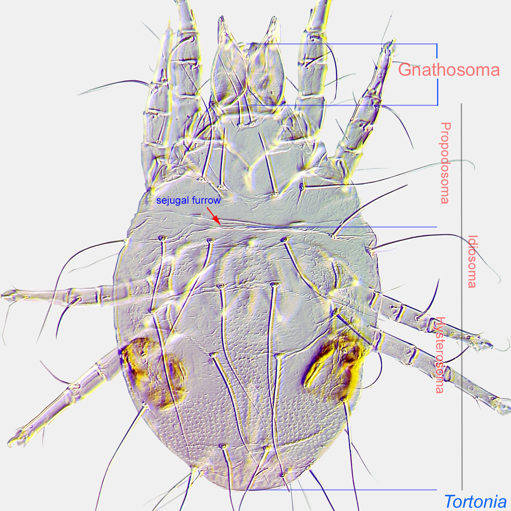
Division of body anterior to the propodosoma bearing two pairs of appendages (palps and chelicerae).
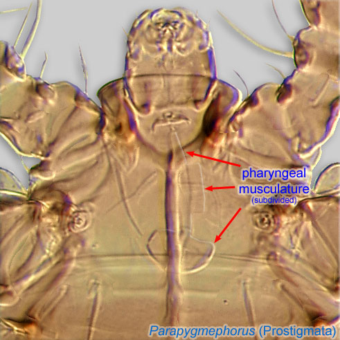
 anteriorly (Figs. 2, 5). Pharynxpharynx:
A muscular ectodermal structure that opens into a buccal cavity or mouth, and serves as a suction pump for ingesting food materials. In certain Heterostigmata pharyngal musculature is subdivided into 2-3 distinct parts.
 not conspicuously enlarged (Figs. 2, 5). Prodorsalprodorsal:
Pertaining to the prodorsum.
shield extends hoodlike over gnathosomagnathosoma:
Division of body anterior to the propodosoma bearing two pairs of appendages (palps and chelicerae).
(Figs. 1, 3). Prodorsalprodorsal:
Pertaining to the prodorsum.
trichobothria present, capitate (Figs. 1, 3). Stigmata situated well posterolaterad of setae v1 (Figs. 1, 3). No hornlike protuberances associated with stigmata (Figs. 1, 3). Setae sc2 well posterior to stigmata (Figs. 1, 3). Dorsal idiosomal setae simplesimple:
Of claws or setae; not modified or not bi- or trifurcate at tip.
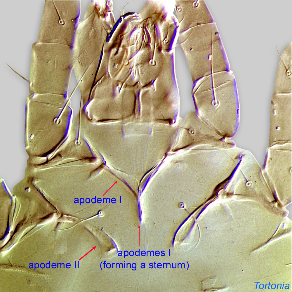
, slightly widened, short (Figs. 1, 4). Metapodosomal venter with 2 pairs of setae (3a and 3b) (3c and 4b absent) (Figs. 2, 6). ApodemesApodeme:
Internal sclerite that serves as an attachment site for muscles. Most commonly used (as "coxal apodeme") to describe elements of coxae fused to the ventral body in Acariformes (coxae are free and not fused to the body in Parasitiformes), and may be variously referred to as ventral, sternal, anterior, or posterior.
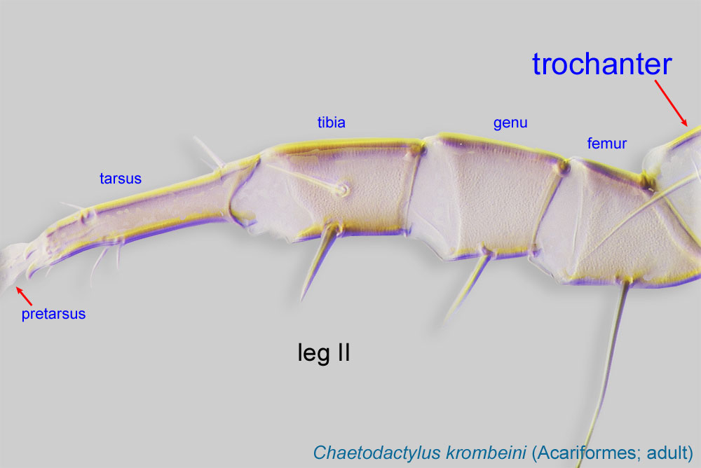
 4 not extending posterolaterally to bases of trochanterstrochanter:
Leg or palp segment (also known as podomere or palpomere) between femur and coxa. In Acariformes this is the most basal movable leg segment (or podomere) forming a joint with the body.
 IV (Figs. 2, 6). Tegulategula:
Lobe-like to acuminate cuticular plate projecting posteriorly between coxae IV in Tarsonemidae. Also used for hinged sclerite covering the wing bases in bees.
short, not elongated (Figs. 2, 6). Ambulacrumambulacrum:
The claws and empodium of the apotele or pretarsus.
I developed (Figs. 1, 3). Claw I present, not enlarged (Figs. 1, 3). Femurfemur:
Leg or palp segment (also known as podomere or palpomere) between genu and trochanter. In ParasitIformes can be subdivided into telofemur and basifemur.
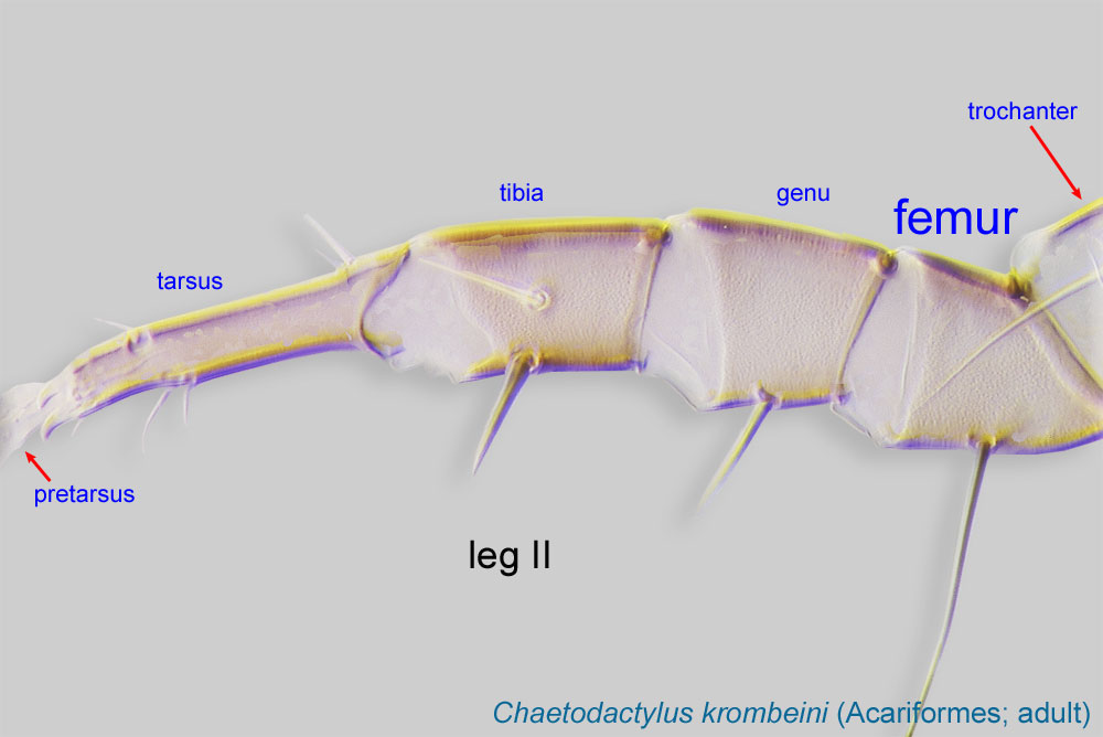 I with four setae, femurfemur:
Leg or palp segment (also known as podomere or palpomere) between genu and trochanter. In ParasitIformes can be subdivided into telofemur and basifemur.
II with three setae (Figs. 2, 5).
Only one species has been described in the genus Crossacarapis.
Crossacarapis and Pseudacarapis differ from Acarapis by their small cheliceraechelicera:
Anterior, paired appendage of the body. Primary organ for food acquisition, adapted for chewing, piercing, tearing, sucking, or filtering.
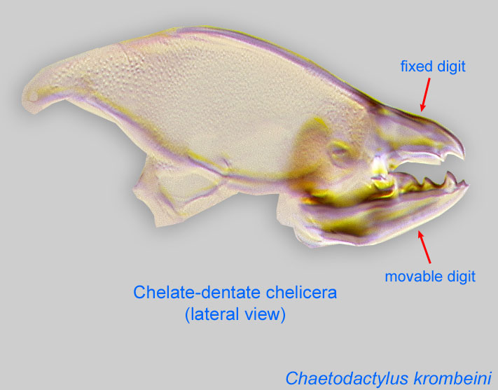 (Fig. 2) (long in Acarapis). Females of Crossacarapis can be distinguished from those of Pseudacarapis by the position of the stigmata well posterior to setae v1 (slightly posterior to stigmata in Pseudacarapis), and by apodemesapodeme:
Internal sclerite that serves as an attachment site for muscles. Most commonly used (as "coxal apodeme") to describe elements of coxae fused to the ventral body in Acariformes (coxae are free and not fused to the body in Parasitiformes), and may be variously referred to as ventral, sternal, anterior, or posterior.
4 not reaching bases of trochanterstrochanter:
Leg or palp segment (also known as podomere or palpomere) between femur and coxa. In Acariformes this is the most basal movable leg segment (or podomere) forming a joint with the body.
IV (reaching in Pseudacarapis).
Brazil
Orchid bee, Euglossa sp.
permanent permanent:
associated exclusively with bees or their close relative, wasps; cannot live without these hosts
(probably)
Based on cheliceral morphology these mites may be pollen feeders or fungivores in the nest, but they could possibly be parasitic on the larval instars of the bees. The short cheliceral stylets could conceivably pierce the thin cuticle of larval bees (Ochoa and OConnor, 1996Ochoa and OConnor, 1996:
Ochoa, R. amp; B. M. OConnor. 1996. A new genus of Tarsonemidae (Acari: Heterostigmata) associated with an orchid bee, Euglossa sp. (Hymenoptera: Apidae) in Brazil. International Journal of Acarology 22: 203-207.).
Authors: P. Klimov, B. OConnor, R. Ochoa, G. Bauchan, A. Redford, J. Scher
Last updated October 2016
tool images at ITP Node
idtools.org


