endoparasites of honey bees; feed on hemolymph in tracheae and air sacs
Acarapis Hirst, 1921
Superorder Acariformes » Order Trombidiformes » Suborder Prostigmata » Infraorder Eleutherengona » Hyporder Heterostigmata » Family Tarsonemidae » Genus Acarapis
Tarsonemus woodi Rennie, 1921
honey bee tracheal mite
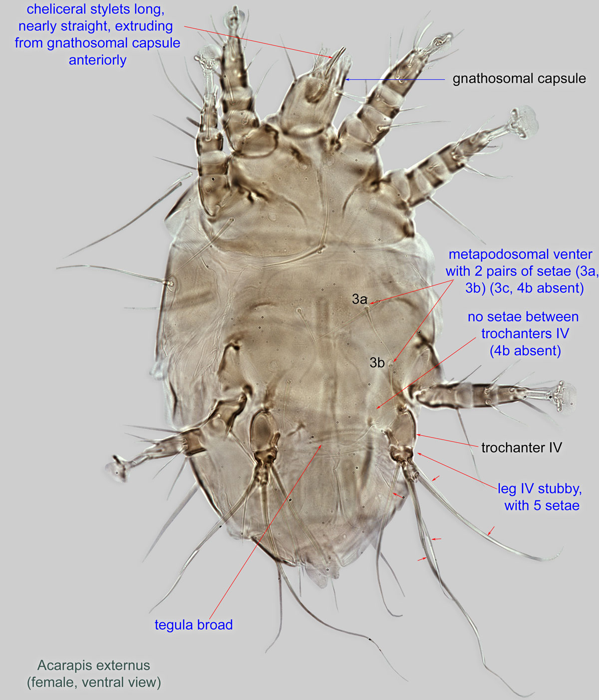
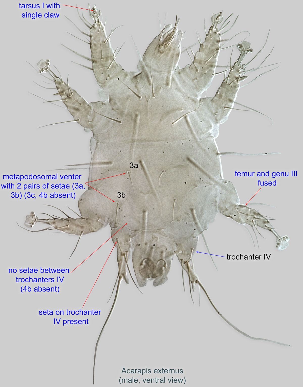
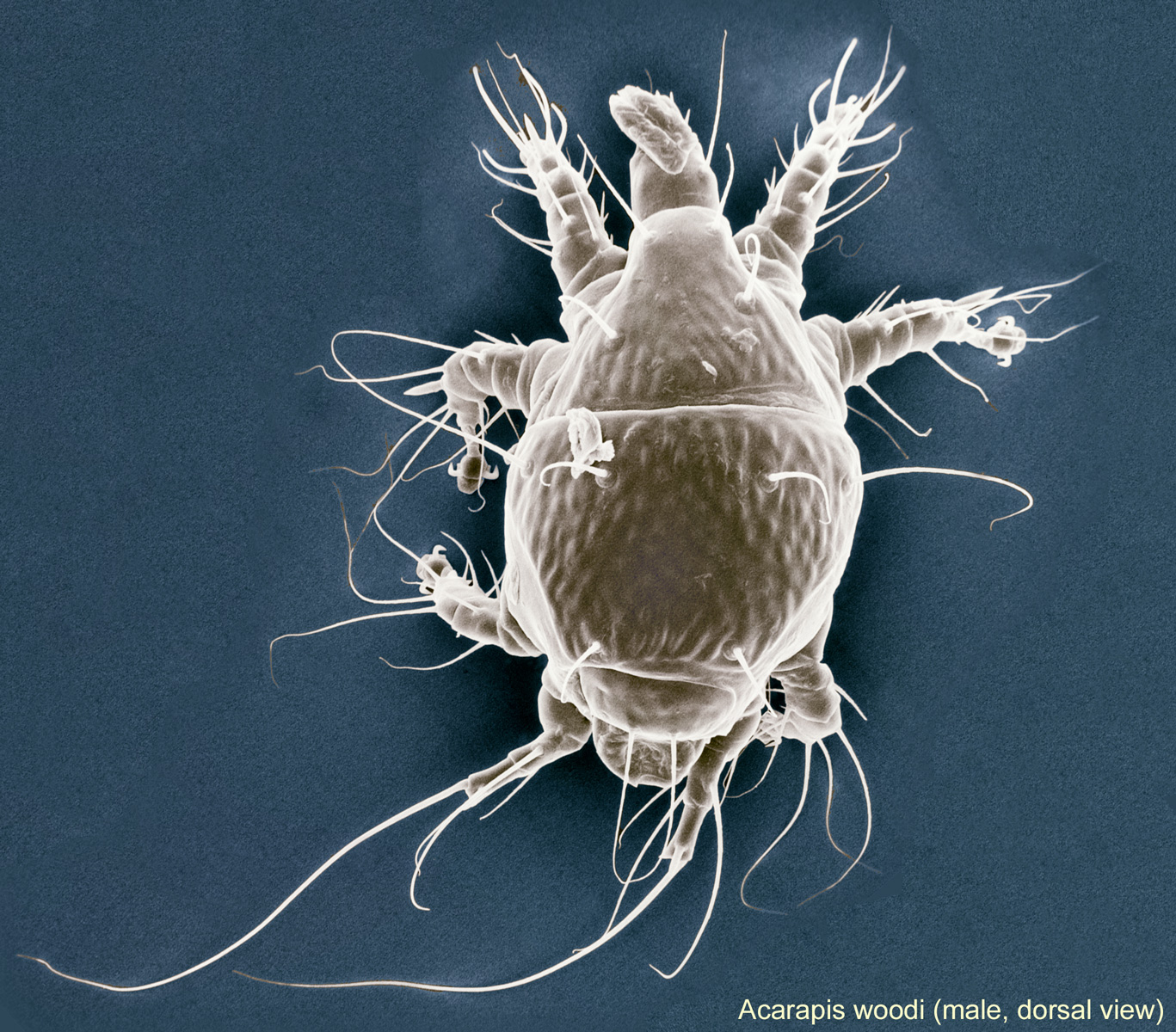
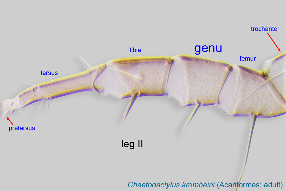
Adult: Metapodosomal venter with 2 pairs of setae (3a, 3b) (3c, 4b absent) (Figs. 2, 4). No setae between trochanterstrochanter:
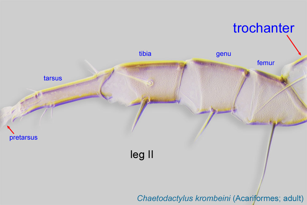
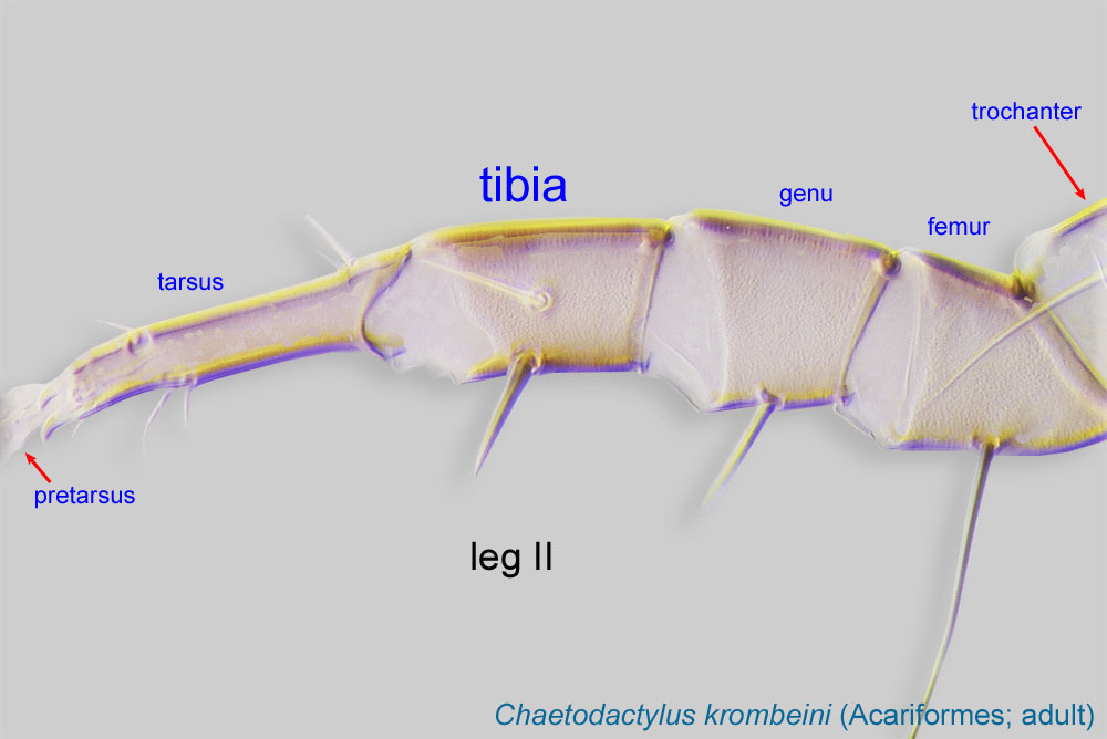
Leg or palp segment (also known as podomere or palpomere) between femur and coxa. In Acariformes this is the most basal movable leg segment (or podomere) forming a joint with the body.
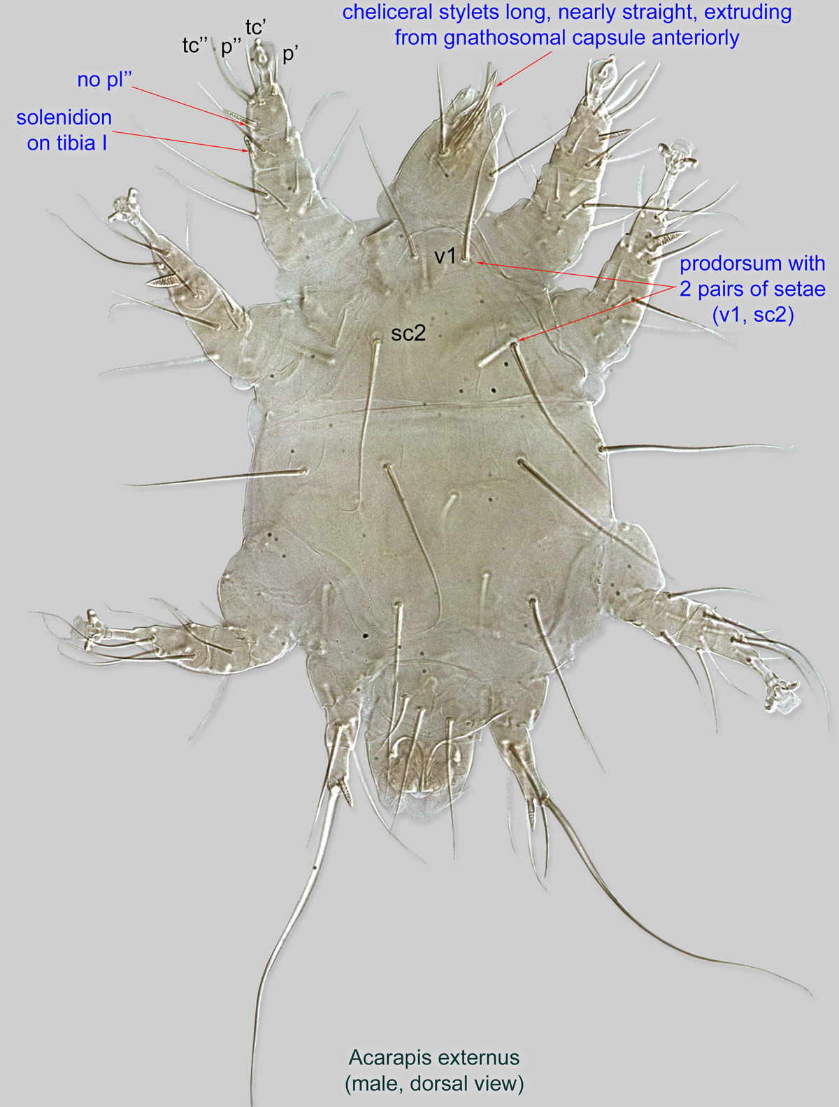
 IV (4b absent) (Figs. 2, 4). Seta pl’’ absent from tarsustarsus:
Terminal segment (also known as podomere or palpomere) of legs or palps. In Parasitoformes it can be subdivided into telotarsus and basitarsus.
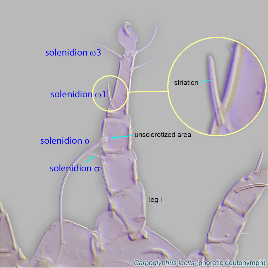
 I (Figs. 1, 3). Solenidionsolenidion:
Thin-walled, terminally rounded or pointed filiform or peglike structure that is not birefringent in polarized light (unlike common setae in Acariformes). Often appears striated because of its internal structure. Found on the palpal tarsus on the gnathosoma and may also occur on the tarsus and tibia, less frequently on the genu, and occasionally on the femur of legs I-IV. In Acariformes, leg solenidia often arise from unsclerotized areas.
 on tibiatibia:
Leg or palp segment (also known as podomere or palpomere) between tarsus and genu.
 I present (Figs. 1, 3). Cheliceral stylets long, nearly straight, extruding from gnathosomal capsule anteriorly (Figs. 2, 3).
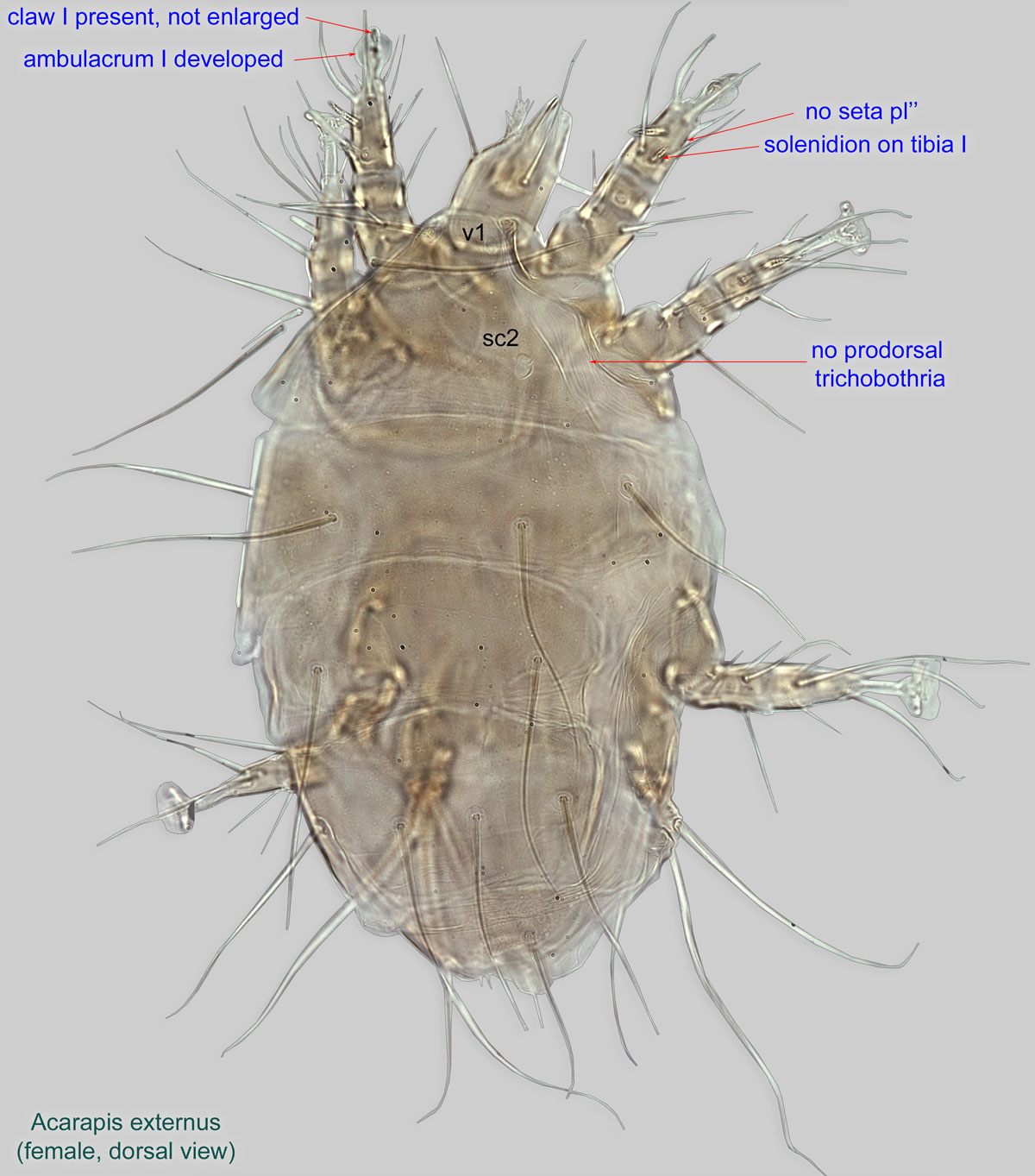
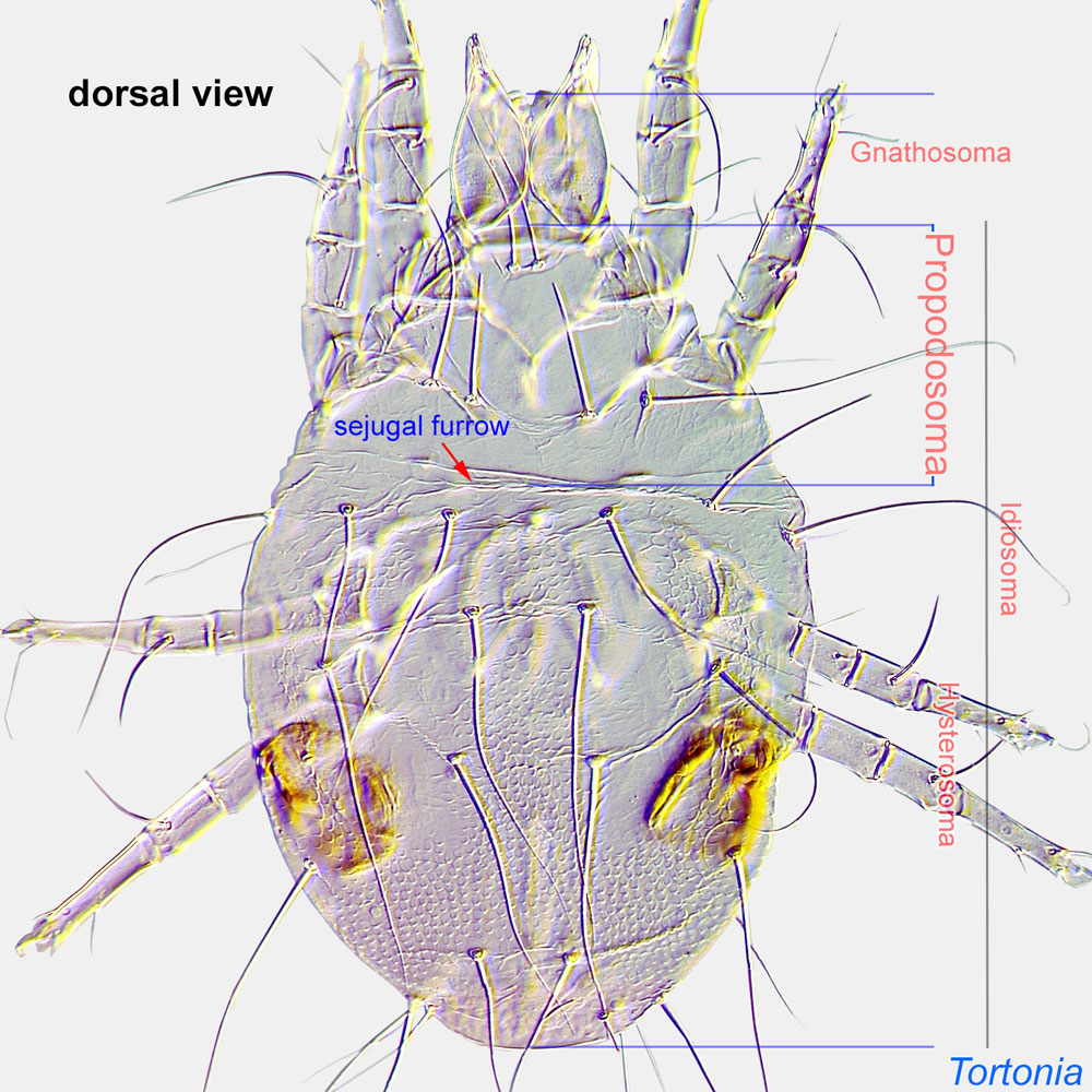
Female: Prodorsalprodorsal:
Pertaining to the prodorsum.
trichobothria absent (Fig. 1). Claw I present, not enlarged (Fig. 1). Ambulacrumambulacrum:
The claws and empodium of the apotele or pretarsus.
I developed (Fig. 1). Leg IV stubby, with 5 setae (Fig. 2). Tegulategula:
Lobe-like to acuminate cuticular plate projecting posteriorly between coxae IV in Tarsonemidae. Also used for hinged sclerite covering the wing bases in bees.
distinct, broad (Fig. 2).
Male: Prodorsumprodorsum:
Dorsal surface of propodosoma.
 with 2 pairs of setae (v1, sc2) (Fig. 3). Femurfemur:
Leg or palp segment (also known as podomere or palpomere) between genu and trochanter. In ParasitIformes can be subdivided into telofemur and basifemur.
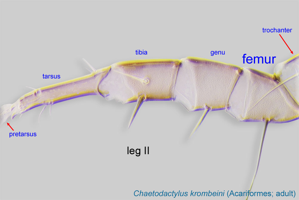 and genugenu:
Leg or palp segment (also known as podomere or palpomere) between tibia and femur.
 III fused (Fig. 4). Tarsustarsus:
Terminal segment (also known as podomere or palpomere) of legs or palps. In Parasitoformes it can be subdivided into telotarsus and basitarsus.
I with single claw (Fig. 4). Seta on trochantertrochanter:
Leg or palp segment (also known as podomere or palpomere) between femur and coxa. In Acariformes this is the most basal movable leg segment (or podomere) forming a joint with the body.
IV present (Fig. 4).
A dichotomous key is available in Delfinado-Baker and Baker, 1982bDelfinado-Baker and Baker, 1982b:
Delfinado-Baker, M. amp; E. W. Baker. 1982b. Notes on honey bee mites of the genus Acarapis Hirst (Acari: Tarsonemidae). International Journal of Acarology 8: 211-226..
This genus is associated with honey bees Apis mellifera, Apis cerana, and Apis dorsata. Acarapis woodi (Rennie, 1921) has been found on all three of these bee species. Acarapis externus Morgenthaler in Morison, 1931 has been found on Apis mellifera and Apis cerana, and Acarapis dorsalis Morgenthaler, 1934 has been found only on Apis mellifera.
permanentpermanent:
associated exclusively with bees or their close relative, wasps; cannot live without these hosts
Acarapis woodi
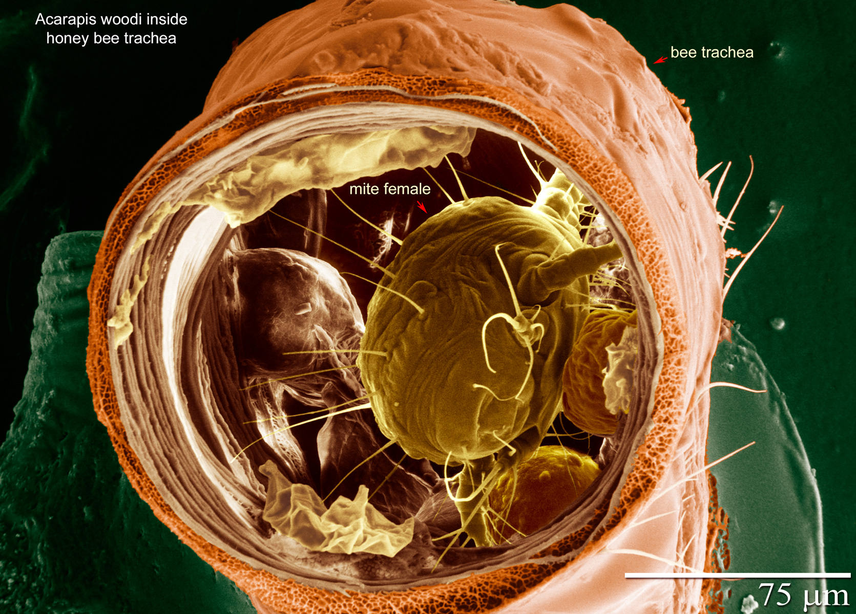
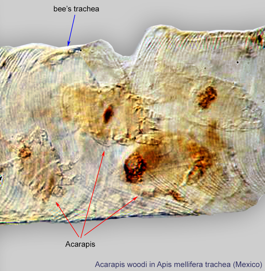
Acarapis woodi mites are usually found in the large tracheal tubes of the mesosoma (Figs. 6, 7). (Detection of the mites requires dissection of the bees.) Development and mating take place in the tracheae and newly mated females migrate from the tracheae of their original host to tracheae in a newly emerged young adult bee. Host-seeking mites attach themselves to bee hairs and then move onto younger bees. Clustering of bees during the winter greatly facilitates cross-infestation. Mite males are seldom found outside the tracheae. Females lay a single large egg at a time, probably one egg per day during the first two weeks of the host's life. The egg hatches into an active feeding larva. Nymphs are pharate, developing inside the larval skin. Adults emerge about 11 days after oviposition. This pattern of development is similar to that of other tarsonemid mites.
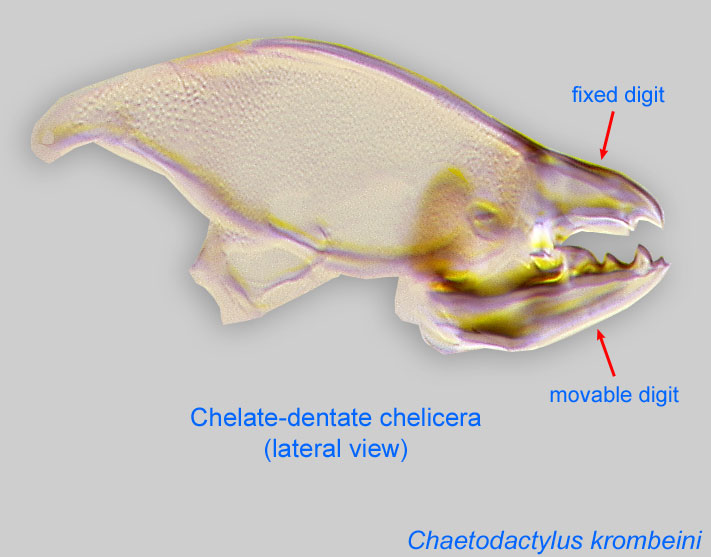
Acarapis woodi mites pierce the bee tracheae and air sacs with their cheliceraechelicera:
Anterior, paired appendage of the body. Primary organ for food acquisition, adapted for chewing, piercing, tearing, sucking, or filtering.
 and feed on the hemolymph, possibly impairing bee respiration. This lowers the bee's ability to use wing muscles. As a consequence, bees cannot keep their cluster warm in winter. Overwintering bees die when crawling out of colonies. In warmer climates, bees will survive, but the mites can pass on harmful viruses. The mite also can weaken drones and queens.
Acarapis woodi is responsible for significant colony losses throughout North America. Reports of losses as great as 90% have been recorded. A heavy mite load causes diminished brood area, smaller bee populations, looser winter clusters, increased honey consumption, lower honey yields, and, ultimately, colony demise. In temperate regions, mite populations increase during winter, when bees are confined to the hive (making it easier for mites to move between bee hosts), and decrease in summer when bee populations are highest. In subtropical climates, the cycle is similar, even though bees are not so confined (after Royce, 1999Royce, 1999:
Royce, L. A. 1999. Biology and epidemiology of honey bee tracheal mites Acarapis woodi. In Acarology 9. Volume 2. Symposia, 309-311. Columbus: Ohio Biological Survey.; Sammataro et al., 2000Sammataro et al., 2000:
Sammataro, D., U. Gerson amp; G. Needham. 2000. Parasitic mites of honey bees: life history, implications, and impact. Annual Review of Entomology. 45: 519-548.).
Two other species, Acarapis dorsalis and A. externus, are found on the European honey bee Apis mellifera, and they also feed on the bee hemolymph (Delfinado-Baker and Baker, 1982bDelfinado-Baker and Baker, 1982b:
Delfinado-Baker, M. amp; E. W. Baker. 1982b. Notes on honey bee mites of the genus Acarapis Hirst (Acari: Tarsonemidae). International Journal of Acarology 8: 211-226.).
Authors: P. Klimov, B. OConnor, R. Ochoa, G. Bauchan, A. Redford, J. Scher
Last updated October 2016
tool images at ITP Node
idtools.org