neutral to beneficial; feeds on harmful microarthropods in hives
Melichares Hering, 1838
Superorder Parasitiformes » Order Mesostigmata » Suborder Monogynaspida » Hyporder Dermanyssiae » Family Melicharidae » Genus Melichares
Melichares agilis Hering, 1838
Garmania. In old literature was confused with Blattisocius.
Female: Dorsal shield entire, not divided, without lateral incisions (Fig. 3). Setae z3 present (Fig. 3). Opisthonotalopisthonotal:
Pertaining to dorsal opisthosoma.
region of dorsal shield with 14–15 pairs of setae (J, S, Z) (Fig. 3). IdiosomaIdiosoma:
Body not including the gnathosoma.
 with 7 or more pairs of marginal setae r-R on soft lateral cuticle (Fig. 3). Metasternal plates absent, st4 situated on soft cuticle (Fig. 5). Adanal setae anterior to hind margin of anus (Figs. 6, 7). Anal shieldanal shield:
In Mesostigmata, a ventral shield bearing the anal opening and circumanal setae (adanal or postanal setae), but without any ventral setae or pores (lyrifissures) on it. If ventral setae are present on shield than referred to as a ventrianal shield.
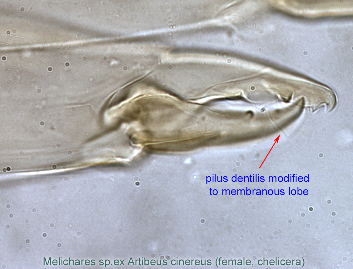
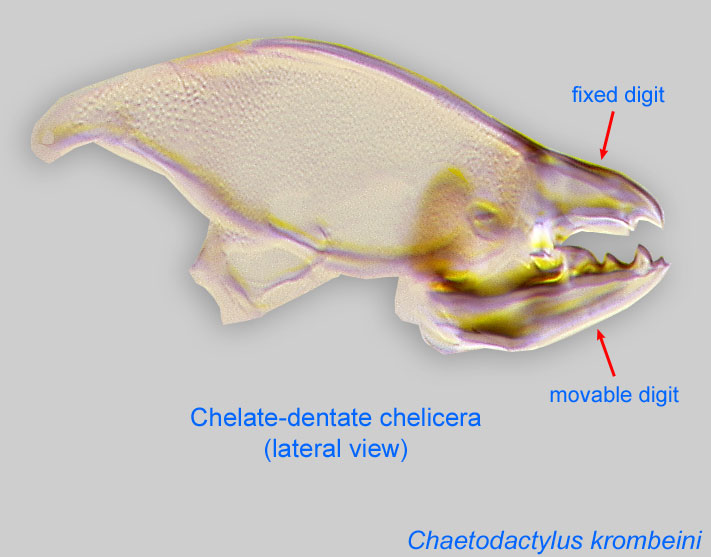
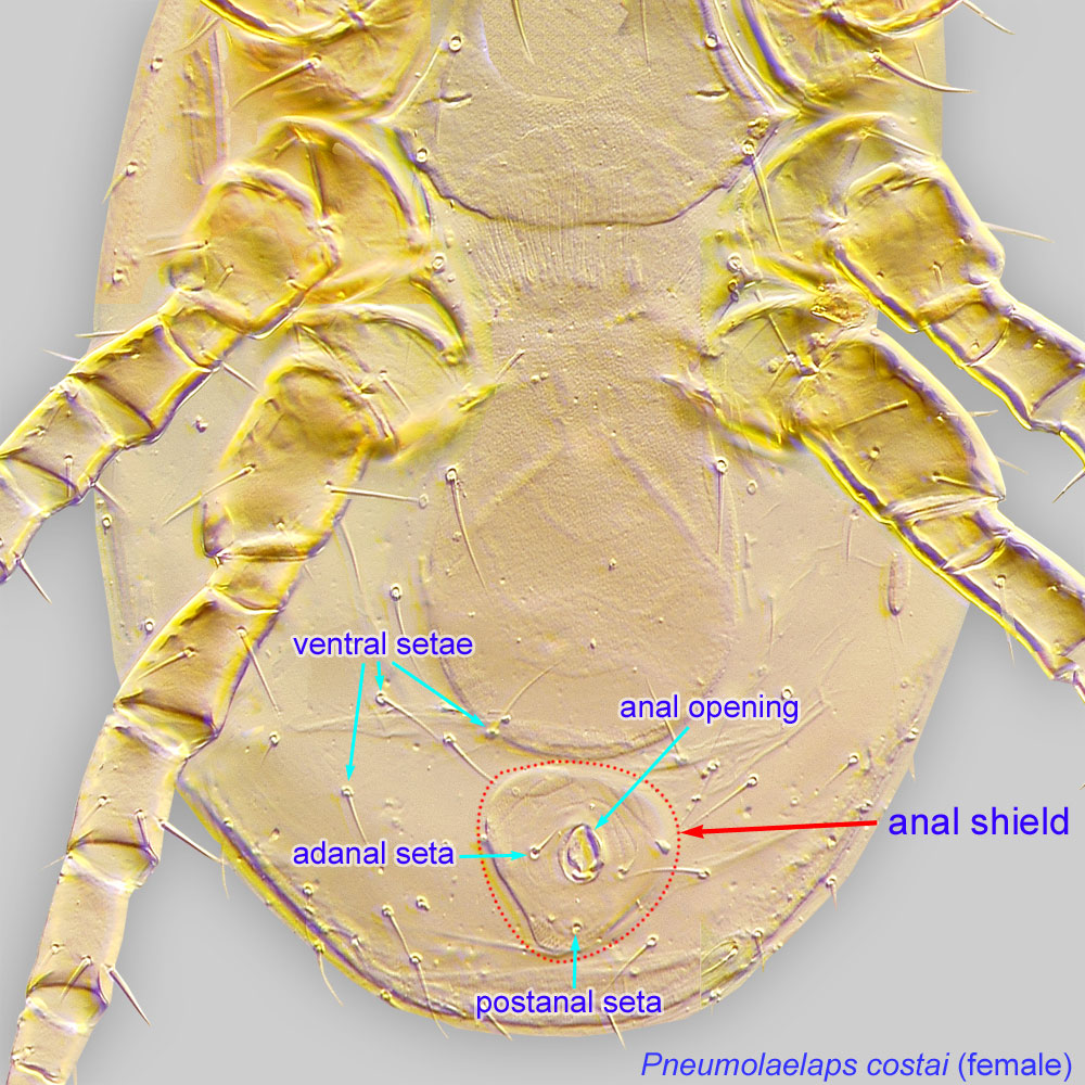 with 3 circumanal setae only (Figs. 6, 7). Epigynial shield rounded posteriorly (Fig. 4). Peritrematic shield at level of stigma less than twice as wide as diameter of stigma (Fig. 8). Peritrematic shield free posteriorly (Figs. 8, 15). Anterior and posterior rows of deutosternal denticles (small, tooth-like processes) different (Fig. 10). Fixed digit of chelicerachelicera:
Anterior, paired appendage of the body. Primary organ for food acquisition, adapted for chewing, piercing, tearing, sucking, or filtering.
 with pilus dentilispilus dentilis:
A seta-like or membranous sensory organ inserted ventrolaterally on the fixed digit of the chelicera of many Mesostigmata.
modified to membranous lobe (sometimes poorly visible) (Fig. 16). Movable digit with ventral mucro (pointed process) near base (Fig. 11). Genugenu:
Leg or palp segment (also known as podomere or palpomere) between tibia and femur.
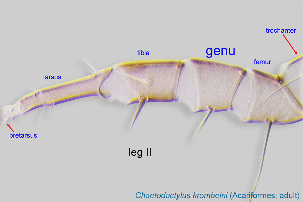 II and III with 11 and 9 setae, respectively (both av1, pv1 present); tibiatibia:
Leg or palp segment (also known as podomere or palpomere) between tarsus and genu.
 II usually with 10 setae (pl2 present) (Fig. 13). Legs II–IV with median lobe of pulvilluspulvillus:
a membranous, pad-like structure associated with the claws in Mesostigmata.
(membranous, pad-like structure) broadly rounded (Fig. 14).
This genus includes two species, but only Melichares agilis has been found in association with bees. This species can be identified using Evans, 1958Evans, 1958:
Evans, G. O. 1958. A revision of the British Aceosejinae (Acarina: Mesostigmata). Proceedings of the Zoological Society of London. 131: 177-229..
Occuring in human-related habitats, Melichares agilis is expected to be a widely distributed, cosmopolitan species. Records from honey bees are from the Western Palaearctic.
European honey bee, Apis mellifera
facultativefacultative:
can complete entire life cycle without bees or their close relative, wasps
Melichares agilis is known as a widely distributed predator of acarid mites in stored food, particularly dried fruits. This species has also been found in hives of the European honey bee Apis mellifera (Haragsim et al., 1987Haragsim et al., 1987:
Haragsim, O., K. Samšiňák amp; E. Vobrázková. 1978. The mites inhabiting the bee hives in ČSR. Zeitschrift für Angewandte Entomologie. 87: 52-67.).
Authors: P. Klimov, B. OConnor, R. Ochoa, G. Bauchan, A. Redford, J. Scher
Last updated October 2016
tool images at ITP Node
idtools.org