potentially harmful; may kill developing bees or may simply prefer living in cells with dead bees
Medeus Volgin, 1974Volgin, 1974:
Volgin, V. I. 1974. New genera and species of acaroid mites (Acariformes, Acaroidea) from Kazakhstan. Entomologicheskoe Obozrenie . 53 : 218-225.
Superorder Acariformes » Order Sarcoptiformes » Suborder Oribatida » Infraorder Desmonomata » Hyporder Astigmata » Family Acaridae » Genus Medeus
Medeus vesparius Volgin, 1974Volgin, 1974:
Volgin, V. I. 1974. New genera and species of acaroid mites (Acariformes, Acaroidea) from Kazakhstan. Entomologicheskoe Obozrenie . 53 : 218-225.
Phoretic phoretic:
Pertaining to phoresy; using another organism (i.e., a host) for dispersal to new habitats. Phoresy can be distinguished from parasitism because feeding typically does not occur during phoresy.
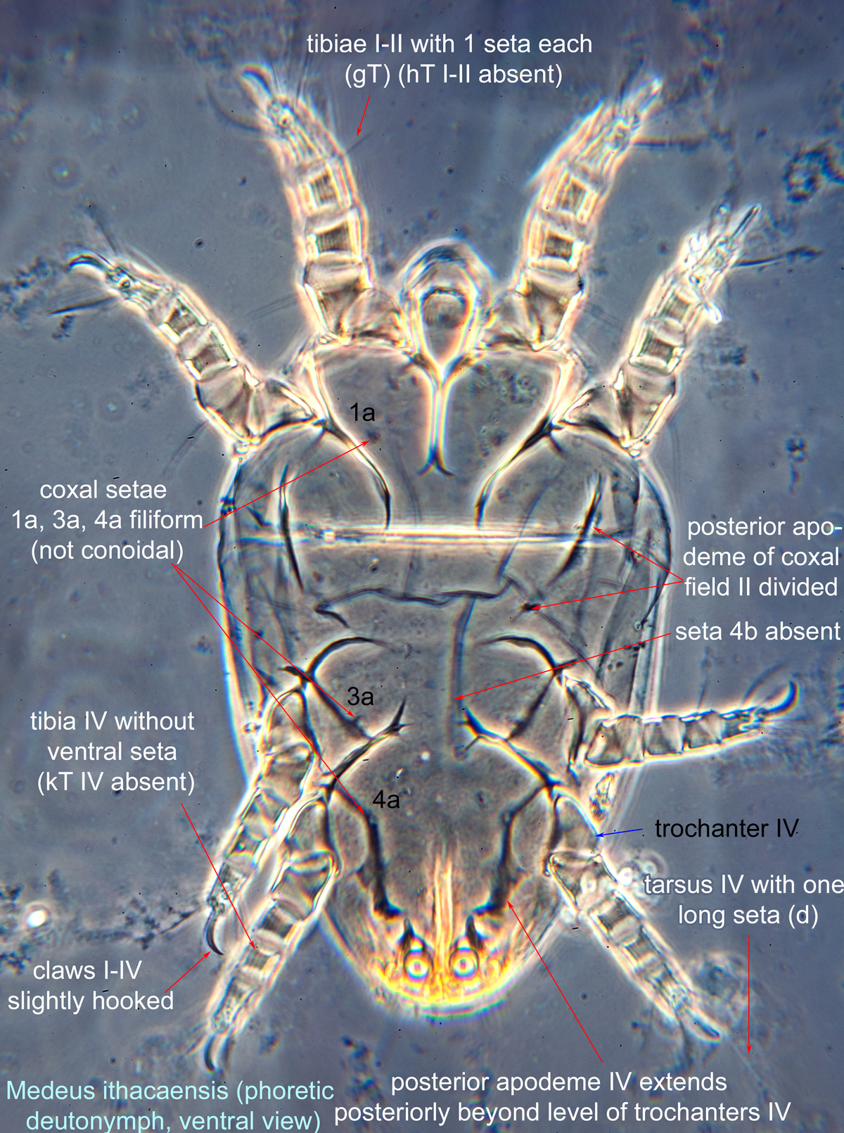
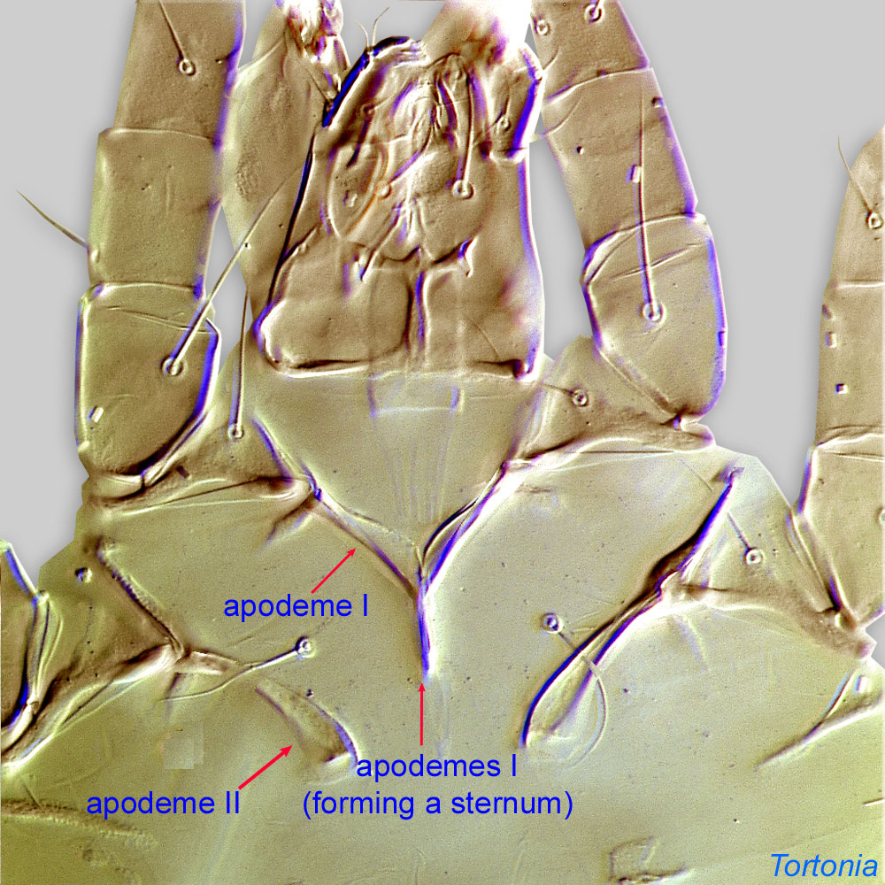
deutonymph: Posterior dorsum with well-developed longitudinal apodemeapodeme:
Internal sclerite that serves as an attachment site for muscles. Most commonly used (as "coxal apodeme") to describe elements of coxae fused to the ventral body in Acariformes (coxae are free and not fused to the body in Parasitiformes), and may be variously referred to as ventral, sternal, anterior, or posterior.
 (Figs. 1, 3). Posterior apodemesapodeme:
Internal sclerite that serves as an attachment site for muscles. Most commonly used (as "coxal apodeme") to describe elements of coxae fused to the ventral body in Acariformes (coxae are free and not fused to the body in Parasitiformes), and may be variously referred to as ventral, sternal, anterior, or posterior.
IV extend posteriorly beyond level of trochanterstrochanter:
Leg or palp segment (also known as podomere or palpomere) between femur and coxa. In Acariformes this is the most basal movable leg segment (or podomere) forming a joint with the body.
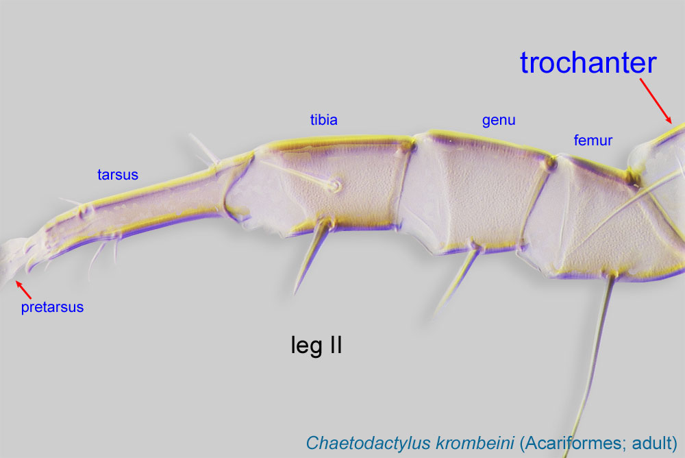 IV (Fig. 2). Tarsustarsus:
Terminal segment (also known as podomere or palpomere) of legs or palps. In Parasitoformes it can be subdivided into telotarsus and basitarsus.
 IV with one long seta (d) (Figs. 1, 2, 7). Coxal setae 1a, 3a, 4a filiform (not conoidal) (Fig 2). Coxal seta 4b absent. Posterior apodemeapodeme:
Internal sclerite that serves as an attachment site for muscles. Most commonly used (as "coxal apodeme") to describe elements of coxae fused to the ventral body in Acariformes (coxae are free and not fused to the body in Parasitiformes), and may be variously referred to as ventral, sternal, anterior, or posterior.
of coxal field II divided (Fig. 2). Claws I-IV slightly hooked (Figs. 2, 5, 6, 8). TarsiTarsus:
Terminal segment (also known as podomere or palpomere) of legs or palps. In Parasitoformes it can be subdivided into telotarsus and basitarsus.
I-II without setae ba I-II (Fig. 5). Setae aa (aa’’) absent from tarsustarsus:
Terminal segment (also known as podomere or palpomere) of legs or palps. In Parasitoformes it can be subdivided into telotarsus and basitarsus.
I (Fig. 5). TibiaeTibia:
Leg or palp segment (also known as podomere or palpomere) between tarsus and genu.
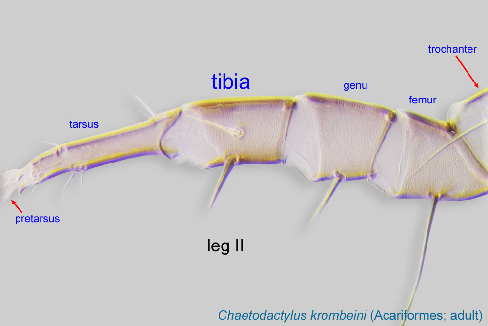 I-II with 1 seta each (gT I-II present, hT I-II absent) (Figs. 2, 6). TibiaeTibia:
Leg or palp segment (also known as podomere or palpomere) between tarsus and genu.
IV without ventral seta (kT IV absent) (Figs. 2, 8).
Adult: Posterior edge of prodorsalprodorsal:
Pertaining to the prodorsum.
sclerite incised (Fig. 13). Coxal seta 4b absent (correlates with character in deutonymphdeutonymph:
Ontogenetic stage between protonymph and tritonymph (or adult, if tritonymph is absent). See <a href="index.cfm?pageID=1720">Life stages page</a> for more details.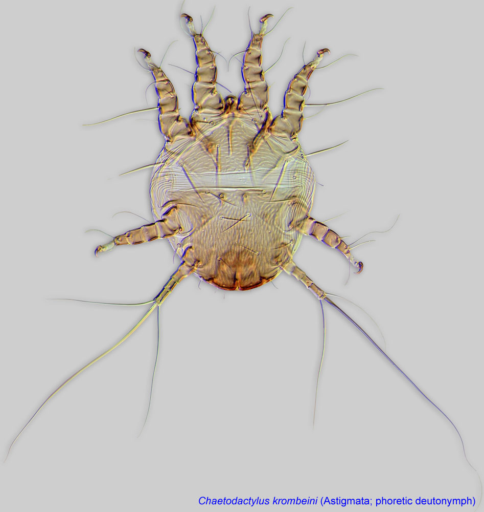 ) (Fig. 10). Setae ve situated nearly at the same level as setae vi, not barbed (Fig. 13). External and internal scapular setae (se, si) situated nearly on the same transverse line (Fig. 13). Grandjean's organGrandjean's organ:
Paired, finger-shaped, lobe-shaped, or otherwise elaborated structure situated on the lateral sides of the propodosoma, typically in association with the podocephalic canal. Its free edges may be strongly fimbriate.
rounded and fimbriate (Fig. 12). Supracoxal setae scx simplesimple:
Of claws or setae; not modified or not bi- or trifurcate at tip.
(Fig. 13). TibiaeTibia:
Leg or palp segment (also known as podomere or palpomere) between tarsus and genu.
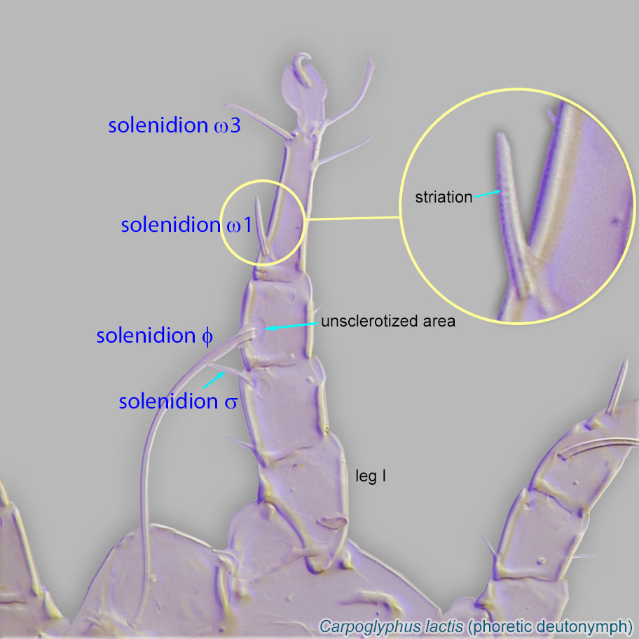
I with solenidionsolenidion:
Thin-walled, terminally rounded or pointed filiform or peglike structure that is not birefringent in polarized light (unlike common setae in Acariformes). Often appears striated because of its internal structure. Found on the palpal tarsus on the gnathosoma and may also occur on the tarsus and tibia, less frequently on the genu, and occasionally on the femur of legs I-IV. In Acariformes, leg solenidia often arise from unsclerotized areas.
 σ' not more than 3 times longer than σ'' (Fig. 14). TibiaeTibia:
Leg or palp segment (also known as podomere or palpomere) between tarsus and genu.
I-II with1 ventral seta (gT I-II present, hT I-II absent) (Fig. 14) (correlates with deutonymphdeutonymph:
Ontogenetic stage between protonymph and tritonymph (or adult, if tritonymph is absent). See <a href="index.cfm?pageID=1720">Life stages page</a> for more details.). TibiaeTibia:
Leg or palp segment (also known as podomere or palpomere) between tarsus and genu.
IV without ventral seta (kT IV absent) (correlates with deutonymphdeutonymph:
Ontogenetic stage between protonymph and tritonymph (or adult, if tritonymph is absent). See <a href="index.cfm?pageID=1720">Life stages page</a> for more details.) (Figs. 15, 16). Tarsal seta ba I present (not correlated with deutonymphdeutonymph:
Ontogenetic stage between protonymph and tritonymph (or adult, if tritonymph is absent). See <a href="index.cfm?pageID=1720">Life stages page</a> for more details.) (Fig. 14). Tarsal seta ba II absent (correlates with deutonymphdeutonymph:
Ontogenetic stage between protonymph and tritonymph (or adult, if tritonymph is absent). See <a href="index.cfm?pageID=1720">Life stages page</a> for more details.) (Fig. 14). Setae aa (aa’’) absent from tarsustarsus:
Terminal segment (also known as podomere or palpomere) of legs or palps. In Parasitoformes it can be subdivided into telotarsus and basitarsus.
I (correlates with deutonymphdeutonymph:
Ontogenetic stage between protonymph and tritonymph (or adult, if tritonymph is absent). See <a href="index.cfm?pageID=1720">Life stages page</a> for more details.) (Fig. 14).
This genus includes two described and one undescribed species. Adults of the two described species (Medeus vesparius and M. ithacaensis) can be distinguished using their original descriptions (OConnor, 1997OConnor, 1997:
OConnor, B. M. 1997. Two new mites (Acari: Acaridae) associated with long-tongued bees (Hymenoptera: Apidae) in North America. Journal of the Kansas Entomological Society . 69 : 15-34.; Volgin, 1974Volgin, 1974:
Volgin, V. I. 1974. New genera and species of acaroid mites (Acariformes, Acaroidea) from Kazakhstan. Entomologicheskoe Obozrenie . 53 : 218-225.). DeutonymphsDeutonymph:
Ontogenetic stage between protonymph and tritonymph (or adult, if tritonymph is absent). See <a href="index.cfm?pageID=1720">Life stages page</a> for more details. are known for M. ithacaensis and the undescribed species.
Nearctic, Palaearctic, and Afrotropical regions
apid bees of the genera Anthophora and rarely, Diadasia
permanentpermanent:
associated exclusively with bees or their close relative, wasps; cannot live without these hosts
disperse on bee hosts.Biological observations are available for Medeus ithacaensis (OConnor, 1997OConnor, 1997:
OConnor, B. M. 1997. Two new mites (Acari: Acaridae) associated with long-tongued bees (Hymenoptera: Apidae) in North America. Journal of the Kansas Entomological Society . 69 : 15-34.): Mites have been found only in cells with moldy provisions or dead bees. It is unknown, however, if the mites cause bee mortality or they simply prefer living in cells without bees (and later find cells with emerging bees in order to disperse). Gut content of the mites contained a mixture of crushed pollen and fungal spores, indicating that these mites can be potentially kleptoparasitic.
Authors: P. Klimov, B. OConnor, R. Ochoa, G. Bauchan, A. Redford, J. Scher
Last updated October 2016
tool images at ITP Node
idtools.org