neutral or beneficial; habitat generalists that feed on microarthropods and fungi
Lasioseius Berlese, 1916
Superorder Parasitiformes » Order Mesostigmata » Suborder Monogynaspida » Hyporder Dermanyssiae » Family Blattisociidae » Genus Lasioseius
Seius musicatus [lapsus pro muricatus] Berlese, 1887 (non Koch, 1839) (=Typhlodromus berlesei Oudemans, 1938)
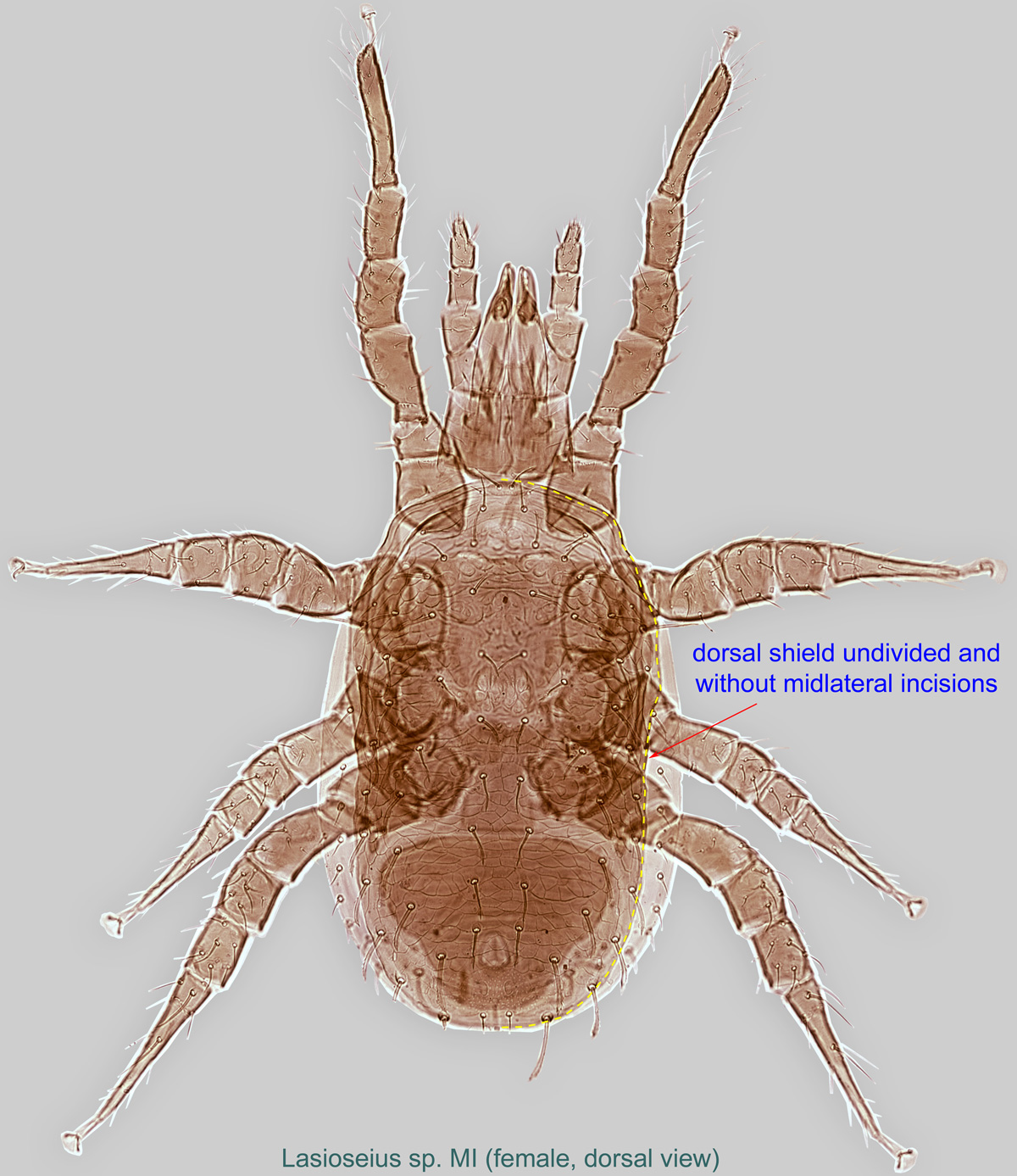
Female: Dorsal shield undivided and without midlateral incisions (Figs. 1). In most species, sternal shieldsternal shield:
A shield in the anterior intercoxal region of parasitiform mites that bears one or more pairs of sternal setae.
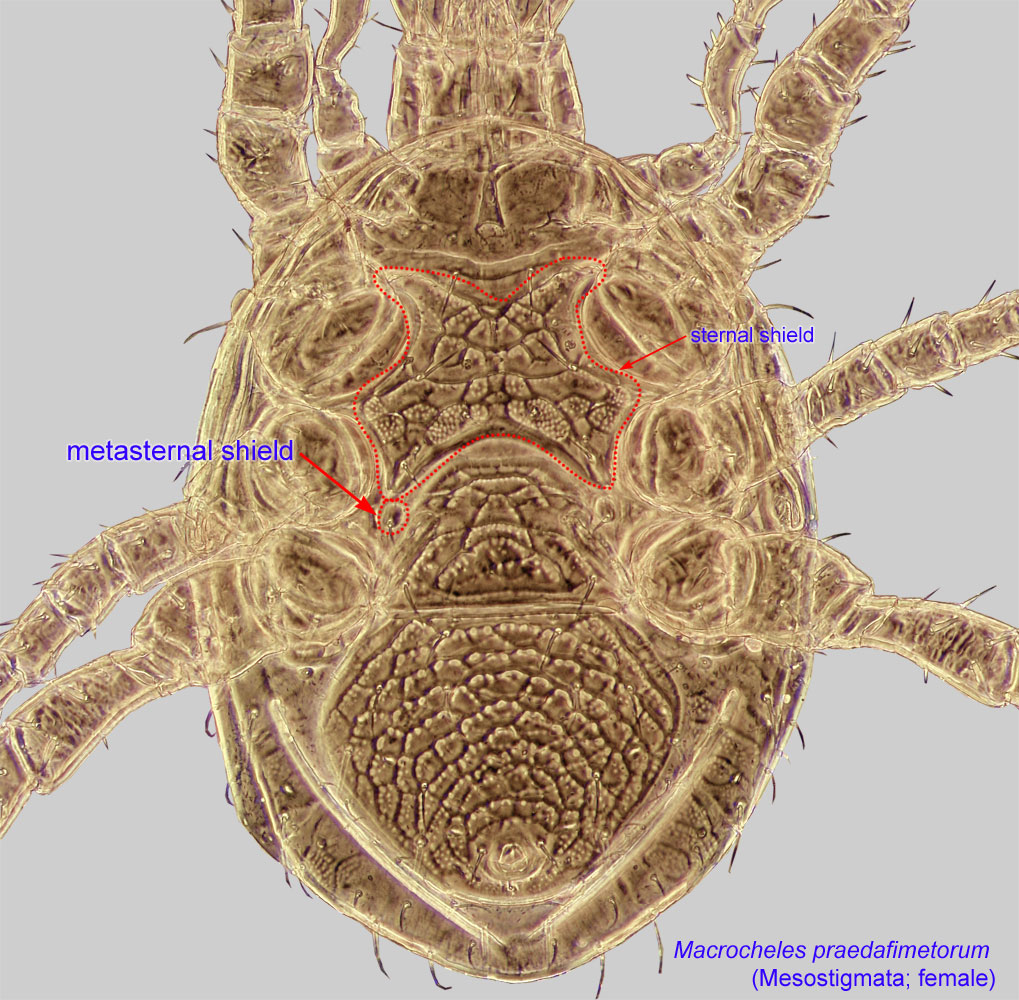
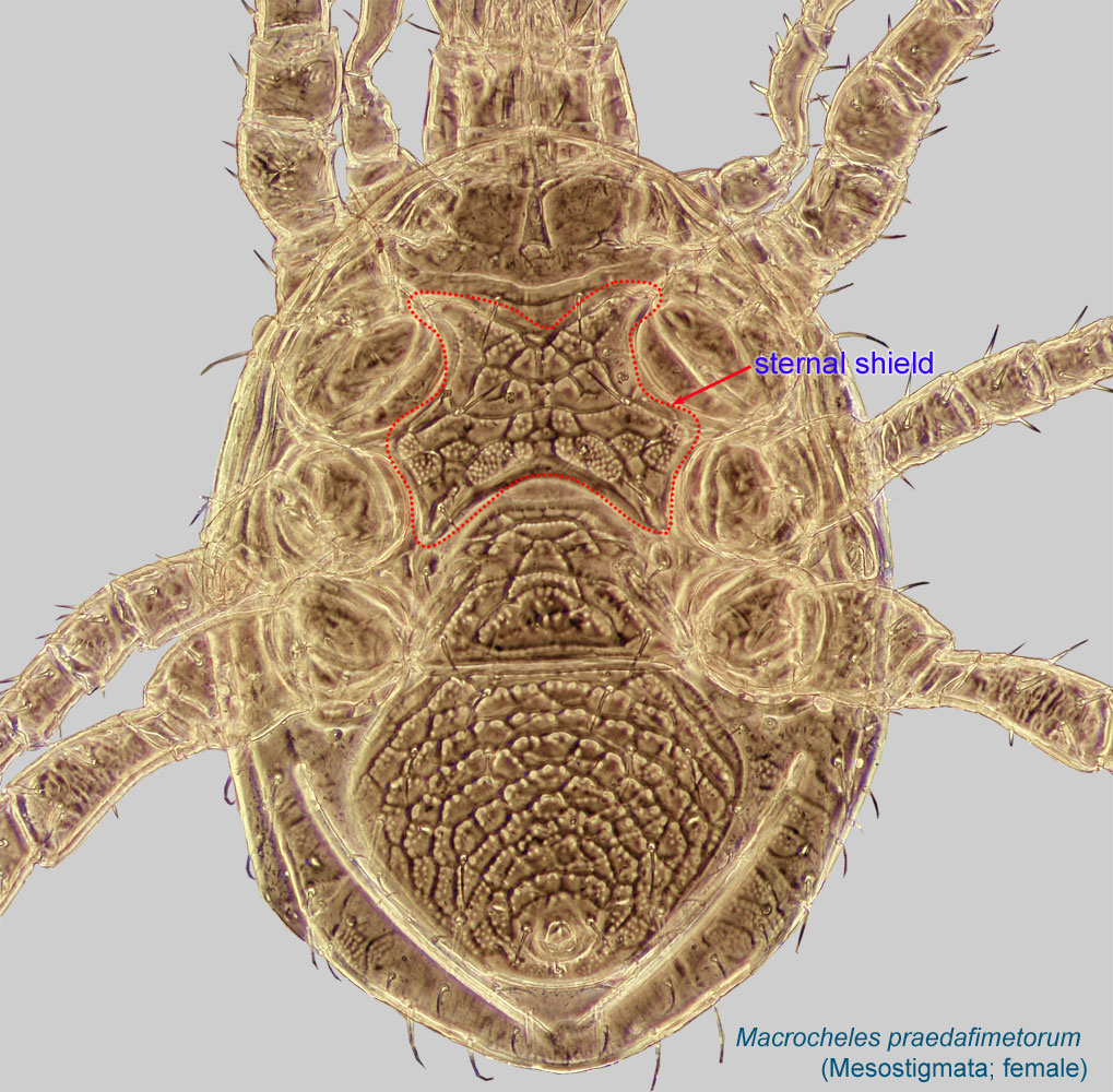 with 3 pairs of setae: st1, st2, st3 (Fig. 3); st4 situated on metasternal shieldsmetasternal shield:
Small, usually teardrop to subtriangular paired shields bearing metasternal setae st4; sometimes fused to the sternal shield or the endopodal shields. Present in Mesostigmata.
 (Figs. 3). Ventrianal shieldventrianal shield:
In Mesostigmata, a ventral shield bearing the anal opening, circumanal (postanal and adanal) setae, and one or more pairs of ventral setae or pores (lyrifissures) (see anal shield); may be narrow or very broad and covering most of the gaster.
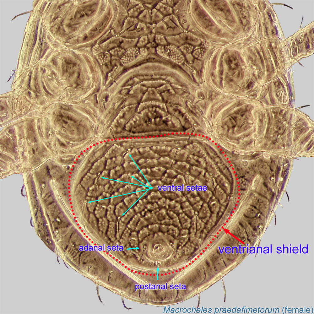 wide, bearing 2 or more pairs of ventral setae (JV and ZV), in addition to the three circumanal setae (Fig. 5). Paranal setae inserted anterior to hind margin of anus (Fig. 5). Peritrematic shield broadly connected to exopodal plate, curving behind coxacoxa:
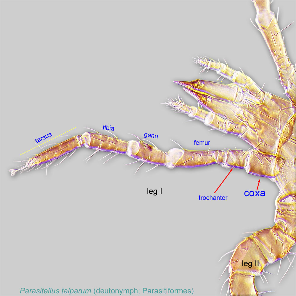
In Parasitiformes, most basal leg segment (or podomere) forming a joint with the body. Areas delimited by coxal apodemes are called coxal fields in Astigmata or coxisternal plates in Prostigmata.
 IV (Fig. 7). Peritrematic shield clearly wider than diameter of stigma at level of stigma (Fig. 7). CorniculiCorniculus:
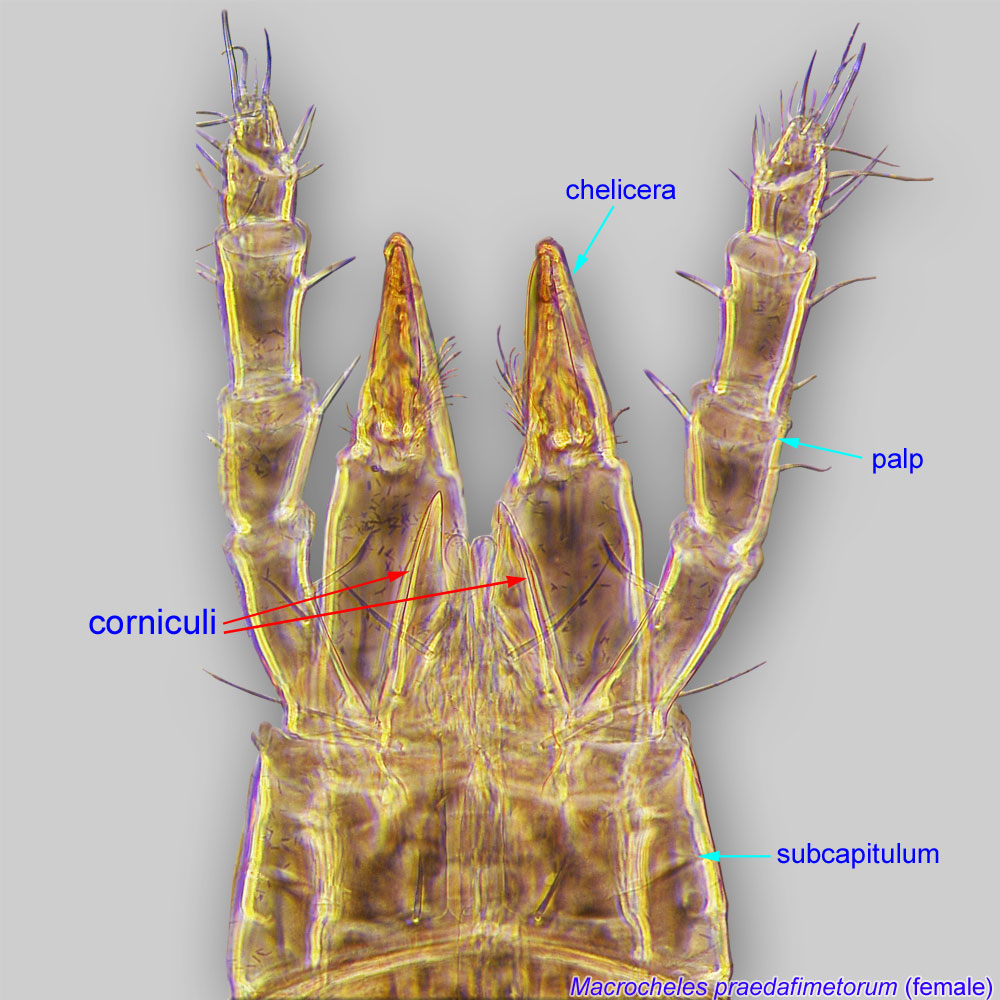
Paired, horn-like process (sometimes toothed, bifurcate, trifurcate, spine-like, spatulate, or membranous) on the subcapitulum of parasitiform mites. These processes usually support the salivary styli. If toothed could be confused with a rutellum, a possibly homologous structure in Acariformes and Opilioacarida.
 widely separated (Figs. 8, 9). Fixed digit with filiform pilus dentilispilus dentilis:
A seta-like or membranous sensory organ inserted ventrolaterally on the fixed digit of the chelicera of many Mesostigmata.
(Fig. 11 ). Tectumtectum:
The leading dorsal, shelf-like projection of the basis capitulum in Mesostigmata. Also known as gnathotectum or epistome.
convex or triramous (Fig. 12). Median lobe of pulvilluspulvillus:
a membranous, pad-like structure associated with the claws in Mesostigmata.
(membranous, pad-like structure) of legs II-IV rounded (Fig. 13).
A dichotomous key is available in Christian and Karg, 2006Christian and Karg, 2006:
Christian, A. amp; W. Karg. 2006. The predatory mite genus Lasioseius Berlese, 1916 (Acari, Gamasina). Abhandlungen und Berichte des Naturkundemuseums Goerlitz.77: 99-250..
Blattisocius. Lasioseius can be separated from Blattisocius by its wider ventrianal shieldventrianal shield:
In Mesostigmata, a ventral shield bearing the anal opening, circumanal (postanal and adanal) setae, and one or more pairs of ventral setae or pores (lyrifissures) (see anal shield); may be narrow or very broad and covering most of the gaster.
(narrower in Blattisocius); metasternal shieldsmetasternal shield:
Small, usually teardrop to subtriangular paired shields bearing metasternal setae st4; sometimes fused to the sternal shield or the endopodal shields. Present in Mesostigmata.
present (absent in Blattisocius); corniculicorniculus:
Paired, horn-like process (sometimes toothed, bifurcate, trifurcate, spine-like, spatulate, or membranous) on the subcapitulum of parasitiform mites. These processes usually support the salivary styli. If toothed could be confused with a rutellum, a possibly homologous structure in Acariformes and Opilioacarida.
widely separated (narrowly separated in Blattisocius); and peritrematic shield clearly wider than diameter of stigma at level of stigma (barely wider in Blattisocius).
Worldwide; found in association with bees in the Palaearctic, Nearctic, Oriental, and Australasian regions.
found in nests of honey bees (Apis) and bumble bees (Bombus)
facultativefacultative:
can complete entire life cycle without bees or their close relative, wasps
Lasioseius is a morphologically diverse genus, with more than 100 described species found on rotting organic substances, under bark, and in forest litter, moss, soil, nests of small mammals and birds, bracket-fungi, tree-holes, stored products, warehouses, and hay. Some species are omnivorous, feeding on microarthropods and on fungi, while others are only predatory (Enkegaard and Brodsgaard, 2000Enkegaard and Brodsgaard, 2000:
Enkegaard, A. amp; H. F. Brodsgaard. 2000. Lasioseius fimetorum: A soil-dwelling predator of glasshouse pests? BioControl (Dordrecht).45: 285-293.; Walter and Lindquist, 1989Walter and Lindquist, 1989:
Walter, D. E. amp; E. E. Lindquist. 1989. Life history and behavior of mites in the genus Lasioseius (Acari: Mesostigmata: Ascidae) from grassland soils in Colorado, with taxonomic notes and description of a new species. Canadian Journal of Zoology.67: 2797-2813.). Several species have been collected in beehives. Among them, one species, Lasioseius penicilliger, has also been found in bumble bee nests. These species are habitat generalists and probably do not have a particular preference for bee nests.
Authors: P. Klimov, B. OConnor, R. Ochoa, G. Bauchan, A. Redford, J. Scher
Last updated October 2016
tool images at ITP Node
idtools.org