lives in honey bee nests and disperses on adult bees, but its effect on bees is unknown
Lasioacarus Kadzhaja and Sevastianov, 1967
Superorder Acariformes » Order Sarcoptiformes » Suborder Oribatida » Infraorder Desmonomata » Hyporder Astigmata » Family Acaridae » Genus Lasioacarus
Lasioacarus nidicolus Kadzhaja and Sevastianov, 1967
Female: With one pair of distinct pits anterolateral to anus (Fig. 11).
Phoretic phoretic:
Pertaining to phoresy; using another organism (i.e., a host) for dispersal to new habitats. Phoresy can be distinguished from parasitism because feeding typically does not occur during phoresy.
deutonymph: Tarsal setae aa I, ba I-II absent (Fig 6). Supracoxal setae scx bifurcate apically (Fig. 5). External conoidal setae of attachment organattachment organ:
Complex unpaired structure in phoretic deutonymphs of Astigmata situated on the posteroventral end of the body that serves for attachment to the host during phoresy. In deutonymphs phoretic on insects, attachment organ consists of a vestigial anal opening and two types of attachment elements: true suckers that create negative pressure and conoids that create adhesive forces. Not to be confused with pedicel.
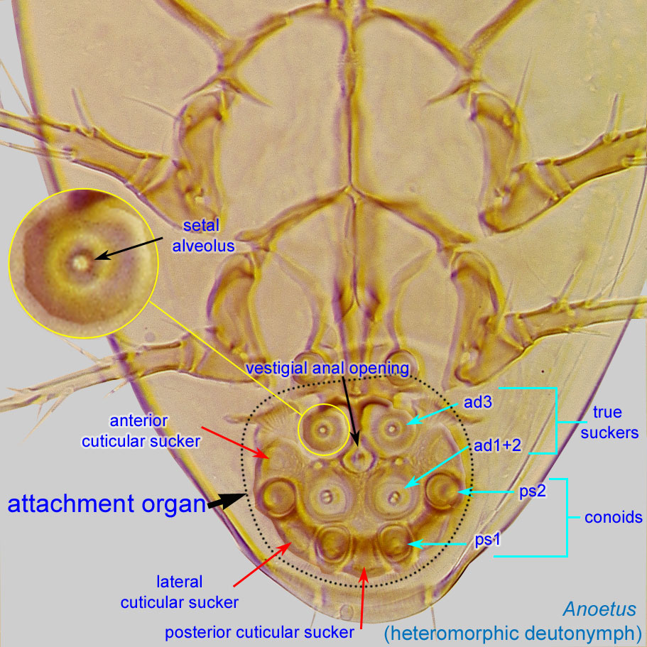 (ps2) lateral to median sucker (ad1+2) (Fig. 4). Sternal apodemeapodeme:
Internal sclerite that serves as an attachment site for muscles. Most commonly used (as "coxal apodeme") to describe elements of coxae fused to the ventral body in Acariformes (coxae are free and not fused to the body in Parasitiformes), and may be variously referred to as ventral, sternal, anterior, or posterior.
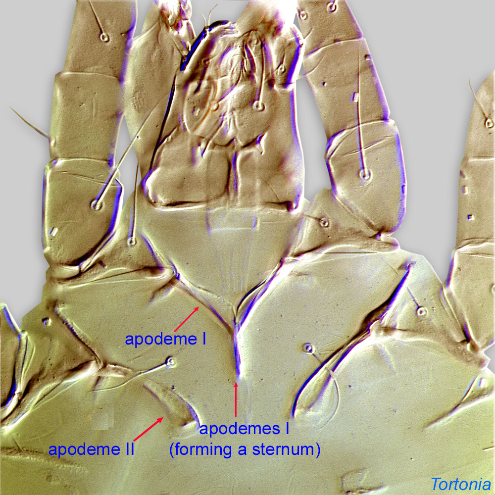 and anterior apodemesapodeme:
Internal sclerite that serves as an attachment site for muscles. Most commonly used (as "coxal apodeme") to describe elements of coxae fused to the ventral body in Acariformes (coxae are free and not fused to the body in Parasitiformes), and may be variously referred to as ventral, sternal, anterior, or posterior.
II not extending to level of coxal apodemesapodeme:
Internal sclerite that serves as an attachment site for muscles. Most commonly used (as "coxal apodeme") to describe elements of coxae fused to the ventral body in Acariformes (coxae are free and not fused to the body in Parasitiformes), and may be variously referred to as ventral, sternal, anterior, or posterior.
III (Fig. 4). GenuaGenu:
Leg or palp segment (also known as podomere or palpomere) between tibia and femur.
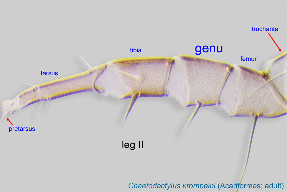 III-IV subequal to tibiaetibia:
Leg or palp segment (also known as podomere or palpomere) between tarsus and genu.
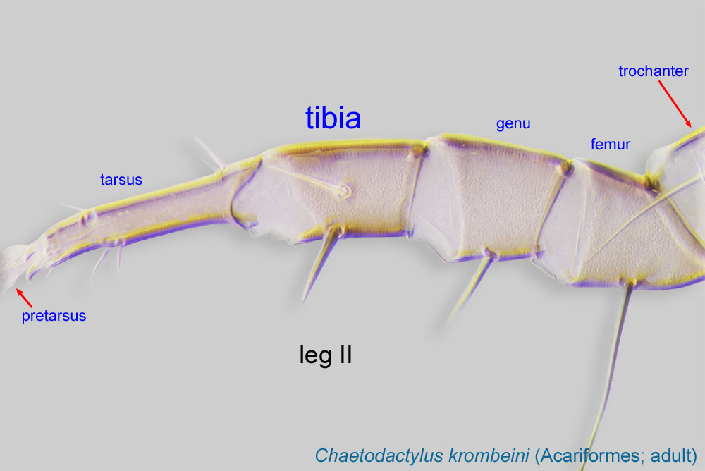 III-IV (Fig. 7). Empodial clawsEmpodial claw:
Claw-like, membranous, or pad-like structure of setal origin. Present only on the pretarsus in Acariformes. In Astigmata, it is the only claw on the pretarsus and often referred to simply as the claw. In the remaining Acariformes, may be accomanied by two lateral claws. Also known as empodium, pretarsal empodium, or central claw.
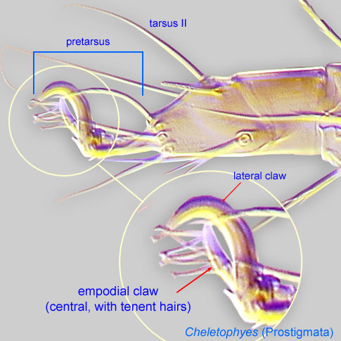 of pretarsipretarsus:
Terminal leg or palpal segment distal to tarsus.
I-IV similar in form, long, at least half the length of tarsustarsus:
Terminal segment (also known as podomere or palpomere) of legs or palps. In Parasitoformes it can be subdivided into telotarsus and basitarsus.
 (Fig. 7). Solenidionsolenidion:
Thin-walled, terminally rounded or pointed filiform or peglike structure that is not birefringent in polarized light (unlike common setae in Acariformes). Often appears striated because of its internal structure. Found on the palpal tarsus on the gnathosoma and may also occur on the tarsus and tibia, less frequently on the genu, and occasionally on the femur of legs I-IV. In Acariformes, leg solenidia often arise from unsclerotized areas.
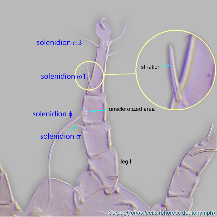 ω2 of tarsustarsus:
Terminal segment (also known as podomere or palpomere) of legs or palps. In Parasitoformes it can be subdivided into telotarsus and basitarsus.
I present (Fig. 6). Tarsal setae e I-II with apical "saucer" slightly expanded (Fig. 6). Posterior setae of genuagenu:
Leg or palp segment (also known as podomere or palpomere) between tibia and femur.
I-II (mG) stout and spine-like, smooth (Fig. 6). TibiaeTibia:
Leg or palp segment (also known as podomere or palpomere) between tarsus and genu.
I-II similar in length (Fig. 6). Internal vertical setae (vi) unbarbed (Fig. 5). External vertical setae (ve) absent (Fig. 5). Bases of internal vertical setae (vi) nearly contiguous (Fig. 5). Posterior setae of tibiatibia:
Leg or palp segment (also known as podomere or palpomere) between tarsus and genu.
I-II (hT I-II) absent (Fig 6). Setae nG III and solenidionsolenidion:
Thin-walled, terminally rounded or pointed filiform or peglike structure that is not birefringent in polarized light (unlike common setae in Acariformes). Often appears striated because of its internal structure. Found on the palpal tarsus on the gnathosoma and may also occur on the tarsus and tibia, less frequently on the genu, and occasionally on the femur of legs I-IV. In Acariformes, leg solenidia often arise from unsclerotized areas.
σ III absent from genugenu:
Leg or palp segment (also known as podomere or palpomere) between tibia and femur.
III (Fig. 7). Coxal setae I-IV 1a, 3a, 4a conoidal Fig. 4). Dorsal hysterosomal setae short and filiform (Fig. 3).
Adult: Tarsal setae aa I, ba I-II absent (correlates with characters in phoreticphoretic:
Pertaining to phoresy; using another organism (i.e., a host) for dispersal to new habitats. Phoresy can be distinguished from parasitism because feeding typically does not occur during phoresy.
deutonymphdeutonymph:
Ontogenetic stage between protonymph and tritonymph (or adult, if tritonymph is absent). See <a href="index.cfm?pageID=1720">Life stages page</a> for more details.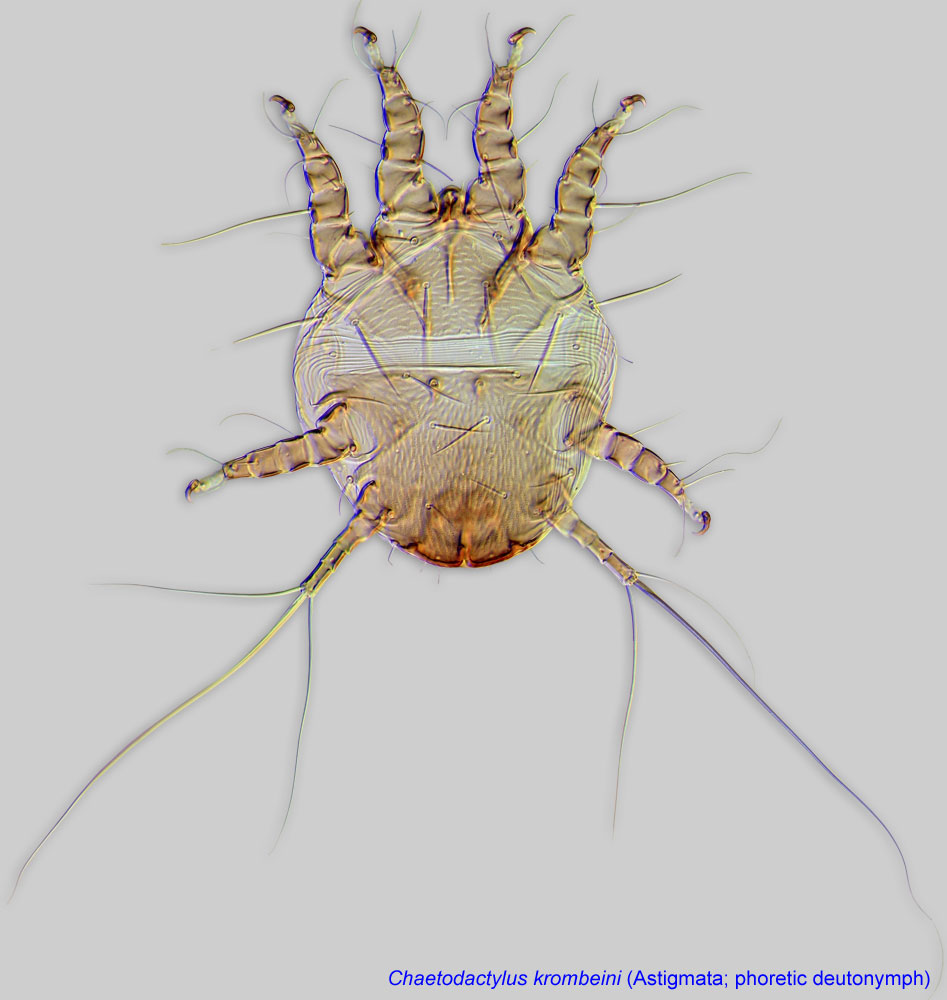 ) (Figs. 12, 13 , 14). Setae ve and si absent (Fig. 10). Supracoxal setae scx present (Fig. 10). Male without sclerotized posterior projection.
This genus includes two described species, Lasioacarus nidicolus and Lasioacarus morsei (originally described as "Kucinia morsei"). These species can be distinguished using published descriptions (El-Banhawy and Abou-Awad, 1990El-Banhawy and Abou-Awad, 1990:
El-Banhawy, E. M. amp; B. A. Abou-Awad. 1990. Description of hypopial stage of a new species of mite associated with the honey bee Apis dorsata F. (Acari: Acaridae). Zoologische Jahrbuecher Abteilung fuer Systematik Oekologie und Geographie der Tiere . 117 : 269-271.; Fain and Chmielewski, 1987Fain and Chmielewski, 1987:
Fain, A. amp; W. Chmielewski. 1987. The phoretic hypopi of two acarid mites described from ants' nest: Tyrophagus formicetorum Volgin, 1948 and Lasioacarus nidicolus Kadzhaja and Sevastianov 1967. Acarologia . 28 : 53-61.; Kadzhaya and Sevastyanov, 1967Kadzhaya and Sevastyanov, 1967:
Kadzhaya, G. S. amp; V. D. Sevastyanov. 1967. New genus and species of the families Acaridae and Saproglyphidae: (Acariformes). Soobshcheniya Akademii Nauk Gruzinskoi SSR . 48 : 195-200.).
Palaearctic and Oriental regions
honey bees (Apis)
permanentpermanent:
associated exclusively with bees or their close relative, wasps; cannot live without these hosts
disperse on adult bees, usually attaching to the hind tibiatibia: and tarsitarsus: (Fig. 15).Lasioacarus nidicolus was found in the nest of an ant, Lasius niger, in Ukraine. Later this species was collected from beehives (Apis mellifera) in Poland as part of a large-scale survey, where its occurrence was 4.9% (Chmielewski, 1991cChmielewski, 1991c:
Chmielewski, W. 1991c. Stored products mites (Acaroidea) in Polish bee hives. In Modern acarology. Volume I: proceedings of the 8 International Congress of Acarology, held in Ceske Budejovice, Czechoslovakia, 6-11 August 1990. , 615-619. The Hague, The Netherlands: SPB Academic Publishing.). In controlled laboratory experiments, the mite preferred beehive debris over bee bread, pollen, comb and wax, and mold (fungi); it did not reproduce on honey, royal jelly, propolispropolis:
A red or brown resinous substance collected by honey bees from tree buds that is used by them to fill crevices and to seal and varnish honeycombs.
, or dead brood bees (Chmielewski, 1991cChmielewski, 1991c:
Chmielewski, W. 1991c. Stored products mites (Acaroidea) in Polish bee hives. In Modern acarology. Volume I: proceedings of the 8 International Congress of Acarology, held in Ceske Budejovice, Czechoslovakia, 6-11 August 1990. , 615-619. The Hague, The Netherlands: SPB Academic Publishing.).
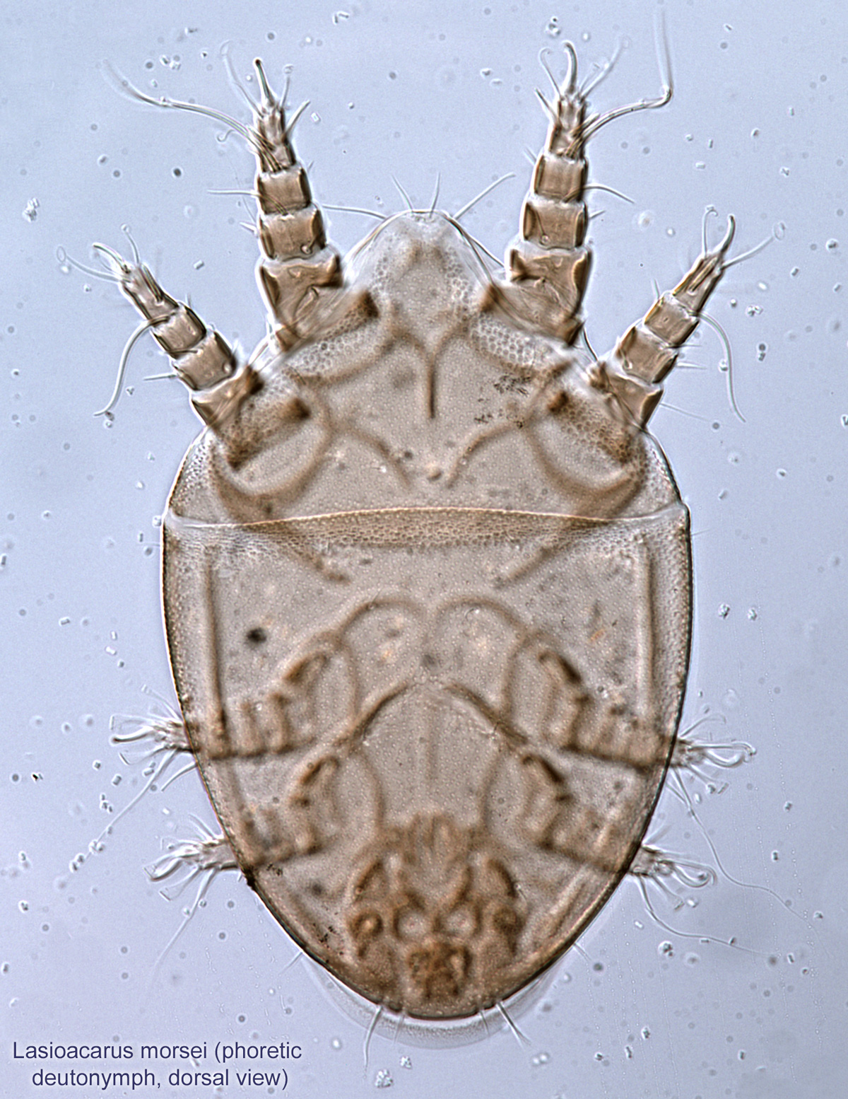
Another species, Lasioacarus morsei, is associated with the giant honey bee Apis dorsata from the Philippines (El-Banhawy and Abou-Awad, 1990El-Banhawy and Abou-Awad, 1990:
El-Banhawy, E. M. amp; B. A. Abou-Awad. 1990. Description of hypopial stage of a new species of mite associated with the honey bee Apis dorsata F. (Acari: Acaridae). Zoologische Jahrbuecher Abteilung fuer Systematik Oekologie und Geographie der Tiere . 117 : 269-271.).
Authors: P. Klimov, B. OConnor, R. Ochoa, G. Bauchan, A. Redford, J. Scher
Last updated October 2016
tool images at ITP Node
idtools.org