

probably neutral to beneficial; feeds on fungi in bee nests
Kerdabania Khaustov, 2009Khaustov, 2009:
Khaustov, A. A. 2009. A description of new genus, Kerdabania gen. n., with four new species (Acari: Heterostigmata: Neopygmephoridae). Acarina . 17 : 171-188.
Superorder Acariformes » Order Trombidiformes » Suborder Prostigmata » Infraorder Eleutherengona » Hyporder Heterostigmata » Family Neopygmephoridae » Genus Kerdabania
Kerdabania magnifica Khaustov, 2009Khaustov, 2009:
Khaustov, A. A. 2009. A description of new genus, Kerdabania gen. n., with four new species (Acari: Heterostigmata: Neopygmephoridae). Acarina . 17 : 171-188.
Formerly was part of Bakerdania and Pygmephorus.
Female: Tergite C not covering or slightly covering prodorsumprodorsum:
Dorsal surface of propodosoma.
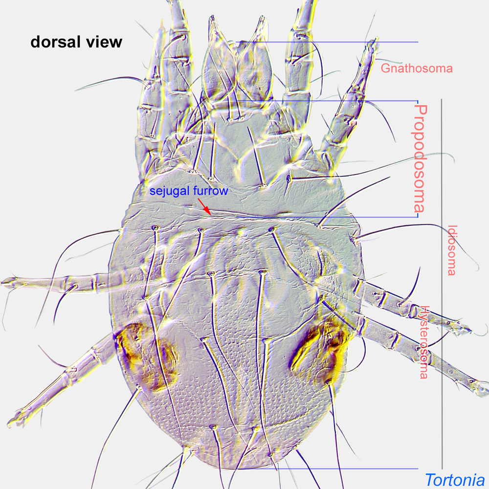 (Fig. 5). Two pairs of propodosomal dorsal setae and clavate bothridial setaebothridial seta:
A modified seta inserted in a cup-like base (bothridium); forms include filiform and capitate. Also known as bothridial sensillum and trichobothrium.
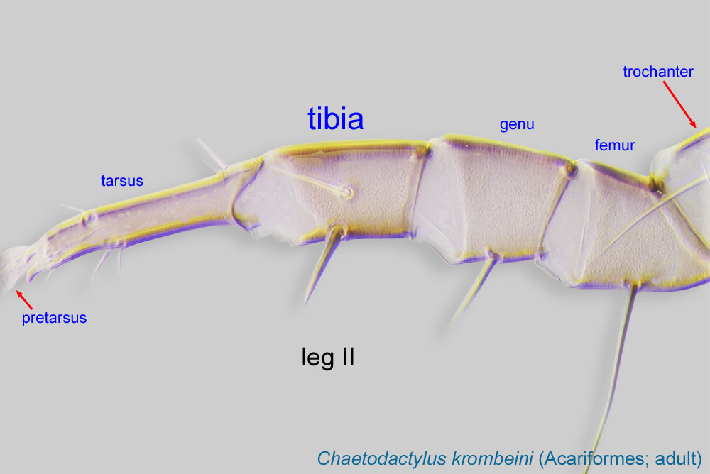
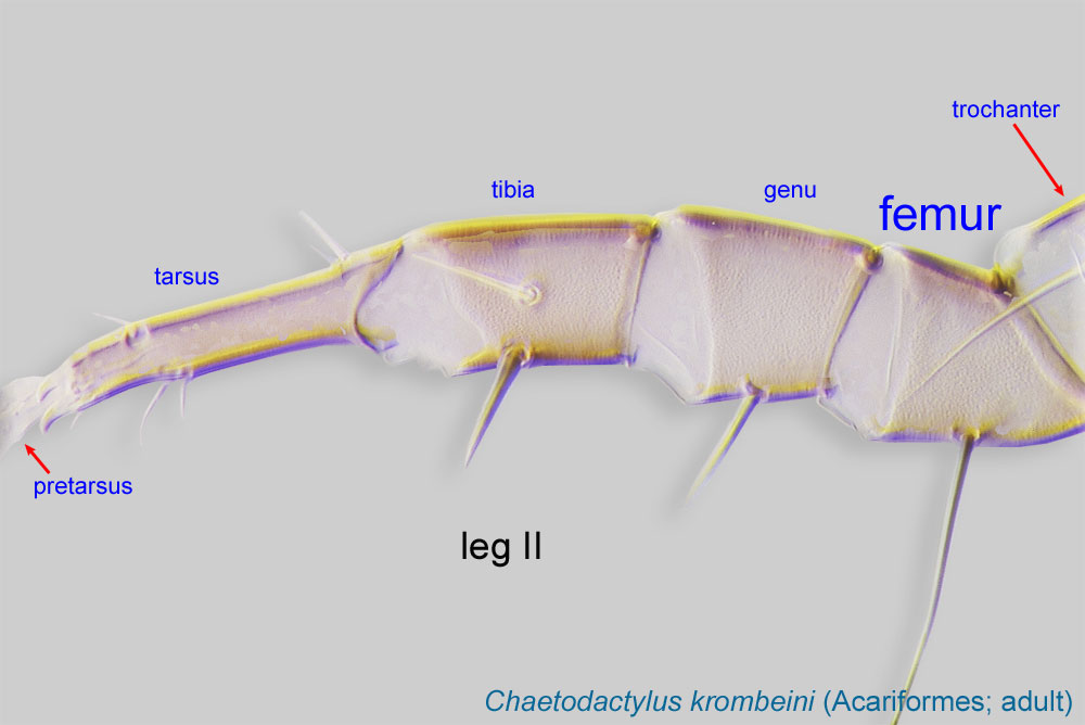
(Fig. 5). Leg I four-segmented, with fused tarsustarsus:
Terminal segment (also known as podomere or palpomere) of legs or palps. In Parasitoformes it can be subdivided into telotarsus and basitarsus.
 and tibiatibia:
Leg or palp segment (also known as podomere or palpomere) between tarsus and genu.
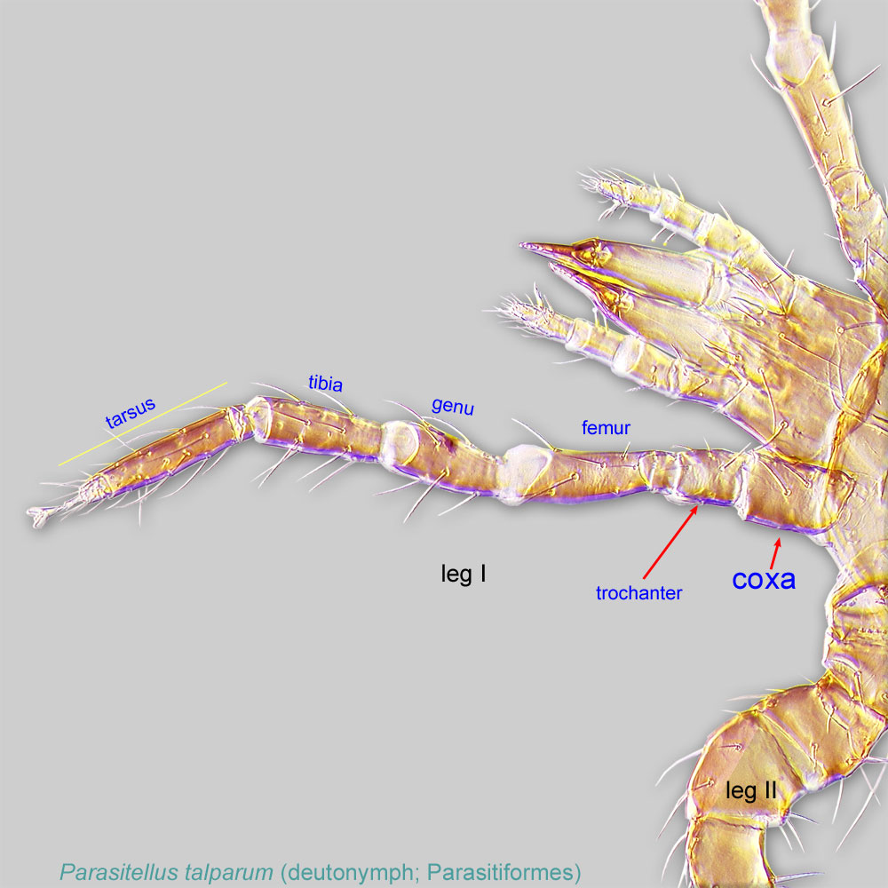
 I, forming tibiotarsus (Figs. 9). Two pairs of setae on each coxaecoxa:
In Parasitiformes, most basal leg segment (or podomere) forming a joint with the body. Areas delimited by coxal apodemes are called coxal fields in Astigmata or coxisternal plates in Prostigmata.
 I-II (Fig. 6). Claw I medium-sized, not striated (Figs. 9, 10). Tarsustarsus:
Terminal segment (also known as podomere or palpomere) of legs or palps. In Parasitoformes it can be subdivided into telotarsus and basitarsus.
IV with claws (Fig. 11). Trochantertrochanter:
Leg or palp segment (also known as podomere or palpomere) between femur and coxa. In Acariformes this is the most basal movable leg segment (or podomere) forming a joint with the body.
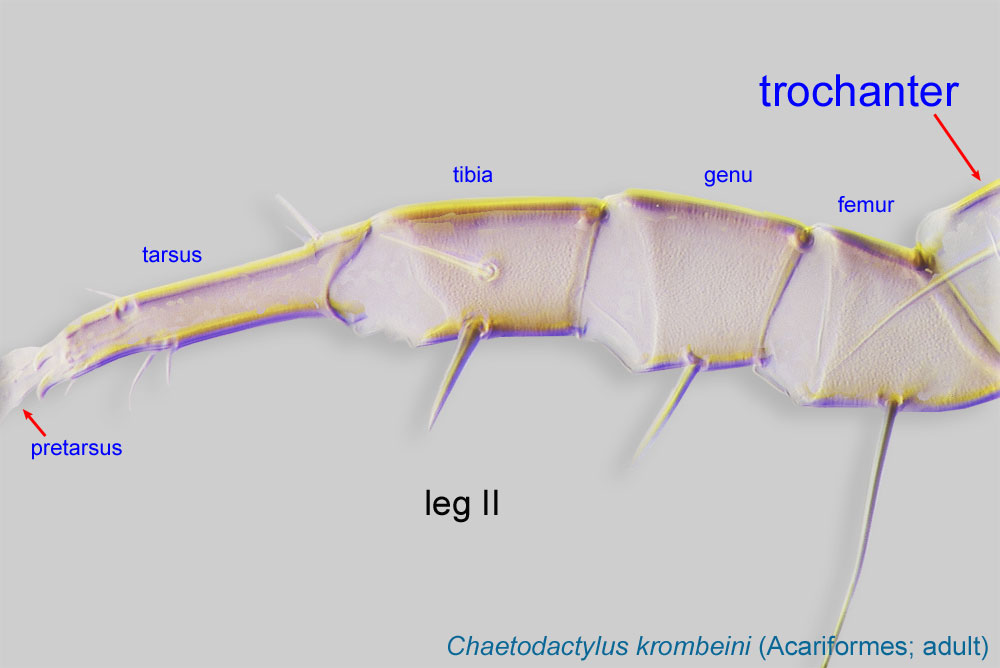 IV subquadrate (not triangular) (Fig. 11). Setae d of femurfemur:
Leg or palp segment (also known as podomere or palpomere) between genu and trochanter. In ParasitIformes can be subdivided into telofemur and basifemur.
 I modified, widened (may be only slightly) (Fig. 9). Pinnaculumpinnaculum:
A subapical, dorsal elevation on tibiotarsus I bearing a distinct cluster of 2-3 rodlike sensory setae. Term is used for Heterostigmata only.
on tibiotarsus I absent (Fig. 9). Gnathosomagnathosoma:
Division of body anterior to the propodosoma bearing two pairs of appendages (palps and chelicerae).
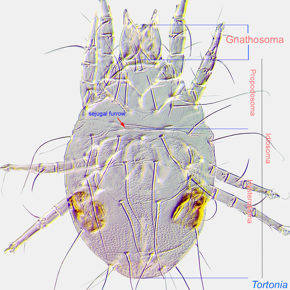 with 1 pair of dorsal setae (Fig. 3). Basal part of gnathosomagnathosoma:
Division of body anterior to the propodosoma bearing two pairs of appendages (palps and chelicerae).
not elongated (Figs. 3, 4). PalpsPalp:
Second (after chelicera) paired appendage of the gnathosoma. Has a sensory function, but may be variously modified for other functions (e.g., raptorial, attachment to host, or filtering).
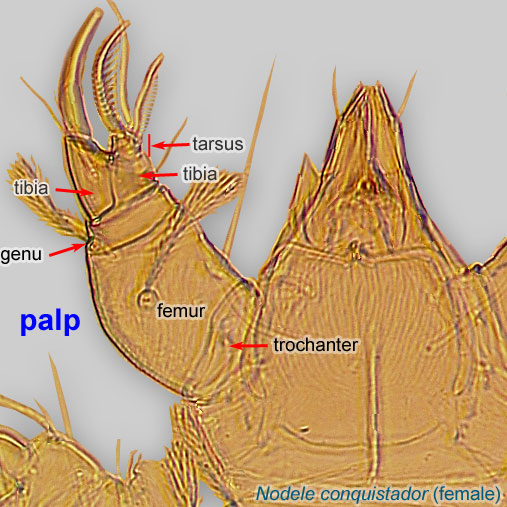 shorter than basal part of gnathosomagnathosoma:
Division of body anterior to the propodosoma bearing two pairs of appendages (palps and chelicerae).
(Fig. 4). Median genital scleritesclerite:
A component section of an exoskeleton; a plate forming the skeleton of an arthropod.
absent (Fig. 8). Posterior margin of posterior sternal plate tripartite (with 3 large lobes) (Fig. 8).
Dichotomous key is available in Khaustov, 2009Khaustov, 2009:
Khaustov, A. A. 2009. A description of new genus, Kerdabania gen. n., with four new species (Acari: Heterostigmata: Neopygmephoridae). Acarina . 17 : 171-188.. Kerdabania quadrata (the only species of Kerdabania found in association with bees) is illustrated in Smiley, 1978Smiley, 1978:
Smiley, R. L. 1978. Taxonomic studies of Pygmephorus species from the western hemisphere, with a key to females and an overview of the current problems for classification (Acari: Pyemotidae and Pygmephoridae). International Journal of Acarology . 4 : 125-160..
The genus Kerdabania is similar to genera Bakerdania and Pseudopygmephorus. From both genera it differs by the presence of only one pair of dorsal gnathosomal setae (2 pairs in Bakerdania and Pseudopygmephorus), by the tripartite posterior margin of its posterior sternal plate (entire in Bakerdania and Pseudopygmephorus). From Pseudopygmephorus the genus Kerdabania differs by its solenidionsolenidion:
Thin-walled, terminally rounded or pointed filiform or peglike structure that is not birefringent in polarized light (unlike common setae in Acariformes). Often appears striated because of its internal structure. Found on the palpal tarsus on the gnathosoma and may also occur on the tarsus and tibia, less frequently on the genu, and occasionally on the femur of legs I-IV. In Acariformes, leg solenidia often arise from unsclerotized areas.
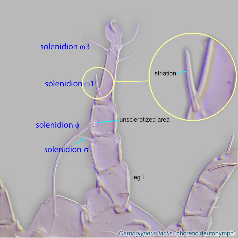 ω1, which is not fused with tibiotarsus (in Pseudopygmephorus solenidionsolenidion:
Thin-walled, terminally rounded or pointed filiform or peglike structure that is not birefringent in polarized light (unlike common setae in Acariformes). Often appears striated because of its internal structure. Found on the palpal tarsus on the gnathosoma and may also occur on the tarsus and tibia, less frequently on the genu, and occasionally on the femur of legs I-IV. In Acariformes, leg solenidia often arise from unsclerotized areas.
ω1 is completely fused with tibiotarsus).
Worldwide, except for Antarctica. The single generalist, Holarctic species, Kerdabania quadrata, has been found in association with bees in the Middle East (Iran) (Smiley, 1978Smiley, 1978:
Smiley, R. L. 1978. Taxonomic studies of Pygmephorus species from the western hemisphere, with a key to females and an overview of the current problems for classification (Acari: Pyemotidae and Pygmephoridae). International Journal of Acarology . 4 : 125-160.; Kamali et al., 2001Kamali et al., 2001:
Kamali, K., H. Ostovan amp; A. Atamehr. 2001. A Catalog of Mites and Ticks (Acari) of Iran. Islamic Azad University Scientific Publication Center. 196 pp.).
honey bee (Apis) hives
facultativefacultative:
can complete entire life cycle without bees or their close relative, wasps
Kerdabania quadrata can develop on only one fungal genus, and it occurs on soil, forest litter, in nests of small mammals and ants, and in beehives. Phoresyphoresy:
Attaching to or boarding another organism (i.e., a host) for dispersal to new habitats. Can be distinguished from parasitism because feeding typically does not occur.
is unknown. One generalist species, Kerdabania quadrata, has been found in a nest of the European honey bee, Apis mellifera, and in other habitats: in litter from under strawstacks, under Sambucus shrubs, in forests, in soil, and from an ant nest (Mahunka, 1981Mahunka, 1981:
Mahunka, S. 1981. The pygmephoroid fauna of the Hortobágy National Park (Acari: Tarsonemida). Natural History of the National Parks of Hungary . 1 : 343-370.). Kerdabania quadrata has also been recorded as a pest of commercially grown mushrooms (Wicht, 1970Wicht, 1970:
Wicht, M. C., Jr. 1970. Three new species of pyemotid mites associated with commercial mushrooms. Acarologia . 12 : 262-268.).
Authors: P. Klimov, B. OConnor, R. Ochoa, G. Bauchan, A. Redford, J. Scher
Last updated October 2016
tool images at ITP Node
idtools.org


