

neutral or beneficial commensals; feed on fungi and waste from developing bee larvae
Imparipes Berlese, 1903
Superorder Acariformes » Order Trombidiformes » Suborder Prostigmata » Infraorder Eleutherengona » Hyporder Heterostigmata » Family Scutacaridae » Genus Imparipes
Imparipes histricinus Berlese, 1903
Female: Tergite C completely covers prodorsumprodorsum:
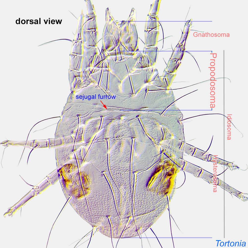
Dorsal surface of propodosoma.
 , forming a characteristic ‘roof’ with striated margin (Fig. 1). Alveolar channel of setae c2 strongly sclerotized (Fig. 3). Setae c1 in central part of tergite C (not on its free striated margin) (Fig. 3). Pharyngeal pump (a muscular wall around pharynxpharynx:
A muscular ectodermal structure that opens into a buccal cavity or mouth, and serves as a suction pump for ingesting food materials. In certain Heterostigmata pharyngal musculature is subdivided into 2-3 distinct parts.
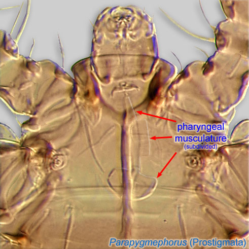 that aids with ingestion) ph2 much larger than ph1 and ph3 (Fig. 5). Claw I not enlarged (Fig. 6). Legs IV with 5 segments (trochanter, femurfemur:
Leg or palp segment (also known as podomere or palpomere) between genu and trochanter. In ParasitIformes can be subdivided into telofemur and basifemur.
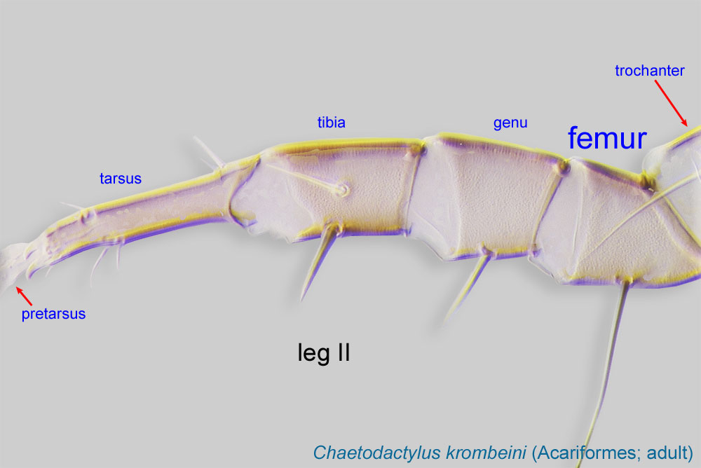 , genugenu:
Leg or palp segment (also known as podomere or palpomere) between tibia and femur.
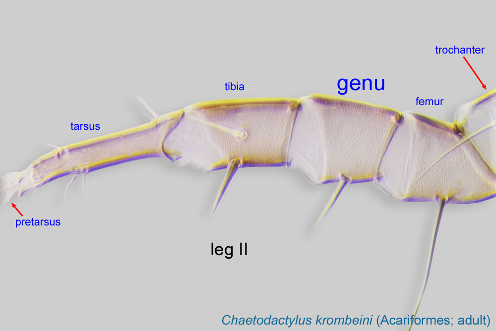 , tibiatibia:
Leg or palp segment (also known as podomere or palpomere) between tarsus and genu.
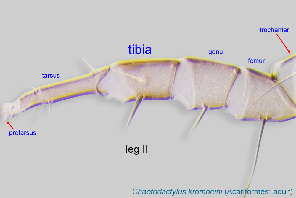 , tarsustarsus:
Terminal segment (also known as podomere or palpomere) of legs or palps. In Parasitoformes it can be subdivided into telotarsus and basitarsus.
 ) and ambulacrumambulacrum:
The claws and empodium of the apotele or pretarsus.
(Fig. 7). Tibiatibia:
Leg or palp segment (also known as podomere or palpomere) between tarsus and genu.
IV with 3 setae (d, l’, v’) (Fig. 7). Tarsustarsus:
Terminal segment (also known as podomere or palpomere) of legs or palps. In Parasitoformes it can be subdivided into telotarsus and basitarsus.
IV abruptly becomes thin distal to point indicated by arrow on Fig. 7. Basal part of gnathosomagnathosoma:
Division of body anterior to the propodosoma bearing two pairs of appendages (palps and chelicerae).
 not elongated (Fig. 2).
Species-rich genus, containing both bee-associated and non bee-associated mites. Except for the key to Palaearctic species (Khaustov, 2008Khaustov, 2008:
Khaustov, A. A. 2008. Mites of the family Scutacaridae of Eastern Palaearctic. Kiev: Akademperiodika. 290 pp.), no keys are available. There are 15 species associated with bees, which should be identified using original descriptions.
Palaearctic, Nearctic, Neotropical, and Afrotropical regions.
Halictidae (Halictus, Lasioglossum, Lasioglossum (Dialictus), Augochlorella, and Nomia), Colletidae (Hylaeus), Andrenidae (Andrena), Megachilidae (Osmia, Megachile), and Apidae (Apis mellifera, Dasypoda, and Bombus)
permanentpermanent:
associated exclusively with bees or their close relative, wasps; cannot live without these hosts
 . The fungus then is used as a food source by the mites developing in the bee nest.
The following is a summary of the biology of Imparipes apicola as described in Cross and Bohart, 1992Cross and Bohart, 1992:
Cross, E. A. amp; G. E. Bohart. 1992. The biology of Imparipes apicola (Acari: Scutacaridae) and its relationships to the alkali bee, Nomia melanderi (Hymenoptera: Halictidae), and to certain fungi in the bee cell ecosystem. Journal of the Kansas Entomological Society 65: 157-173..
Female mites are phoreticphoretic:
Pertaining to phoresy; using another organism (i.e., a host) for dispersal to new habitats. Phoresy can be distinguished from parasitism because feeding typically does not occur during phoresy.
on bee hosts, dropping off into new cells as they are constructed and provisioned. In the nest, this species is a fungivorousfungivorous:
Feeding on fungi.
commensal, completing its life cycle in cells containing diseased or healthy bees. In cells containing healthy bees, foundress mites normally initiate ovogenesis only at the time of larval bee defecation. Fungus of the genus Ascosphaera is of fundamental importance to Imparipes apicola. This fungus is ingested by the developing bee larva and germinates in the gut, where it develops without harming the bee, and is passed in the feces, stringing the fecal pellets together in characteristic fashion. Its presence probably cues the initiation of ovogenesis in mite females, and it is also a primary source of mite food. Female mites overwinter in the bee cell and attach to emerging bees the following summer.
The mite egg hatches into the larva (the only immature stage). After molting, larvae develop ino either feeding females or non-feeding males, although most of the feeding occurs during the larval stage. In cells with healthy bees, mite larvae have usually been found on the fecal mass, where they appeared to be feeding, but in certain cells showing disease, especially those containing soupy pollen, they have been found more commonly in the upper portions of the cell. In healthy cells, they often appeared to feed from the sparse mycelia growing from the feces and along the cell walls, and especially from the thick strands characteristic of Ascosphaera.
Authors: P. Klimov, B. OConnor, R. Ochoa, G. Bauchan, A. Redford, J. Scher
Last updated October 2016
tool images at ITP Node
idtools.org


