

probably commensal or somewhat beneficial; feeds on fungi and decomposing organic matter in nest
Forcellinia Oudemans, 1924Oudemans, 1924:
Oudemans, A. C. 1924. Acarologische Aanteekehingen LXXVII. Entomologische Berichten (Amsterdam). 6: 317-336.
Superorder Acariformes » Order Sarcoptiformes » Suborder Oribatida » Infraorder Desmonomata » Hyporder Astigmata » Family Acaridae » Genus Forcellinia
Tyroglyphus wasmanni Moniez, 1892
Dorylacarus Mahunka, 1979 and Ocellacarus Mahunka, 1979; probably Hungaroglyphus Mahunka, 1962 and Paraforcellinia Kadzhaja, 1973
Phoretic phoretic:
Pertaining to phoresy; using another organism (i.e., a host) for dispersal to new habitats. Phoresy can be distinguished from parasitism because feeding typically does not occur during phoresy.
deutonymph: Conoidal setae 4a bilobed (Fig. 3), but see the "Similar genera" topic below.
Phoretic phoretic:
Pertaining to phoresy; using another organism (i.e., a host) for dispersal to new habitats. Phoresy can be distinguished from parasitism because feeding typically does not occur during phoresy.
deutonymph: Tarsal setae aa I present, very long. Tarsal seta ba I absent, setae ba II present. SolenidiaSolenidion:
Thin-walled, terminally rounded or pointed filiform or peglike structure that is not birefringent in polarized light (unlike common setae in Acariformes). Often appears striated because of its internal structure. Found on the palpal tarsus on the gnathosoma and may also occur on the tarsus and tibia, less frequently on the genu, and occasionally on the femur of legs I-IV. In Acariformes, leg solenidia often arise from unsclerotized areas.
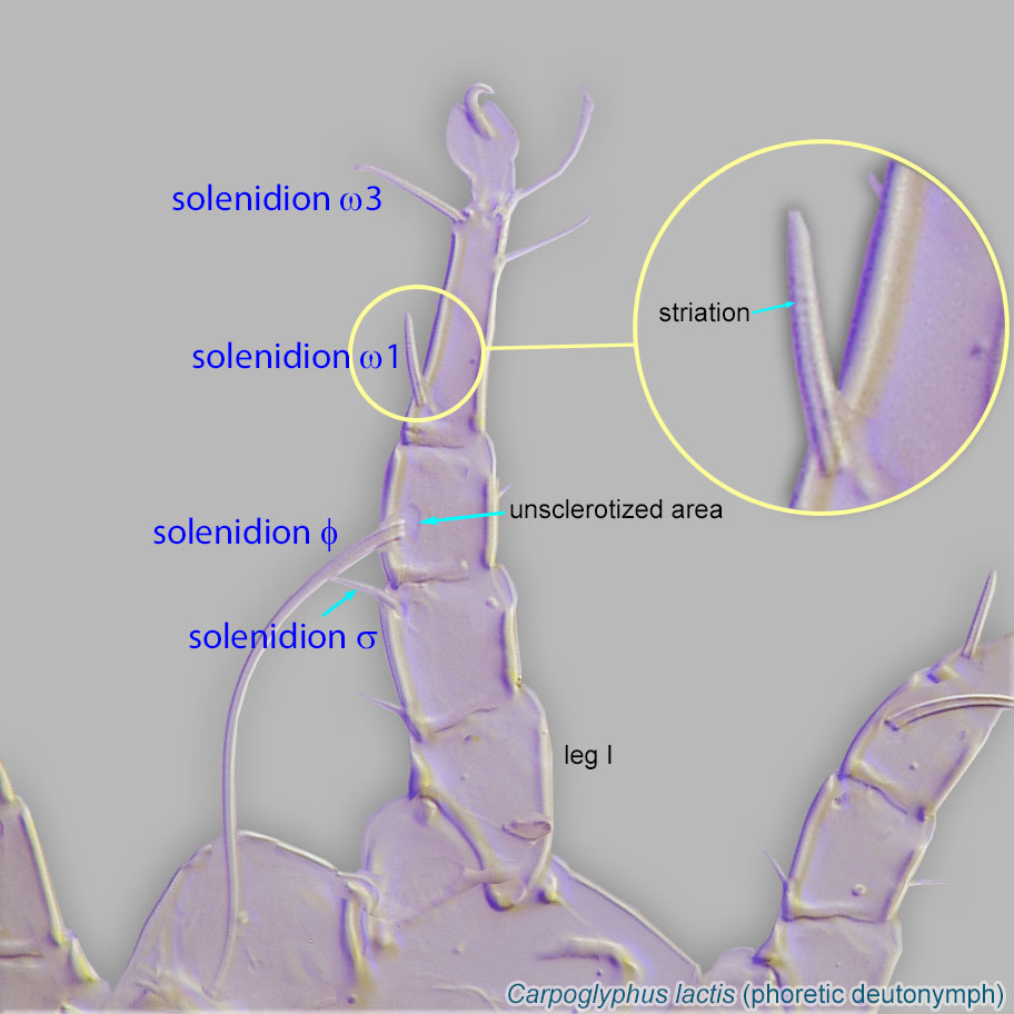 ω1 and ω3 closely associated on tarsustarsus:
Terminal segment (also known as podomere or palpomere) of legs or palps. In Parasitoformes it can be subdivided into telotarsus and basitarsus.
 I. Solenidionsolenidion:
Thin-walled, terminally rounded or pointed filiform or peglike structure that is not birefringent in polarized light (unlike common setae in Acariformes). Often appears striated because of its internal structure. Found on the palpal tarsus on the gnathosoma and may also occur on the tarsus and tibia, less frequently on the genu, and occasionally on the femur of legs I-IV. In Acariformes, leg solenidia often arise from unsclerotized areas.
ω2 present. External conoidal setae ps2 of attachment organattachment organ:
Complex unpaired structure in phoretic deutonymphs of Astigmata situated on the posteroventral end of the body that serves for attachment to the host during phoresy. In deutonymphs phoretic on insects, attachment organ consists of a vestigial anal opening and two types of attachment elements: true suckers that create negative pressure and conoids that create adhesive forces. Not to be confused with pedicel.
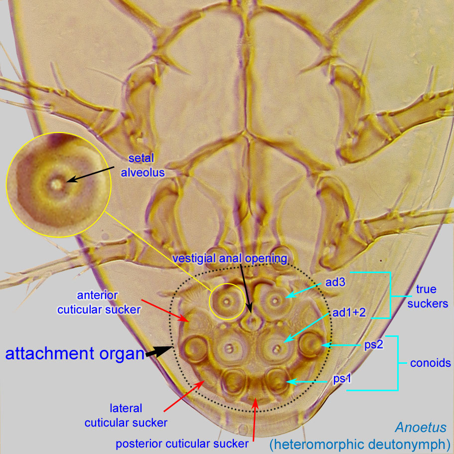 clearly anterior to median sucker (ad1+2) (Fig. 3). Sternal apodemeapodeme:
Internal sclerite that serves as an attachment site for muscles. Most commonly used (as "coxal apodeme") to describe elements of coxae fused to the ventral body in Acariformes (coxae are free and not fused to the body in Parasitiformes), and may be variously referred to as ventral, sternal, anterior, or posterior.
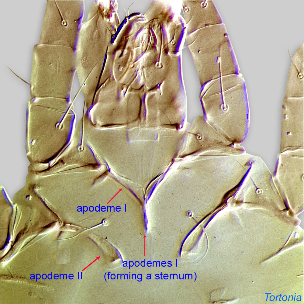 and anterior apodemesapodeme:
Internal sclerite that serves as an attachment site for muscles. Most commonly used (as "coxal apodeme") to describe elements of coxae fused to the ventral body in Acariformes (coxae are free and not fused to the body in Parasitiformes), and may be variously referred to as ventral, sternal, anterior, or posterior.
II extending to level of coxal apodemesapodeme:
Internal sclerite that serves as an attachment site for muscles. Most commonly used (as "coxal apodeme") to describe elements of coxae fused to the ventral body in Acariformes (coxae are free and not fused to the body in Parasitiformes), and may be variously referred to as ventral, sternal, anterior, or posterior.
III (Fig. 3). Setae of coxal fields 1a, 3a, and 4a conoidal (Fig. 3). Empodial clawsEmpodial claw:
Claw-like, membranous, or pad-like structure of setal origin. Present only on the pretarsus in Acariformes. In Astigmata, it is the only claw on the pretarsus and often referred to simply as the claw. In the remaining Acariformes, may be accomanied by two lateral claws. Also known as empodium, pretarsal empodium, or central claw.
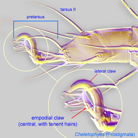 of pretarsipretarsus:
Terminal leg or palpal segment distal to tarsus.
I-IV similar in form. Setae gT I-II and hT I-II smooth. GenuaGenu:
Leg or palp segment (also known as podomere or palpomere) between tibia and femur.
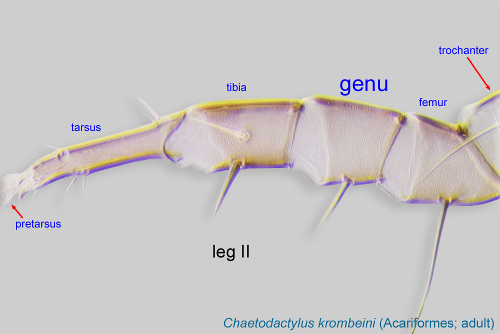 III-IV subequal to tibiaetibia:
Leg or palp segment (also known as podomere or palpomere) between tarsus and genu.
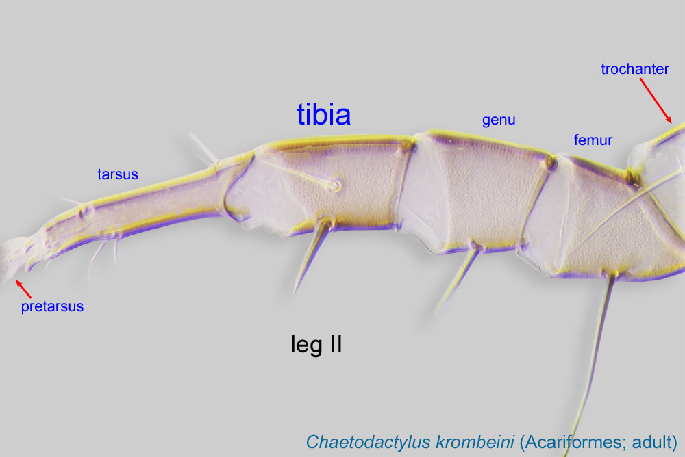 III-IV. Genugenu:
Leg or palp segment (also known as podomere or palpomere) between tibia and femur.
III without a short dorsal solenidionsolenidion:
Thin-walled, terminally rounded or pointed filiform or peglike structure that is not birefringent in polarized light (unlike common setae in Acariformes). Often appears striated because of its internal structure. Found on the palpal tarsus on the gnathosoma and may also occur on the tarsus and tibia, less frequently on the genu, and occasionally on the femur of legs I-IV. In Acariformes, leg solenidia often arise from unsclerotized areas.
(σ).
Adult: Genugenu:
Leg or palp segment (also known as podomere or palpomere) between tibia and femur.
I with solenidionsolenidion:
Thin-walled, terminally rounded or pointed filiform or peglike structure that is not birefringent in polarized light (unlike common setae in Acariformes). Often appears striated because of its internal structure. Found on the palpal tarsus on the gnathosoma and may also occur on the tarsus and tibia, less frequently on the genu, and occasionally on the femur of legs I-IV. In Acariformes, leg solenidia often arise from unsclerotized areas.
σ' no more than three times longer than σ'' (Fig. 6). Setae ve barbed, subequal to vi (Forcellinia and Hungaroglyphus) (Fig. 6) or 40-50% of the length of vi (Paraforcellinia). Setae ve situated near anterior lateral corners of prodorsalprodorsal:
Pertaining to the prodorsum.
sclerite, so setae ve and vi are both situated on the same transverse level (Fig. 6). Setae si subequal with se or shorter (Fig. 6). Dorsal setae flattened and pectinate (Fig. 6). At least some anterior hysterosomal setae rounded at tips (Fig. 6).
A dichotomous key to phoreticphoretic:
Pertaining to phoresy; using another organism (i.e., a host) for dispersal to new habitats. Phoresy can be distinguished from parasitism because feeding typically does not occur during phoresy.
deutonymphsdeutonymph:
Ontogenetic stage between protonymph and tritonymph (or adult, if tritonymph is absent). See <a href="index.cfm?pageID=1720">Life stages page</a> for more details.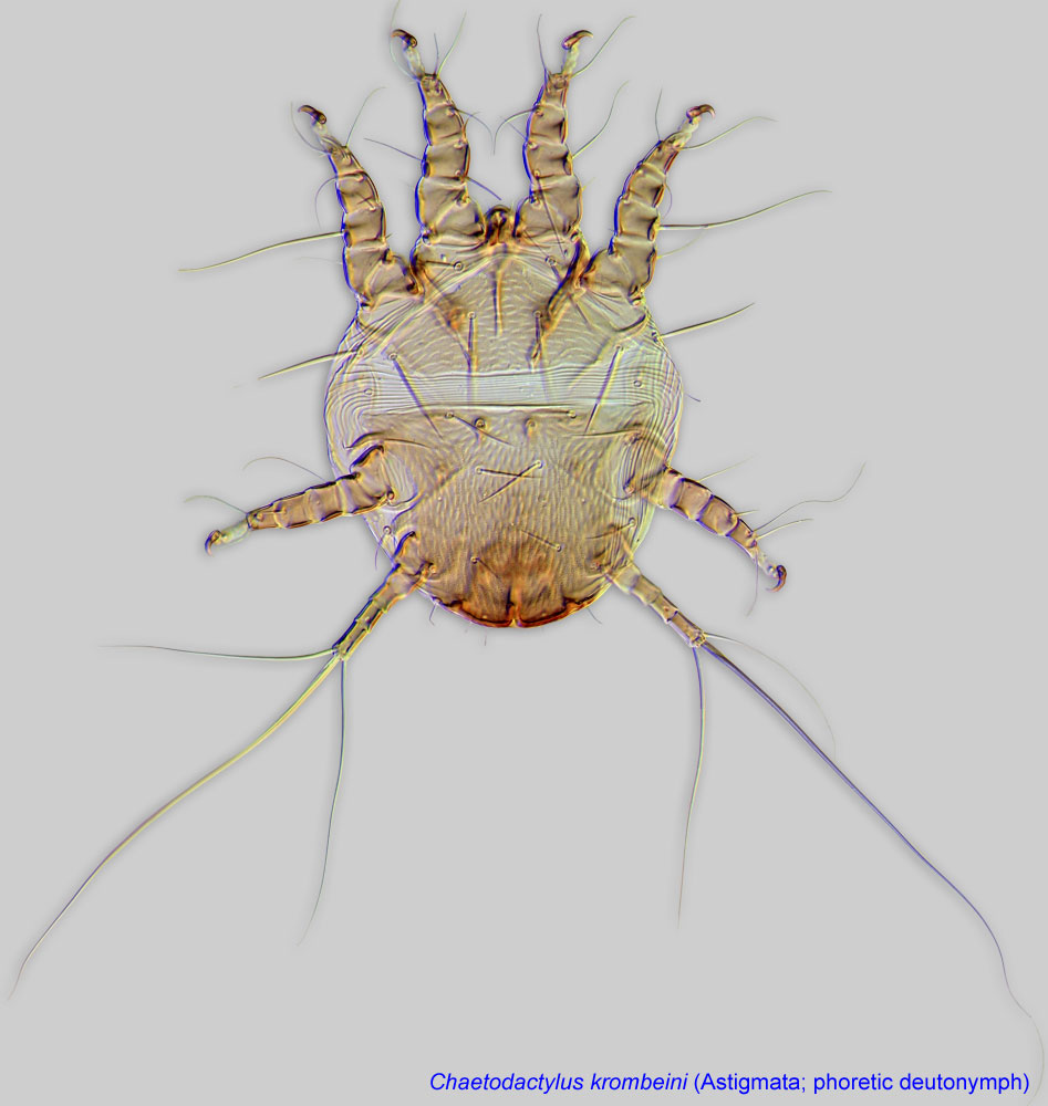 , available in Fain, 1987Fain, 1987:
Fain, A. 1987. Observations on the hypopi of the genus Forcellinia Oudemans, 1924 (Acari, Acaridae). Bulletin de l'Institut Royal des Sciences Naturelles de Belgique Entomologie . 57 : 111-120., can be used to identify Forcellinia galleriella recorded from ants and beehives. Later this species was split into Forcellinia galleriella (in strict sense) and Forcellinia faini, also found in bee hives. Delfinado-Baker and Baker, 1989Delfinado-Baker and Baker, 1989:
Delfinado-Baker, M. amp; E. W. Baker. 1989. A new mite in beehives, Forcellinia faini, n. sp. (Acari, Acaridae). American Bee Journal.129: 127-128. can be used to distinguish these two species.
Phoretic phoretic:
Pertaining to phoresy; using another organism (i.e., a host) for dispersal to new habitats. Phoresy can be distinguished from parasitism because feeding typically does not occur during phoresy.
deutonymphsdeutonymph:
Ontogenetic stage between protonymph and tritonymph (or adult, if tritonymph is absent). See <a href="index.cfm?pageID=1720">Life stages page</a> for more details. of Tyrophagus and Forcellinia are morphologically indistinguishable. Adults of Tyrophagus have internal scapular setae (si) distinctly longer than se (si and se subequal or si shorter in adults of Forcellinia).
Cosmopolitan; records from bees are from the Nearctic, Palaearctic, Oriental, and Australian regions.
Some species develop large population sizes in nests of honey bees (Apis) and stingless bees (Meliponini).
facultative facultative:
can complete entire life cycle without bees or their close relative, wasps
(Forcellinia galleriella) or probably permanentpermanent:
associated exclusively with bees or their close relative, wasps; cannot live without these hosts
(Forcellinia faini)
have been found on ants but not on bees.Species of Forcellinia are primarily associated with ants (Formicidae), living in their nests as feeding stages and dispersing on adult ants as phoreticphoretic:
Pertaining to phoresy; using another organism (i.e., a host) for dispersal to new habitats. Phoresy can be distinguished from parasitism because feeding typically does not occur during phoresy.
deutonymphsdeutonymph:
Ontogenetic stage between protonymph and tritonymph (or adult, if tritonymph is absent). See <a href="index.cfm?pageID=1720">Life stages page</a> for more details.. For example, F. wasmanni feeds on dead ants and organic detritus in the lower level of ants' nests. Some species develop large population sizes in nests of honey bees (Apis) and stingless bees (Meliponini). They may occasionally occur in house dust (Forcellinia galleriella) (Andrews et al., 1995Andrews et al., 1995:
Andrews, J. R. H., J. Crane, L. Phillips amp; B. Robertson. 1995. House dust mites, asthma and skin prick testing in a group of Wairoa school children. New Zealand Entomologist . 18 : 71-75.) and stored products such as dried milk (Forcellinia breviseta), in forest litter or under bark (Forcellinia diamesa), and in sweepings from grain stores and rotting potatoes. Feeding habits are unknown, but they presumably feed on decomposing organic matter and fungi. It is unlikely, although not entirely impossible, that Forcellinia species may attack pollen. Most bees store pollen in sealed cells, and it appears that Forcellinia lacks adaptations to either penetrate the sealed cells or sneak into them before they are sealed.
Two described and one undescribed species are known from nests of honey bees: Forcellinia galleriella (Apis mellifera), Forcellinia faini (Apis mellifera and Apis cerana), Forcellinia sp. (beehives in Hong Kong) (Bowman and Ferguson, 1985Bowman and Ferguson, 1985:
Bowman, C. E. amp; C. A. Ferguson. 1985. Forcellinia galleriella , a mite new to British beehives. Bee World . 66 : 51-53.; Delfinado-Baker and Baker, 1989Delfinado-Baker and Baker, 1989:
Delfinado-Baker, M. amp; E. W. Baker. 1989. A new mite in beehives, Forcellinia faini, n. sp. (Acari, Acaridae). American Bee Journal.129: 127-128.). Forcellinia galleriella probably opportunistically invaded beehives from an ant nest. The second species (F. faini) is probably more specific to bees (there are several repetitive records from different countries), although its potential associations with ants cannot be ruled out. In Thailand, these mites were very common in beehive debris of Apis cerana but were never found in close associations with the bees (Fain and Gerson, 1990Fain and Gerson, 1990:
Fain, A. amp; U. Gerson. 1990. Notes on two astigmatic mites (Acari) living in beehives in Thailand. Acarologia . 31 : 381-384.).
In Costa Rica and Brazil, we observed that Forcellinia was common in two nests of stingless bees, probably feeding on fungi growing in abundance in old nest material. In both cases, an active ant nest was present inside or immediately nearby the bee nest, suggesting that occurrence of Forcellinia in nests of meliponine stingless bees is attributable to mites' spill-over from ant nests rather than to their long-term associations with bees. We hypothesize that since phoreticphoretic:
Pertaining to phoresy; using another organism (i.e., a host) for dispersal to new habitats. Phoresy can be distinguished from parasitism because feeding typically does not occur during phoresy.
deutonymphsdeutonymph:
Ontogenetic stage between protonymph and tritonymph (or adult, if tritonymph is absent). See <a href="index.cfm?pageID=1720">Life stages page</a> for more details. of Forcellinia have never been found on adult bees to date, it is likely that dispersal of these mites to beehives can be accomplished by ants entering bee nests in search for food but not by bees themselves.
Authors: P. Klimov, B. OConnor, R. Ochoa, G. Bauchan, A. Redford, J. Scher
Last updated October 2016
tool images at ITP Node
idtools.org


