probably mutualistic; feeds on harmful mites or other small arthropods in bee nests
Cheletophyes Oudemans, 1914
Superorder Acariformes » Order Trombidiformes » Suborder Prostigmata » Infraorder Eleutherengona » Hyporder Raphignathina » Family Cheyletidae » Genus Cheletophyes
Cheletophyes vitzthumi Oudemans, 1914
Female: Hysteronotalhysteronotal:
Pertaining to dorsal hysterosoma.
shield situated at posterior end of idiosomaidiosoma:
Body not including the gnathosoma.
 very far from propodonotalpropodonotal:
Pertaining to dorsal propodosoma (prodorsum).
shield (Fig. 1).
Female: IdiosomaIdiosoma:
Body not including the gnathosoma.
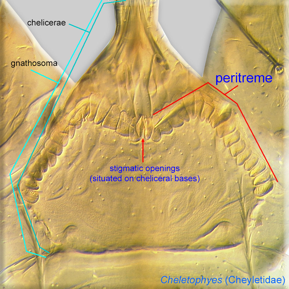
rhomboid, not elongate (Fig. 1). Palptarsus with 2 comb-like setae, acm and sul (Figs. 3, 4). Palpfemur with 4 setae (vF’,vF’’,dF, l’’Ge) (Fig. 4). Seta dGe on palpgenu rod-like or filiform (not lanceolate or fan-like) (Fig. 3). PeritremesPeritreme:
Paired, tubular, elaborated extensions of a tracheal system associated with stigmatic openings. Can be chambered, arch-like, and situated on the bases of chelicerae as in Cheyletidae (Prostigmata) or, in Mesostigmata, linear and situated on the lateral sides of the body.
 M-shaped (Fig. 3). Tarsal seta vs posterior to level of solenidionsolenidion:
Thin-walled, terminally rounded or pointed filiform or peglike structure that is not birefringent in polarized light (unlike common setae in Acariformes). Often appears striated because of its internal structure. Found on the palpal tarsus on the gnathosoma and may also occur on the tarsus and tibia, less frequently on the genu, and occasionally on the femur of legs I-IV. In Acariformes, leg solenidia often arise from unsclerotized areas.
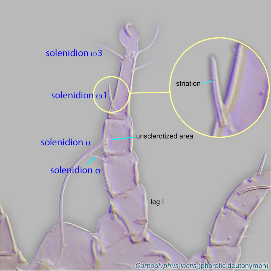 ω1 (Fig. 1). Setae se situated off propodonotalpropodonotal:
Pertaining to dorsal propodosoma (prodorsum).
shield (Fig. 1). Setae f2 lateral, not medial (Fig. 1).
A dichotomous key is available in Fain and Bochkov, 2001Fain and Bochkov, 2001:
Fain, A. amp; A. V. Bochkov. 2001. A review of some genera of cheyletid mites (Acari: Prostigmata) with descriptions of new species. Acarina.9: 47-95.. Species described after 2001 should be identified using their original descriptions.
Oriental, Afrotropical, Australian, and Neotropical regions
large carpenter bees (Xylocopa)
permanentpermanent:
associated exclusively with bees or their close relative, wasps; cannot live without these hosts
All stages of these mites are predators of different microarthropods that feed on provisions in the bees' nests (Eickwort, 1994Eickwort, 1994:
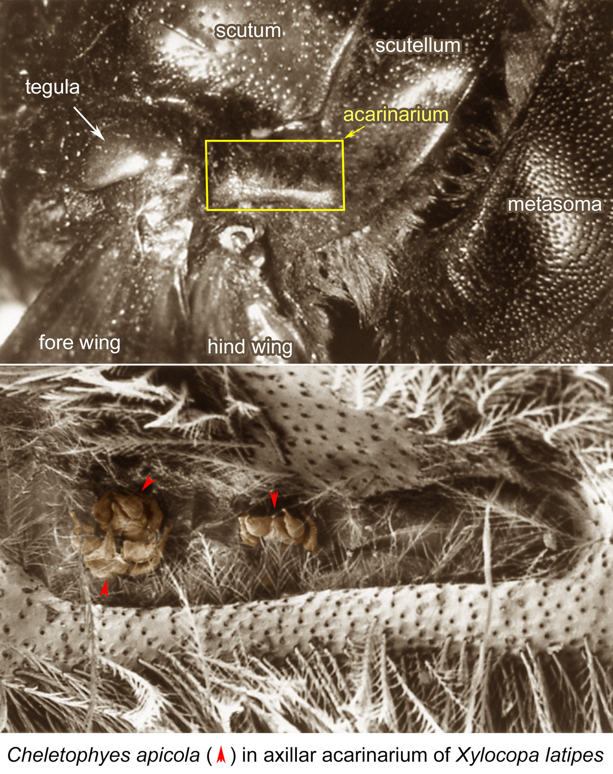
Eickwort, G. C. 1994. Evolution and life-history patterns of mites associated with bees. In Mites: Ecological and Evolutionary Analyses of Life-History Patterns, ed. M. A. Houck, 218-251. New York: Chapman amp; Hall.). In the Old World, females of Cheletophyes disperse in mesosomal (axillar) acarinariaacarinarium:
A specialized morphological structure that facilitates retention of mites on the body of an organism, typically a bee or wasp.
of adult female bees (Figs. 5, 6). AcarinariaAcarinarium:
A specialized morphological structure that facilitates retention of mites on the body of an organism, typically a bee or wasp.
are absent in male bees. According to the hypothesis of OConnor (1993), the relationships between Cheletophyes and their bee hosts are mutualistic, and the bees have developed mesosomal acarinariaacarinarium:
A specialized morphological structure that facilitates retention of mites on the body of an organism, typically a bee or wasp.
to transfer these predaceous mites that control nest kleptoparasiteskleptoparasite:
An animal that takes prey or other food from another animal that has caught, collected, or otherwise prepared the food, including stored food. Both kleptoparasitic bees and kleptoparasitic mites feed on food provisioned in the host bee nest. Kleptoparasitic bees do not make their own nests; they stealthily deposit eggs in the nest of a bee host and can act as phoretic hosts for mites only because they deliver them to nests of actual bee hosts. Variant spelling: cleptoparasite.
. Despite the fact that the mites do not directly depend on resources provided by the bees, they are highly specific to their hosts (Klimov et al., 2006Klimov et al., 2006:
Klimov, P. B., A. V. Bochkov amp; B. M. OConnor. 2006. Host specificity and multivariate diagnostics of cryptic species in predacious cheyletid mites of the genus Cheletophyes (Acari: Cheyletidae) associated with large carpenter bees. Biological Journal of the Linnean Society of London . 87 : 45-58.). One species, Cheletophyes panamensis, has been recorded for the Neotropical region (Klompen et al., 1984Klompen et al., 1984:
Klompen, J. S. H., E. Mendez amp; F. S. Lukoschus. 1984. A new species of the genus Cheletophyes Oudemans, 1914 (Prostigmata: Cheyletidae) from the nest of a carpenter bee in Panama. Acarologia . 25 : 249-251.). This species has been found in the nest of its host only. Large carpenter bees from the Neotropical region lack an acarinariumacarinarium:
A specialized morphological structure that facilitates retention of mites on the body of an organism, typically a bee or wasp.
. So far, phoreticphoretic:
Pertaining to phoresy; using another organism (i.e., a host) for dispersal to new habitats. Phoresy can be distinguished from parasitism because feeding typically does not occur during phoresy.
specimens of Ch. panamensis have not been discovered on their host's bodies, although it is likely this species can disperse on the bodies of adult female bees.
Authors: P. Klimov, B. OConnor, R. Ochoa, G. Bauchan, A. Redford, J. Scher
Last updated October 2016
tool images at ITP Node
idtools.org