at least one pest species; feeds on pollen stored in honeycombs, honey, royal jelly, and other nest materials; may cause colony collapse
Carpoglyphus Robin, 1869 (sometimes, the year is cited as 1860, however, Carpoglyphus Robin, 1860Robin, 1860:
Robin, C. 1860. Mémoire zoologique et anatomique sur diverses espèces d'Acariens de la famille des Sarcoplides. Bulletin de la Société Impériale Des Naturalistes de Moscou . 33 : 184-293 + plates 1-8. is not an available name; Robin, 1860Robin, 1860:
Robin, C. 1860. Mémoire zoologique et anatomique sur diverses espèces d'Acariens de la famille des Sarcoplides. Bulletin de la Société Impériale Des Naturalistes de Moscou . 33 : 184-293 + plates 1-8. cites some collective characters for "Carpoglyphus" and other genera of mites, which does not constitute a valid description, and, thus, the name Carpoglyphus Robin, 1860Robin, 1860:
Robin, C. 1860. Mémoire zoologique et anatomique sur diverses espèces d'Acariens de la famille des Sarcoplides. Bulletin de la Société Impériale Des Naturalistes de Moscou . 33 : 184-293 + plates 1-8. (nom. nud.) is unavailable (ICZN Art. 12.1)).
Superorder Acariformes » Order Sarcoptiformes » Suborder Oribatida » Infraorder Desmonomata » Hyporder Astigmata » Family Carpoglyphidae » Genus Carpoglyphus
"Carpoglyphus passularum (Robin, 1869 ex Hering, 1838)" (=Acarus passularum sensu Robin, 1869 non Hering, 1838; =Acarus lactis Linnaeus, 1767).
Dichotomiopus Fain and Camerik, 1978 (OConnor, unpublished synonymy, types studied)
prune mite, dried fruit mite, driedfruit mite, sugar mite; all names apply to Carpoglyphus lactis
Phoretic phoretic:
Pertaining to phoresy; using another organism (i.e., a host) for dispersal to new habitats. Phoresy can be distinguished from parasitism because feeding typically does not occur during phoresy.
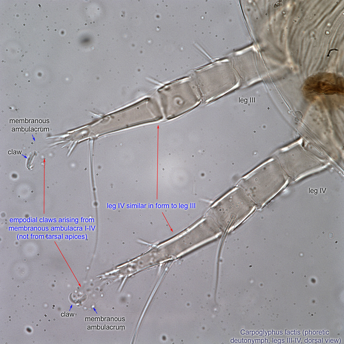
deutonymph: Empodial clawsEmpodial claw:
Claw-like, membranous, or pad-like structure of setal origin. Present only on the pretarsus in Acariformes. In Astigmata, it is the only claw on the pretarsus and often referred to simply as the claw. In the remaining Acariformes, may be accomanied by two lateral claws. Also known as empodium, pretarsal empodium, or central claw.
 arising from membranous ambulacraambulacrum:
The claws and empodium of the apotele or pretarsus.
I-IV (not from tarsal apices) (Figs. 4, 5). Leg IV with empodial clawempodial claw:
Claw-like, membranous, or pad-like structure of setal origin. Present only on the pretarsus in Acariformes. In Astigmata, it is the only claw on the pretarsus and often referred to simply as the claw. In the remaining Acariformes, may be accomanied by two lateral claws. Also known as empodium, pretarsal empodium, or central claw.
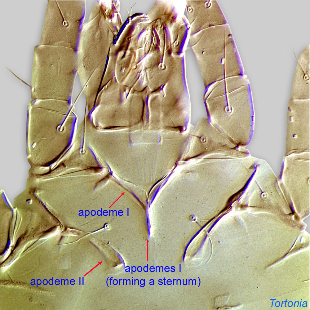
present (Fig. 5). Leg IV generally similar in form to leg III (Figs. 2, 5). Posterior median ventral apodemeapodeme:
Internal sclerite that serves as an attachment site for muscles. Most commonly used (as "coxal apodeme") to describe elements of coxae fused to the ventral body in Acariformes (coxae are free and not fused to the body in Parasitiformes), and may be variously referred to as ventral, sternal, anterior, or posterior.
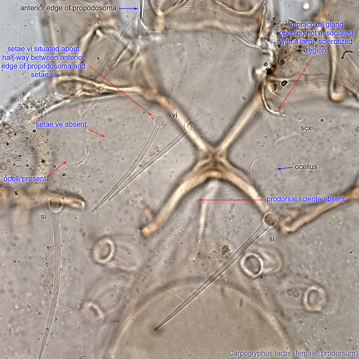
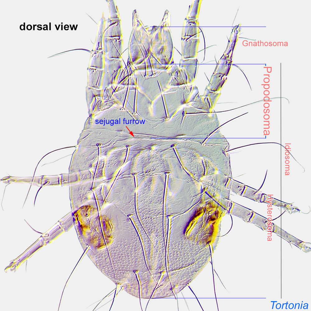
 absent (Fig. 2). Ocelli present, widely separated on propodosomapropodosoma:
Anterior part of idiosoma, in front of sejugal furrow.
 (Fig. 3).
Adult: Prodorsumprodorsum:
Dorsal surface of propodosoma.
with setae ve absent (Fig. 8). Prodorsalprodorsal:
Pertaining to the prodorsum.
sclerite absent (Fig. 8). Setae vi situated about half-way between anterior edge of propodosomapropodosoma:
Anterior part of idiosoma, in front of sejugal furrow.
and setae si (Fig. 8). Ocelli present (Fig. 8). Supracoxal glandsupracoxal gland:
Paired glands serving for maintenance of water balance in Astigmata. These glands are usually invisible on typical microscopic preparations. However, their presence can be detected by the well-sclerotized supracoxal gland openings, which are situated on supracoxal sclerites.
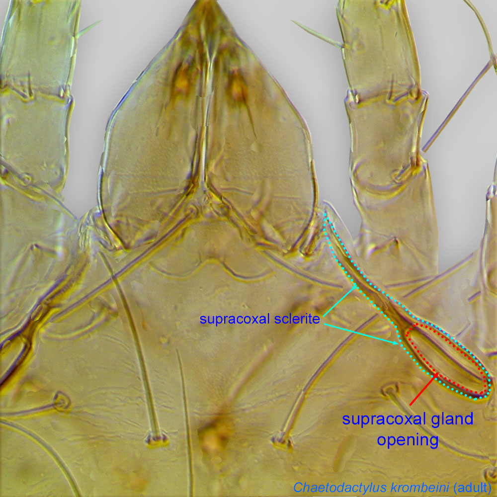 opening not associated with a large sclerotized region (Fig. 8). TarsiTarsus:
Terminal segment (also known as podomere or palpomere) of legs or palps. In Parasitoformes it can be subdivided into telotarsus and basitarsus.
 I-II elongate (Fig. 11). PretarsiPretarsus:
Terminal leg or palpal segment distal to tarsus.
similar on all legs (Fig. 11). PretarsiPretarsus:
Terminal leg or palpal segment distal to tarsus.
with long, thin condylophorescondylophore:
In Acariformes, pretarsal paired sclerotized structures arising from the distal end of the tarsus and forming a joint with lateral claws (in Endeostigmata, Oribatida and Trombidiformes) and the empodial claw. Not to be confused with vertical sclerites of Parasitiformes.
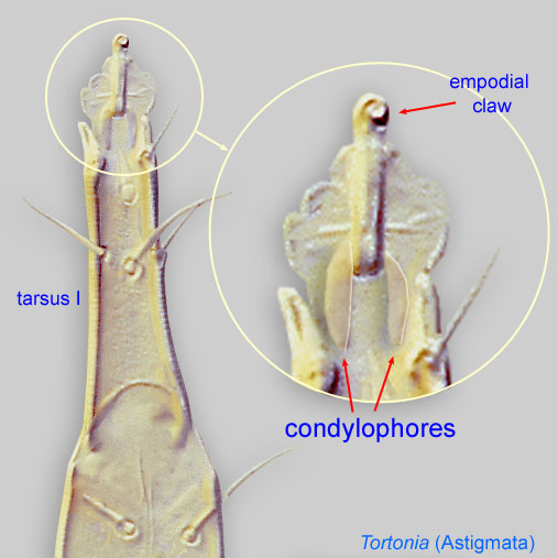 (Figs. 11, 13). Empodial clawsEmpodial claw:
Claw-like, membranous, or pad-like structure of setal origin. Present only on the pretarsus in Acariformes. In Astigmata, it is the only claw on the pretarsus and often referred to simply as the claw. In the remaining Acariformes, may be accomanied by two lateral claws. Also known as empodium, pretarsal empodium, or central claw.
present (Figs. 11, 13). Dorsal setae smooth, not heavily barbed (Fig. 6). Males without paranal suckers or sucker-like setae on tarsustarsus:
Terminal segment (also known as podomere or palpomere) of legs or palps. In Parasitoformes it can be subdivided into telotarsus and basitarsus.
IV (Fig. 13). Coxal apodemesapodeme:
Internal sclerite that serves as an attachment site for muscles. Most commonly used (as "coxal apodeme") to describe elements of coxae fused to the ventral body in Acariformes (coxae are free and not fused to the body in Parasitiformes), and may be variously referred to as ventral, sternal, anterior, or posterior.
I fused medially with coxal apodemesapodeme:
Internal sclerite that serves as an attachment site for muscles. Most commonly used (as "coxal apodeme") to describe elements of coxae fused to the ventral body in Acariformes (coxae are free and not fused to the body in Parasitiformes), and may be variously referred to as ventral, sternal, anterior, or posterior.
II closing coxal fields I in both sexes (Fig. 9). CondylophoresCondylophore:
In Acariformes, pretarsal paired sclerotized structures arising from the distal end of the tarsus and forming a joint with lateral claws (in Endeostigmata, Oribatida and Trombidiformes) and the empodial claw. Not to be confused with vertical sclerites of Parasitiformes.
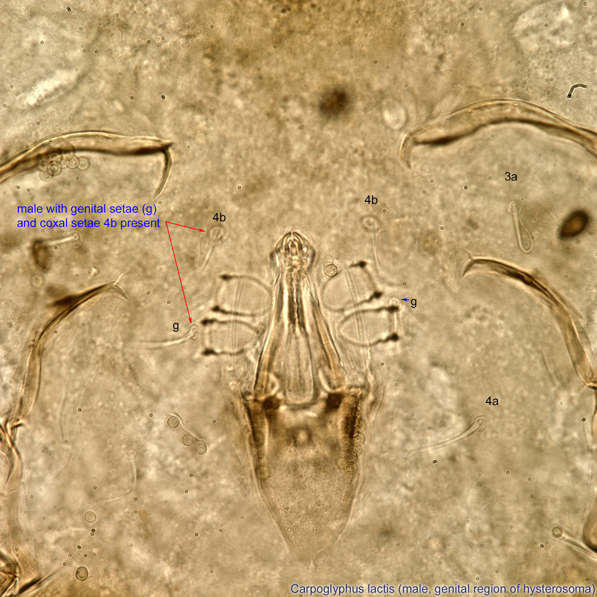
asymmetrical in male (Fig. 12). Male with genital setae (g) and coxal setae 4b present (Fig. 10).
A dichotomous key to adults (males and females) is available in Fain and Rack, 1987Fain and Rack, 1987:
Fain, A. amp; G. Rack. 1987. Notes on the mites living in the flowers of Espeletia spp. (Asteraceae) in Colombia. 1. Carpoglyphus sturmi sp. n. (Acari, Carpoglyphidae). Entomologische Mitteilungen aus dem Zoologischen Staatsinstitut und Zoologischen Museum Hamburg . 9 : 9-19.. This key can be used to identify the two species found in associations with bees: Carpoglyphus lactis and Carpoglyphus munroi. One non bee-associated species, Carpoglyphus wardleorum, was described later (Clark, 2010Clark, 2010:
Clark, J. M. 2010. A new species of Carpoglyphus (Astigmatina: Carpoglyphidae) from the bark of black beech ( Nothofagus ) honeydew in New Zealand. International Journal of Acarology . 36 : 453-459.).
Cosmopolitain. Mites from honey bees have been reported from the Holarctic, Oriental, and Australian regions.
Feeding stages live in beehives of honey bees (Apis) and probably in nests of stingless bees (Meliponini).
facultativefacultative:
can complete entire life cycle without bees or their close relative, wasps
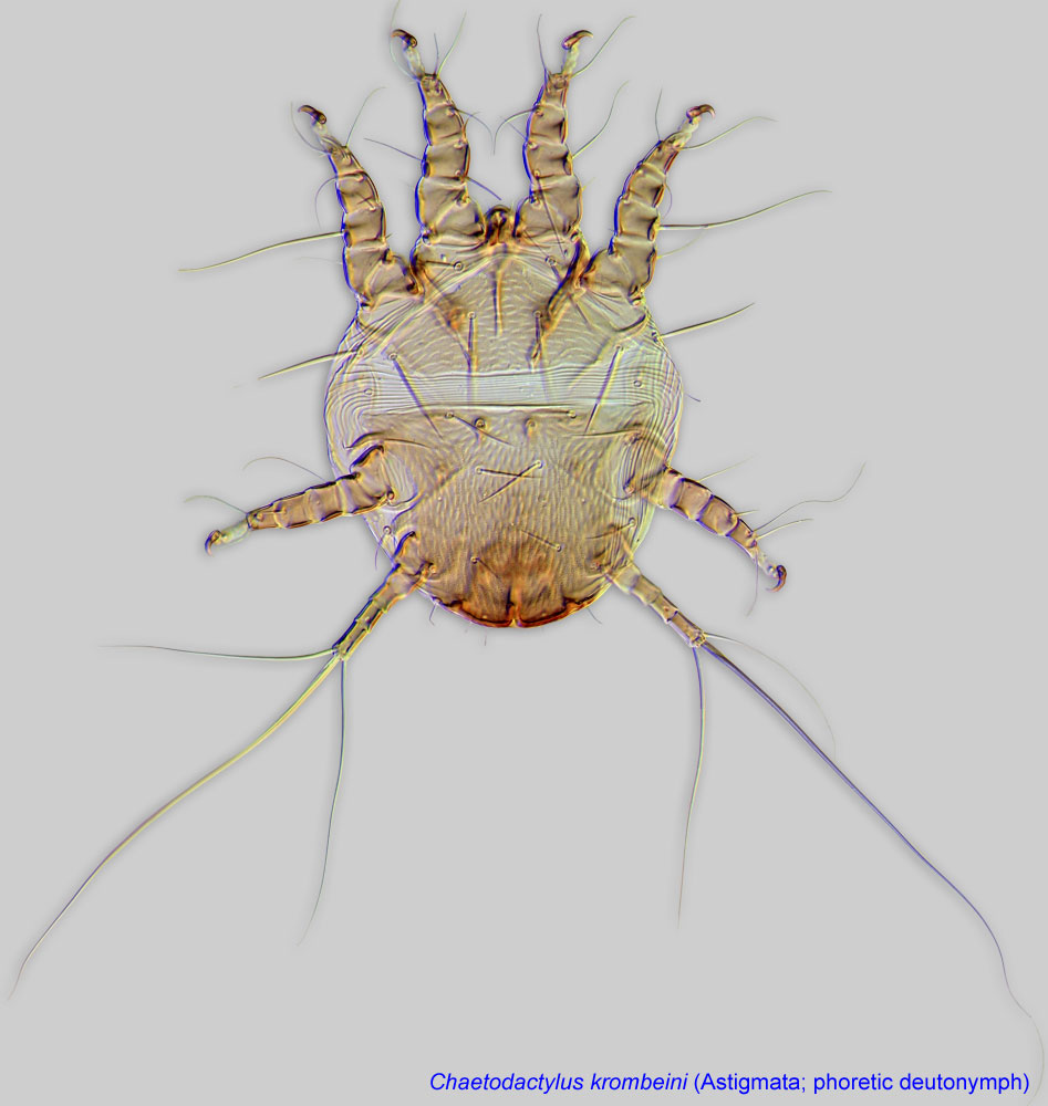 disperse on adult insect hosts to suitable habitats. Phoresyphoresy:
Carpoglyphus lactis is a relatively common species in beehives, occurring in the debris on the bottom boards of beehives, honeycombs, dead bees, honey, and especially on the bee bread (bee pollen with added honey and bee secretions that is stored in brood cells). This mite can penetrate the brood cells and make burrows in the stored pollen, consuming it and causing the pollen and the debris to spill from the cells. The infested bee bread, mixed with large quantities of dead and live mites, turns to a golden-brown or yellow powdery material covering honeycombs and bottom boards of beehives. The mite damage is especially severe in stored overwintering nests. For example, 250 honeycombs were destroyed by these mites over one winter in a single storage area in Germany (Zander, 1947Zander, 1947:
Zander, E. 1947. Handbuch der Bienenkunde in Einzeldarstellungen. vol 2. Krankheiten und Schädlinge der erwachsenen Bienen. Stuttgart: Verlag Eugen Ulmer. 147 pp.). A similar case was reported in the USA (Alabama), where stored honeycombs were found heavily infested after winter storage (Baker and Delfinado, 1978Baker and Delfinado, 1978:
Baker, E. W. amp; M. D. Delfinado. 1978. Notes on the driedfruit mite Carpoglyphus lactis (Acarina: Carpoglyphidae) infesting honey bee combs. Journal of Apicultural Research . 17 : 52-54.). Weak bee colonies are more susceptible to mite attacks (Zander, 1947Zander, 1947:
Zander, E. 1947. Handbuch der Bienenkunde in Einzeldarstellungen. vol 2. Krankheiten und Schädlinge der erwachsenen Bienen. Stuttgart: Verlag Eugen Ulmer. 147 pp.), while healthy bee colonies usually can clean up the infested pollen (Baker and Delfinado, 1978Baker and Delfinado, 1978:
Baker, E. W. amp; M. D. Delfinado. 1978. Notes on the driedfruit mite Carpoglyphus lactis (Acarina: Carpoglyphidae) infesting honey bee combs. Journal of Apicultural Research . 17 : 52-54.).
Furthermore, a case of collapse of a managed colony of a stingless bee, Tetragonula iridipennis, was reported in India (Vijayakumar et al., 2013Vijayakumar et al., 2013:
Vijayakumar, K., Muthuraman, M. and Jayaraj, R. 2013. Infestation of Carpoglyphus lactis (Linnaeus) (Acari: Carpoglyphidae) on Trigona iridipennis (Apidae: Meliponinae) from India. Scholarly Journal of Agricultural Science . 3 : 25-28.). Although the mite illustrated in this paper belongs to the family Cheyletidae, the yellowish carpet of dust on the bottom of the nest (Figs. 2A,B) may represent an astigmatid mite (Carpoglyphus).
Aside from beehives, Carpoglyphus lactis is also found in old honeycombs, wine barrels in cellars, dried sweet fruits, canned fruits, fruits preserved in sugar, fermenting pulp, dairy products (milk and cheese), and stored honey. It often infests products following substantial development of yeast. In the field, this species is found in fermenting tree sap flows, burrows of moles, and as phoreticphoretic:
Pertaining to phoresy; using another organism (i.e., a host) for dispersal to new habitats. Phoresy can be distinguished from parasitism because feeding typically does not occur during phoresy.
deutonymphsdeutonymph:
Ontogenetic stage between protonymph and tritonymph (or adult, if tritonymph is absent). See <a href="index.cfm?pageID=1720">Life stages page</a> for more details. on butterflies, moths, and scarabaeid beetles (e.g., Gnorimus).
When large numbers of C. lactis mites are ingested with infested food or beverages, they can cause dysentery. These mites also cause dermatitis to handlers of infested materials, such as dried plums (O'Donovan, 1922O'Donovan, 1922:
O'Donovan, W. J. 1922. Dermatitis due to Carpoglyphus passularum . British Journal of Dermatology and Syphilis . 34 : 297-298.), and occupational allergies in the biological pesticide industry (Krop et al., 2012Krop et al., 2012:
Krop, E., R. Houba, J. Spithoven, S. de Wind, J. Rooyackers amp; D. Heederik. 2012. Occupational allergy to mites and moths in a biological pesticides company. Allergy . 67 : 73-73.). However, they may be beneficial in the wine industry, as mite alarm pheromones enhance the aroma of pale and dry wines aged under flor yeasts (Marin et al., 2009Marin et al., 2009:
Marin, J., R. Ocete, M. Pedroza, A. Zalacain, C. de Miguel, M. A. Lopez amp; M. R. Salinas. 2009. Influence of the mite Carpoglyphus lactis (L) on the aroma of pale and dry wines aged under flor yeasts. Journal of Food Composition and Analysis . 22 : 745-750.).
The second mite species found in beehives, Carpoglyphus munroi, is relatively rare and has been recorded from beehives from the Czech Republic.
Authors: P. Klimov, B. OConnor, R. Ochoa, G. Bauchan, A. Redford, J. Scher
Last updated October 2016
tool images at ITP Node
idtools.org