probably beneficial; provides sanitary services by feeding on fungi on leaves that bees use as cell lining material
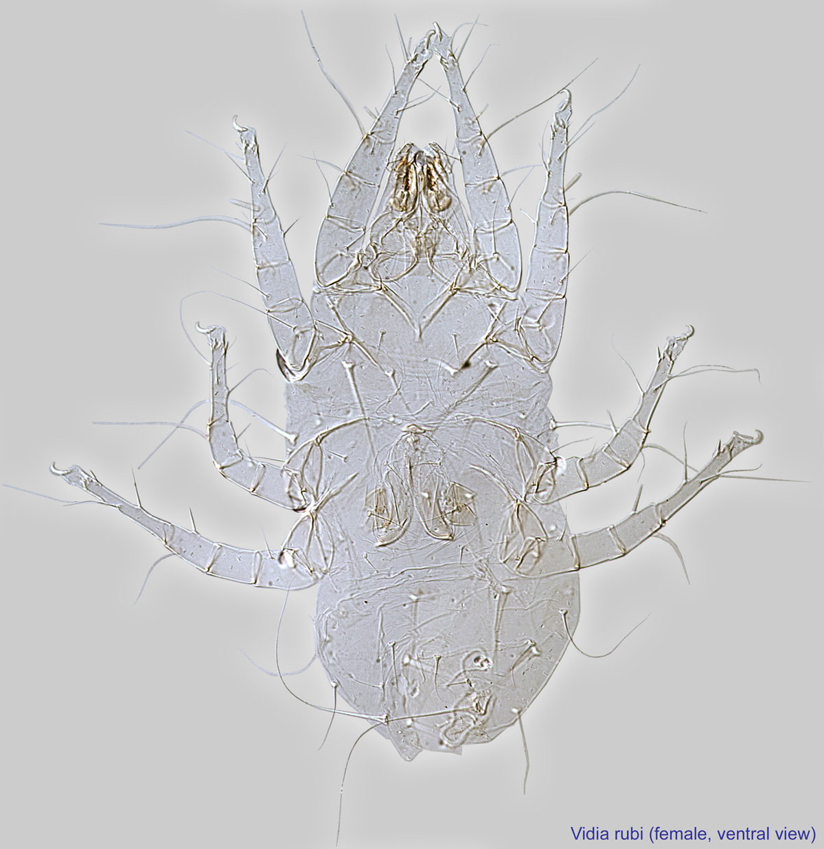
Vidia Oudemans, 1905
Superorder Acariformes » Order Sarcoptiformes » Suborder Oribatida » Infraorder Desmonomata » Hyporder Astigmata » Family Winterschmidtiidae » Genus Vidia
Vidia undulata Oudemans, 1905
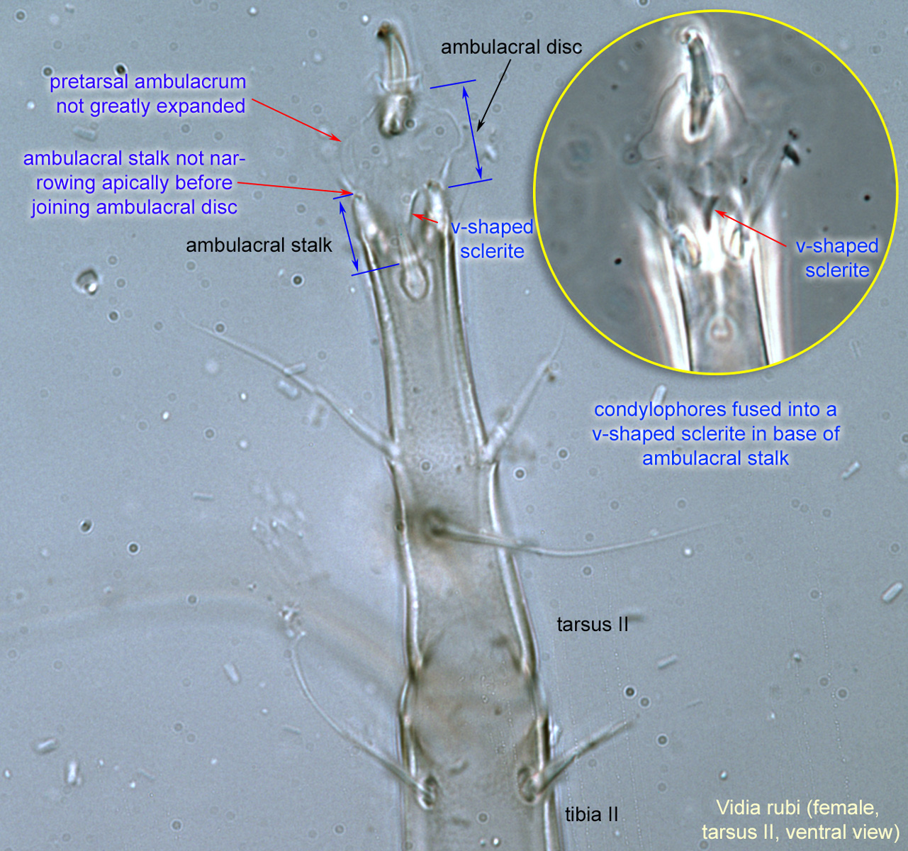
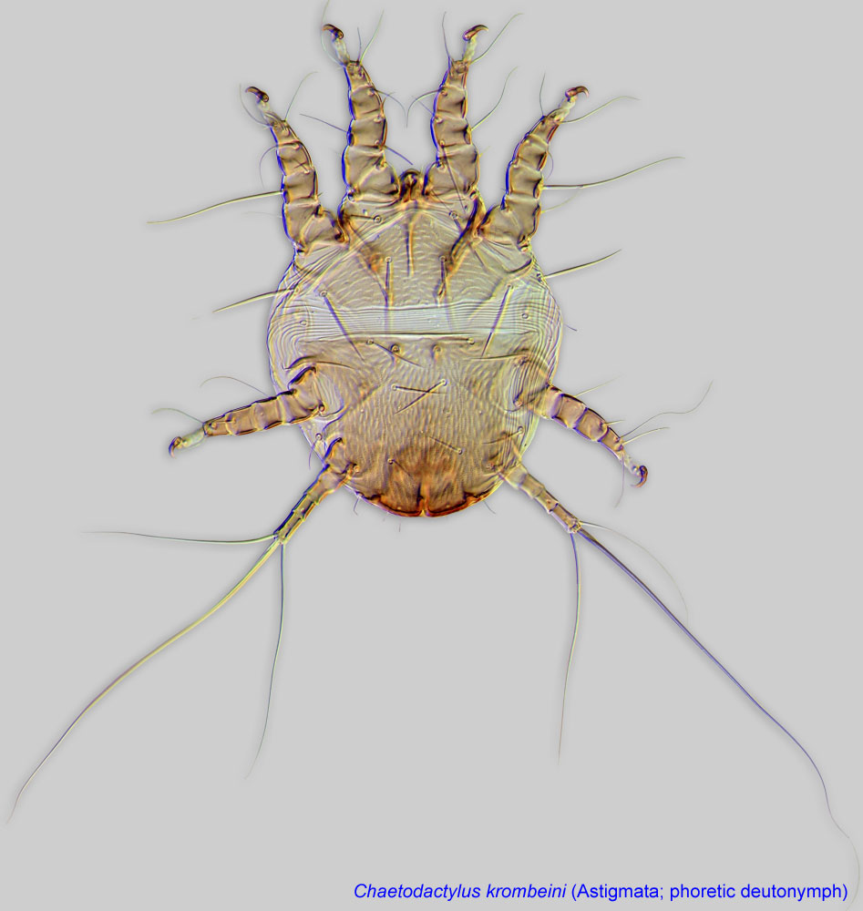
Adult: Condylophores fused into a V-shaped scleritesclerite:
A component section of an exoskeleton; a plate forming the skeleton of an arthropod.
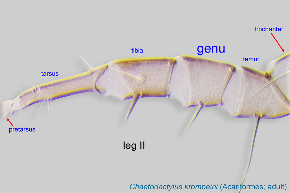
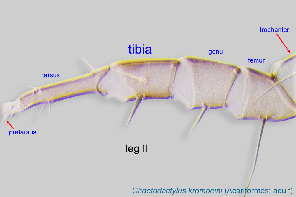
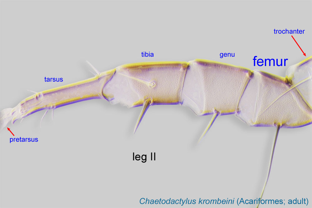
in base of ambulacral stalk (Fig 13). Genu genu:
Leg or palp segment (also known as podomere or palpomere) between tibia and femur.
 III without a dorsal solenidionsolenidion:
Thin-walled, terminally rounded or pointed filiform or peglike structure that is not birefringent in polarized light (unlike common setae in Acariformes). Often appears striated because of its internal structure. Found on the palpal tarsus on the gnathosoma and may also occur on the tarsus and tibia, less frequently on the genu, and occasionally on the femur of legs I-IV. In Acariformes, leg solenidia often arise from unsclerotized areas.
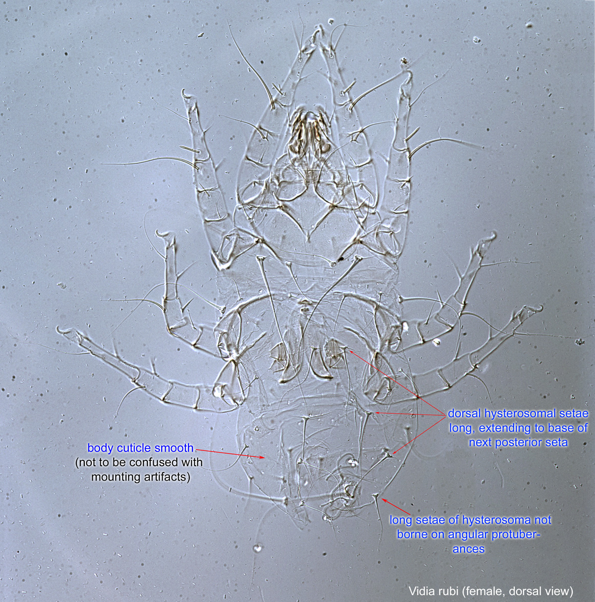
 (Fig. 14). Ambulacral stalk not narrowing apically before joining ambulacral disc (Fig 13). Dorsal hysterosomal setae long, extending to base of next posterior seta (Fig 8). Body cuticle smooth (Fig. 8). Tarsustarsus:
Terminal segment (also known as podomere or palpomere) of legs or palps. In Parasitoformes it can be subdivided into telotarsus and basitarsus.
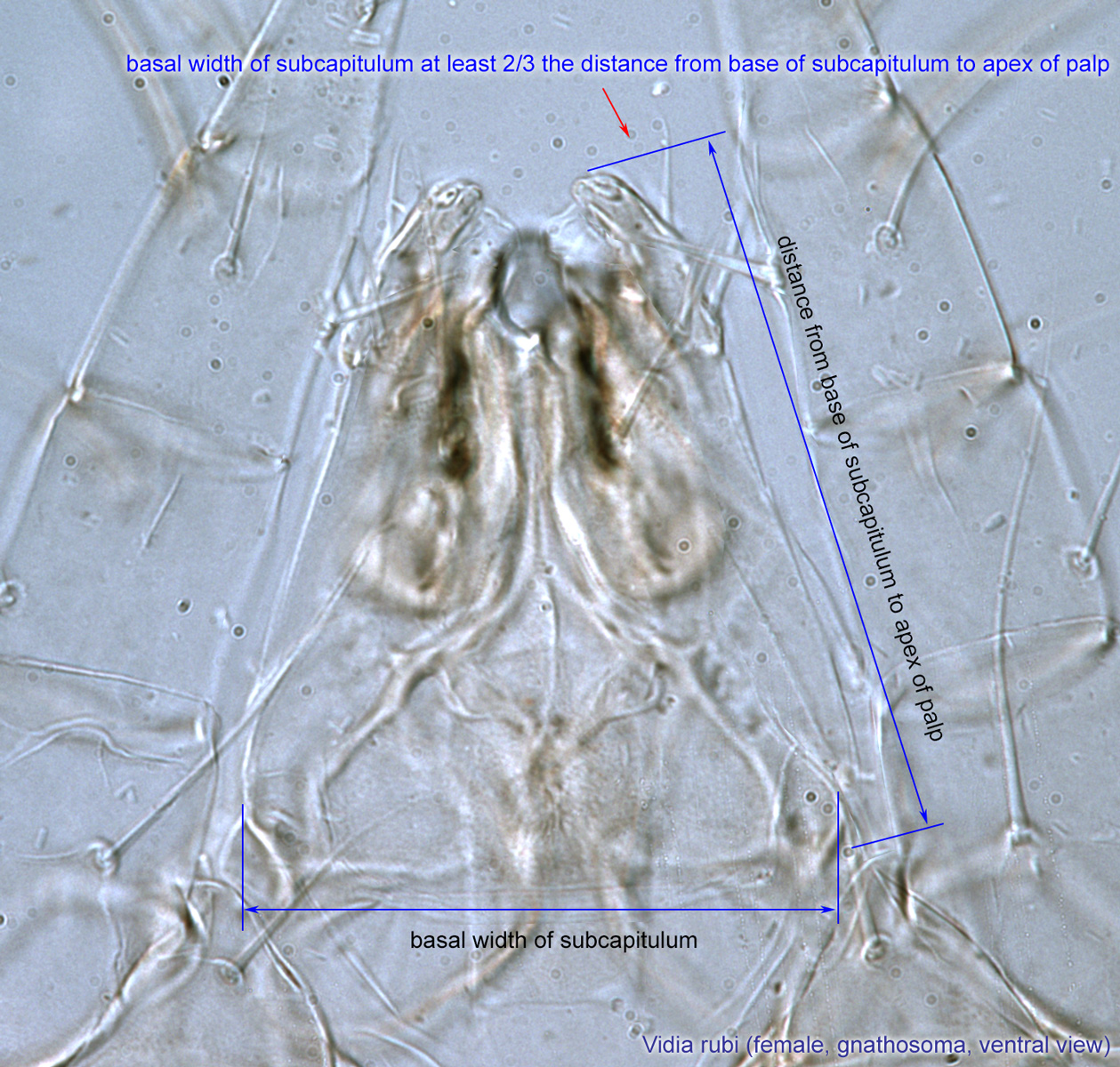
 I less than 4 times longer than it is wide (Figs. 12). Basal width of subcapitulumsubcapitulum:
Ventral faces of the fused palpcoxae.
at least 2/3 the distance from base of subcapitulumsubcapitulum:
Ventral faces of the fused palpcoxae.
to apex of palp (Fig 10).
Phoretic phoretic:
Pertaining to phoresy; using another organism (i.e., a host) for dispersal to new habitats. Phoresy can be distinguished from parasitism because feeding typically does not occur during phoresy.
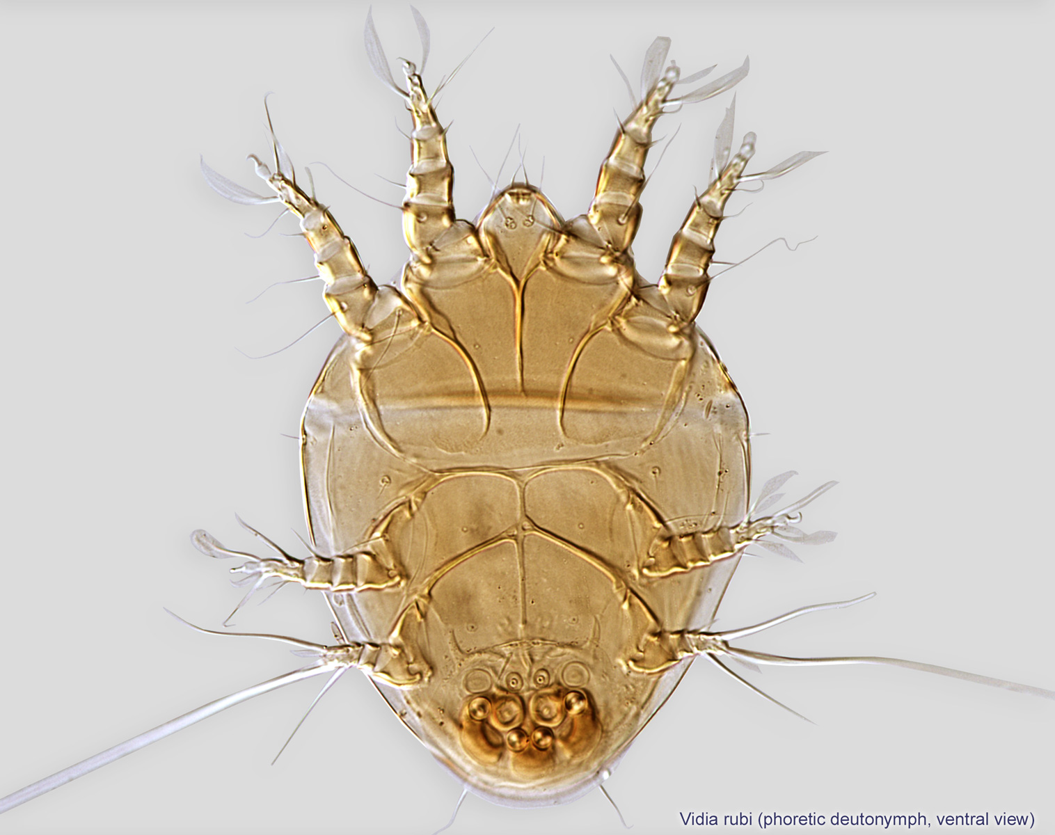
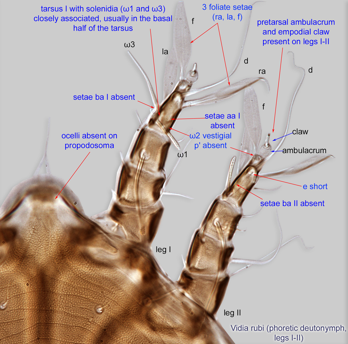
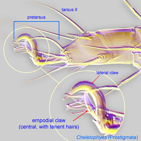
deutonymph: Setae ba I-II and aa I absent (correlates with character in adult) (Fig. 5). Pretarsal ambulacrumambulacrum:
The claws and empodium of the apotele or pretarsus.
and empodial clawempodial claw:
Claw-like, membranous, or pad-like structure of setal origin. Present only on the pretarsus in Acariformes. In Astigmata, it is the only claw on the pretarsus and often referred to simply as the claw. In the remaining Acariformes, may be accomanied by two lateral claws. Also known as empodium, pretarsal empodium, or central claw.
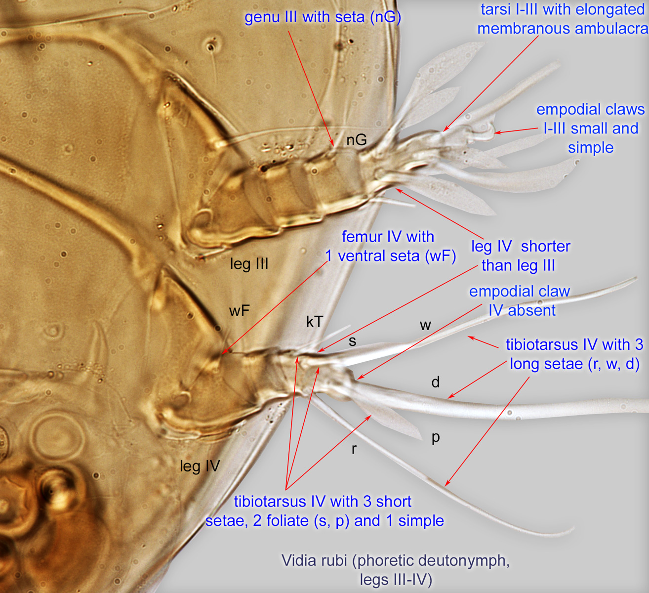
 present on legs I-II (Fig. 5). TarsiTarsus:
Terminal segment (also known as podomere or palpomere) of legs or palps. In Parasitoformes it can be subdivided into telotarsus and basitarsus.
I-III with elongated membranous ambulacraambulacrum:
The claws and empodium of the apotele or pretarsus.
(Fig. 7). Empodial clawsEmpodial claw:
Claw-like, membranous, or pad-like structure of setal origin. Present only on the pretarsus in Acariformes. In Astigmata, it is the only claw on the pretarsus and often referred to simply as the claw. In the remaining Acariformes, may be accomanied by two lateral claws. Also known as empodium, pretarsal empodium, or central claw.
I-III small and simplesimple:
Of claws or setae; not modified or not bi- or trifurcate at tip.
(Fig. 7). Empodial clawempodial claw:
Claw-like, membranous, or pad-like structure of setal origin. Present only on the pretarsus in Acariformes. In Astigmata, it is the only claw on the pretarsus and often referred to simply as the claw. In the remaining Acariformes, may be accomanied by two lateral claws. Also known as empodium, pretarsal empodium, or central claw.
IV absent (Fig. 7). Leg IV shorter than leg III (Fig. 7). Tibiotarsus IV with 3 long apical setae (Fig. 7) and 3 short (2 foliate, 1 simplesimple:
Of claws or setae; not modified or not bi- or trifurcate at tip.
) (Fig. 7). Tarsustarsus:
Terminal segment (also known as podomere or palpomere) of legs or palps. In Parasitoformes it can be subdivided into telotarsus and basitarsus.
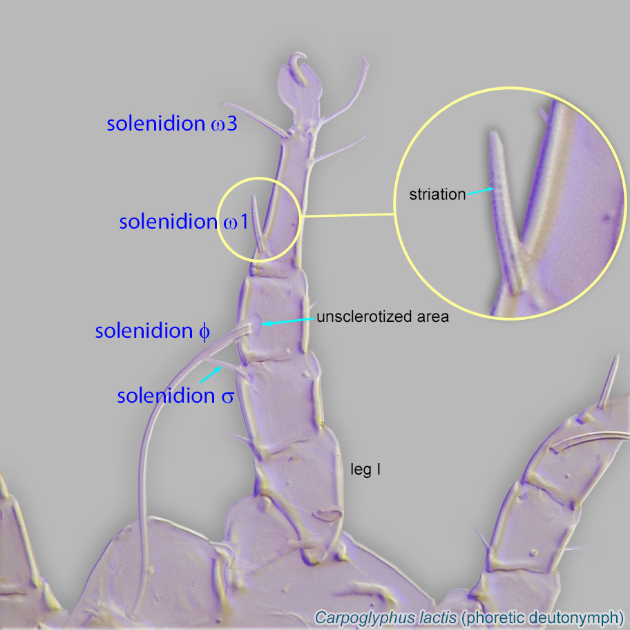
I with solenidiasolenidion:
Thin-walled, terminally rounded or pointed filiform or peglike structure that is not birefringent in polarized light (unlike common setae in Acariformes). Often appears striated because of its internal structure. Found on the palpal tarsus on the gnathosoma and may also occur on the tarsus and tibia, less frequently on the genu, and occasionally on the femur of legs I-IV. In Acariformes, leg solenidia often arise from unsclerotized areas.
(ω1 and ω3) closely associated, usually in the basal half of the tarsustarsus:
Terminal segment (also known as podomere or palpomere) of legs or palps. In Parasitoformes it can be subdivided into telotarsus and basitarsus.
(Fig. 5). Solenidionsolenidion:
Thin-walled, terminally rounded or pointed filiform or peglike structure that is not birefringent in polarized light (unlike common setae in Acariformes). Often appears striated because of its internal structure. Found on the palpal tarsus on the gnathosoma and may also occur on the tarsus and tibia, less frequently on the genu, and occasionally on the femur of legs I-IV. In Acariformes, leg solenidia often arise from unsclerotized areas.
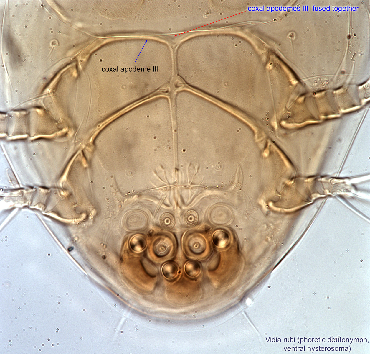
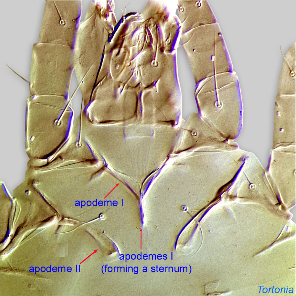
ω2 vestigial, alveolar (Fig. 5). Coxal apodemesapodeme:
Internal sclerite that serves as an attachment site for muscles. Most commonly used (as "coxal apodeme") to describe elements of coxae fused to the ventral body in Acariformes (coxae are free and not fused to the body in Parasitiformes), and may be variously referred to as ventral, sternal, anterior, or posterior.
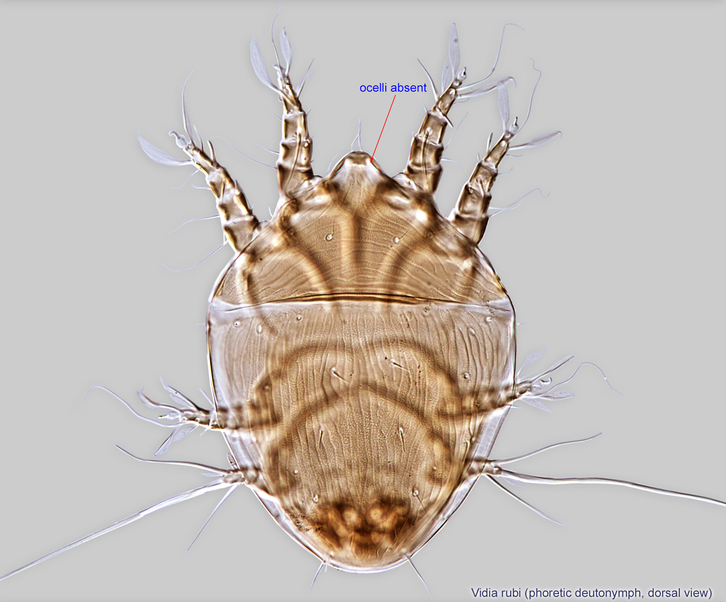
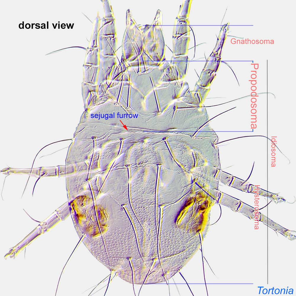
 III fused together (Fig. 4). Ocelli absent on propodosomapropodosoma:
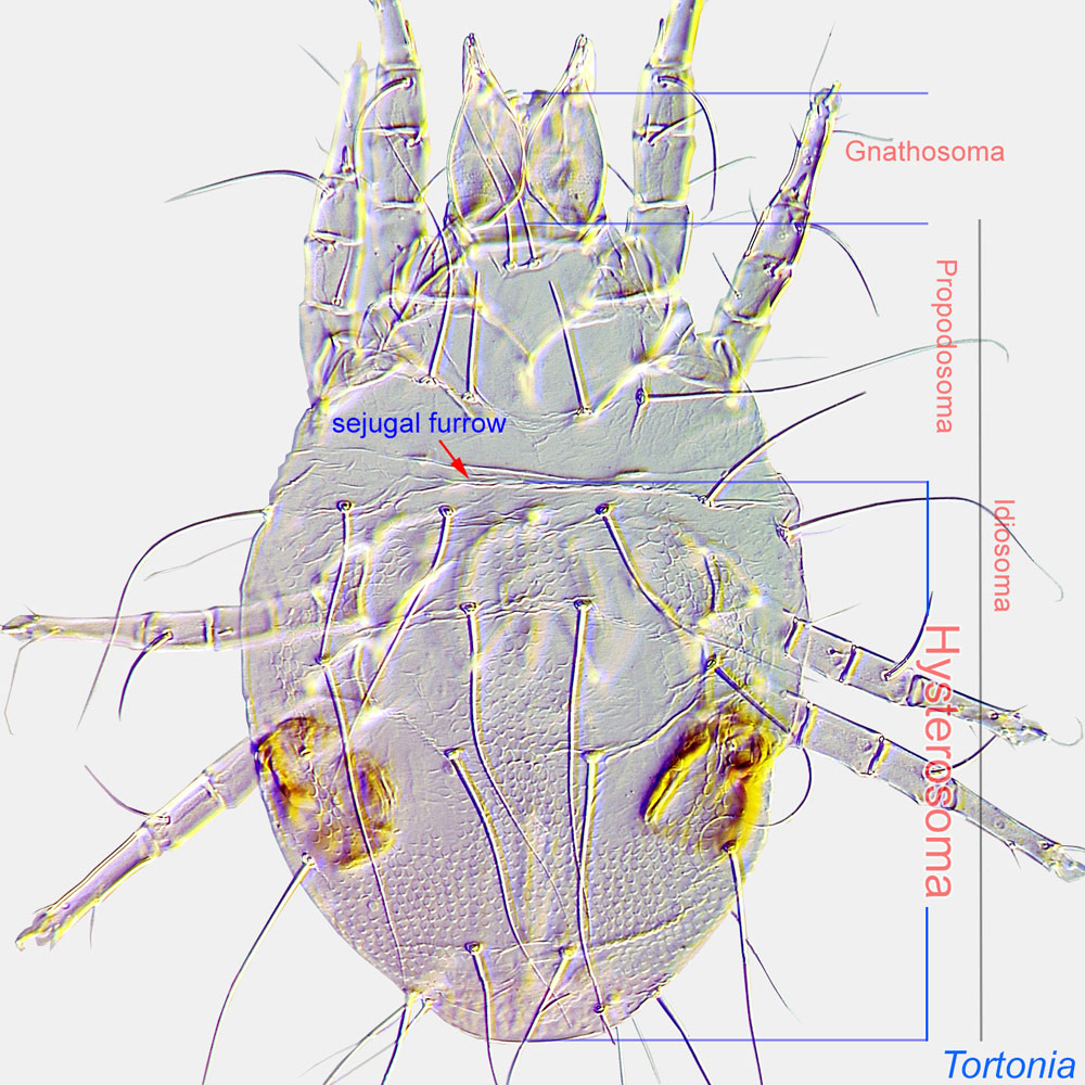
Anterior part of idiosoma, in front of sejugal furrow.
 (Figs. 1, 5). TarsiTarsus:
Terminal segment (also known as podomere or palpomere) of legs or palps. In Parasitoformes it can be subdivided into telotarsus and basitarsus.
I-II with 6 setae (e, f, d, ra, la, wa; p' absent), of which 3 foliate (ra, la, f), e short (Figs. 5, 6). TibiaeTibia:
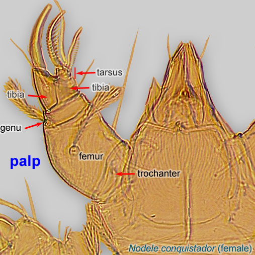
Leg or palp segment (also known as podomere or palpomere) between tarsus and genu.
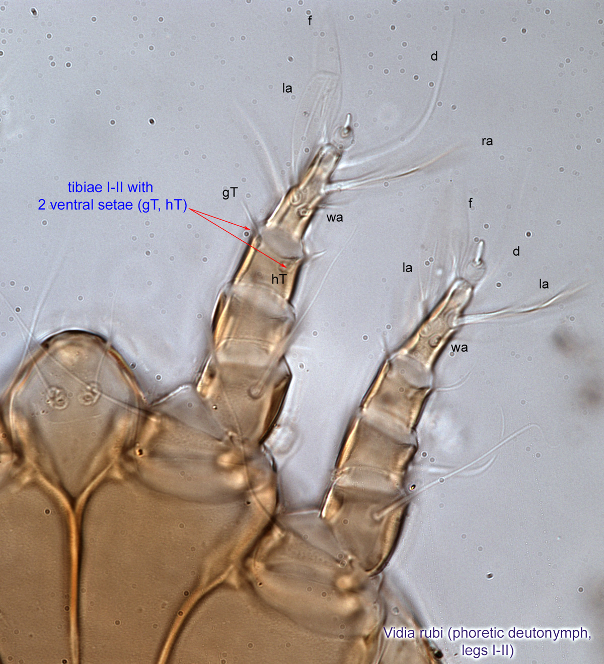
 I-II with 2 ventral seta (Fig. 6). Femurfemur:
Leg or palp segment (also known as podomere or palpomere) between genu and trochanter. In ParasitIformes can be subdivided into telofemur and basifemur.
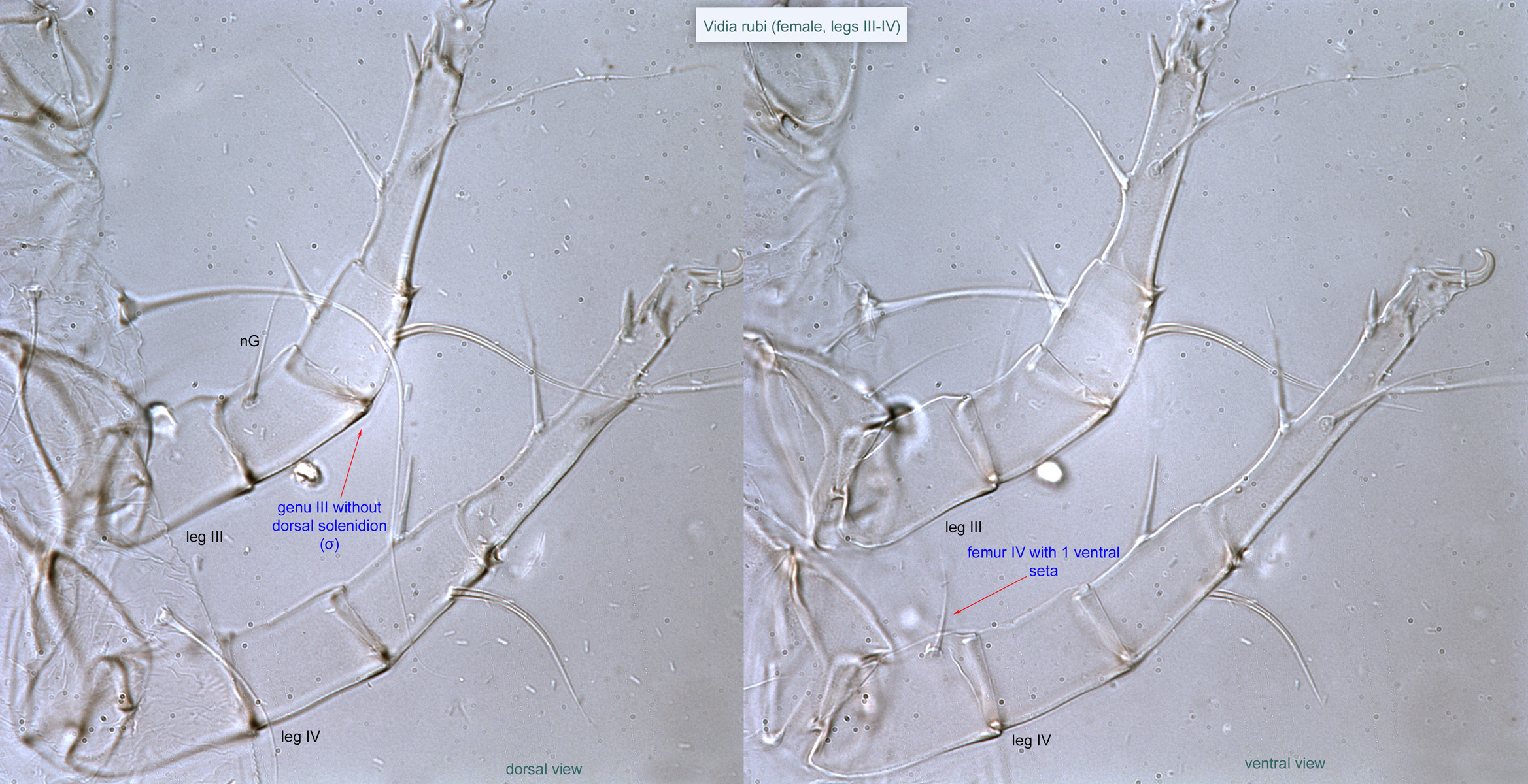
 IV with 1 ventral seta (wF) (Fig. 7). Genugenu:
Leg or palp segment (also known as podomere or palpomere) between tibia and femur.
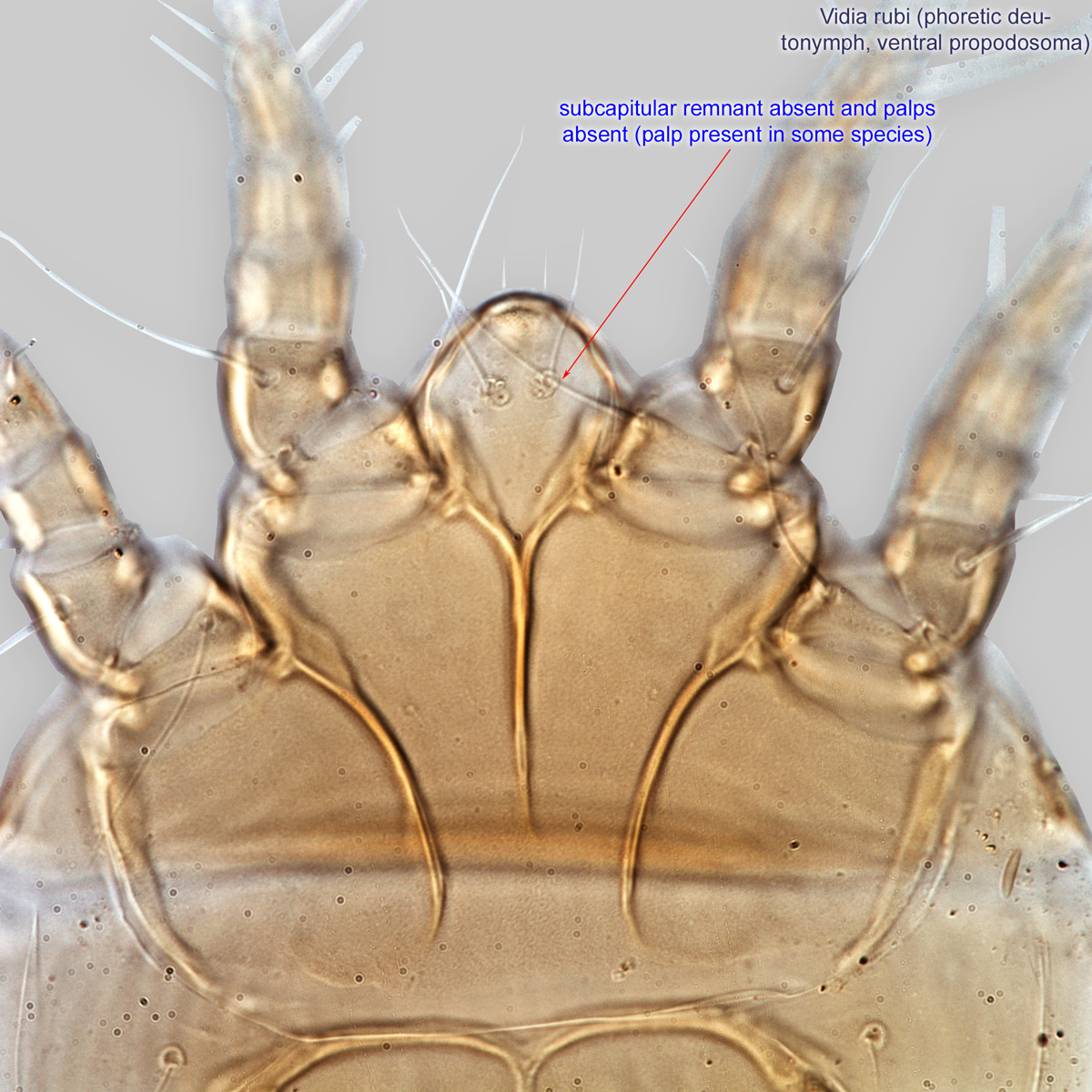
III with seta nG (Fig. 7). Subcapitular remnant absent (Fig. 3). PalpsPalp:
Second (after chelicera) paired appendage of the gnathosoma. Has a sensory function, but may be variously modified for other functions (e.g., raptorial, attachment to host, or filtering).
 present or absent (Fig. 3).
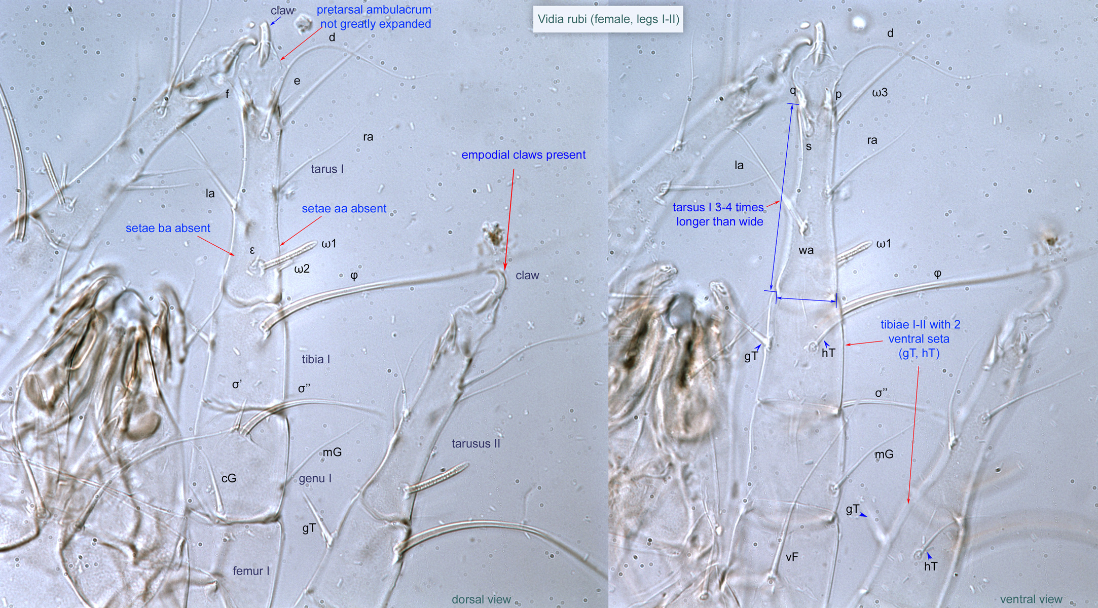
Adult: Pretarsal ambulacrumambulacrum:
The claws and empodium of the apotele or pretarsus.
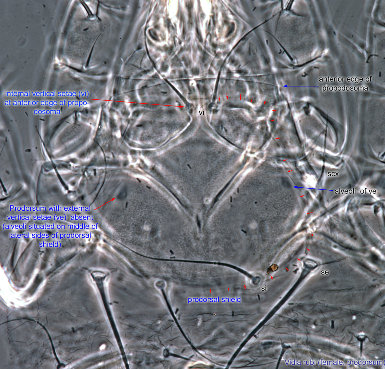
not greatly expanded (Figs. 12, 13). Prodorsumprodorsum:
Dorsal surface of propodosoma.
with external vertical setae (ve) absent (alveoli situated on middle of lateral sides of prodorsalprodorsal:
Pertaining to the prodorsum.
shield) (Fig 11). Internal vertical setae (vi) at anterior edge of propodosomapropodosoma:
Anterior part of idiosoma, in front of sejugal furrow.
(Fig. 11). Empodial clawsEmpodial claw:
Claw-like, membranous, or pad-like structure of setal origin. Present only on the pretarsus in Acariformes. In Astigmata, it is the only claw on the pretarsus and often referred to simply as the claw. In the remaining Acariformes, may be accomanied by two lateral claws. Also known as empodium, pretarsal empodium, or central claw.
present (Fig 12). Femurfemur:
Leg or palp segment (also known as podomere or palpomere) between genu and trochanter. In ParasitIformes can be subdivided into telofemur and basifemur.
IV with 1 ventral seta (Fig. 14). TarsiTarsus:
Terminal segment (also known as podomere or palpomere) of legs or palps. In Parasitoformes it can be subdivided into telotarsus and basitarsus.
long, at least three times as long as wide (Fig. 12-14). Male without obvious striation pattern on hysterosomahysterosoma:
Division of body posterior to the sejugal furrow, bearing legs III and IV.
 . TibiaeTibia:
Leg or palp segment (also known as podomere or palpomere) between tarsus and genu.
I-II with 2 ventral seta (Fig. 12, 13). Long setae of hysterosomahysterosoma:
Division of body posterior to the sejugal furrow, bearing legs III and IV.
not borne on angular protuberances (Fig. 8).
The two Palaearctic species, Vidia lineata and Vidia undulata, can be identified using descriptions and illustrations from Fain, 1972Fain, 1972:
Fain, A. 1972. Notes sur les hypopes des Saproglyphidae (Acarina: Sarcoptiformes) 2. Redefinition des genres. Acarologia. 14: 225-249.. Nearctic species (Vidia hirsuta, Vidia latimanus, Vidia rubi, and Vidia texana) can be identified using OConnor and Eickwort, 1988OConnor and Eickwort, 1988:
OConnor, B. M. amp; G. C. Eickwort. 1988. Morphology, ontogeny and systematics of the genus Vidia (Acari: Winterschmidtiidae). Acarologia. 29: 147-174.. Trichotarsus bomborum Leonardi, 1900Leonardi, 1900:
Leonardi, G. 1900. Storia naturale degli Acari Insetticoli. Bollettino della Societa Entomologica Italiana. 32: 1-76. probably belongs in Vidia. No key to species exists and many undescribed species have been collected.
Cosmopolitan except for Antarctica: Holarctic, Neotropical, Afrotropical, Oriental, and Australian regions.
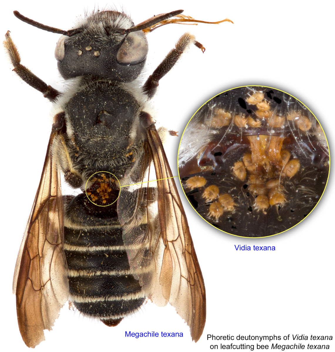
This genus is primarily associated with leafcutting and resin bees (Megachile), but a few records are available from Hylaeus, bumble bees (Bombus), and honey bees (Apis).
permanentpermanent:
associated exclusively with bees or their close relative, wasps; cannot live without these hosts
 (non-feeding stage) disperse on adult bees from one nest to another. Kleptoparasitic bees (Coelioxys) are also used as transport.
Observations on Vidia rubi (OConnor and Eickwort, 1988OConnor and Eickwort, 1988:
OConnor, B. M. amp; G. C. Eickwort. 1988. Morphology, ontogeny and systematics of the genus Vidia (Acari: Winterschmidtiidae). Acarologia. 29: 147-174.), associated with ground-nesting Megachile rubi, suggest that Vidia does not harm the bee directly or indirectly, but instead stays on the leaves that Megachile bees use for nest cell walls, where the mites presumably feed on fungi. The mites do not occur on the bees' provision masses nor on the young bee larvae. In contrast, large numbers of Vidia texana have been found on prepupae of Megachile texana, which was originally interpreted as evidence for parasitism (Eickwort et al., 1981Eickwort et al., 1981:
Eickwort, G. C., R. W. Matthews amp; J. Carpenter. 1981. Observations on the nesting behavior of Megachile rubi and M. texana with a discussion of the significance of soil nesting in the evolution of megachilid bees (Hymenoptera: Megachilidae). Journal of the Kansas Entomological Society. 54: 557-570.). However, it was later noted that mites aggregate on bee prepupae as they prepare to form the phoreticphoretic:
Pertaining to phoresy; using another organism (i.e., a host) for dispersal to new habitats. Phoresy can be distinguished from parasitism because feeding typically does not occur during phoresy.
stage and disperse on the adult bee (OConnor and Eickwort, 1988OConnor and Eickwort, 1988:
OConnor, B. M. amp; G. C. Eickwort. 1988. Morphology, ontogeny and systematics of the genus Vidia (Acari: Winterschmidtiidae). Acarologia. 29: 147-174.).
The following account on the biology of Vidia is based on observations of Vidia rubi and Vidia texana (OConnor and Eickwort, 1988OConnor and Eickwort, 1988:
OConnor, B. M. amp; G. C. Eickwort. 1988. Morphology, ontogeny and systematics of the genus Vidia (Acari: Winterschmidtiidae). Acarologia. 29: 147-174.). DeutonymphsDeutonymph:
Ontogenetic stage between protonymph and tritonymph (or adult, if tritonymph is absent). See <a href="index.cfm?pageID=1720">Life stages page</a> for more details. disembark from adult female bees when cells are provisioned or during or following oviposition. They crawl onto the leaves that form the cell's walls and make their way between tightly pressed leaf pieces, where they molt to tritonymphstritonymph:
Ontogenetic stage between the deutonymph and adult. Sometimes this stage is absent and deutonymph transforms directly to the adult stage. See <a href="index.cfm?pageID=1720">Life stages page</a> for more details.
. TritonymphsTritonymph:
Ontogenetic stage between the deutonymph and adult. Sometimes this stage is absent and deutonymph transforms directly to the adult stage. See <a href="index.cfm?pageID=1720">Life stages page</a> for more details.
(presumably) feed there and molt into adults, which mate. Female mites oviposit in the same location and larvae, protonymphsprotonymph:
Ontogenetic stage between larva and deutonymph. See <a href="index.cfm?pageID=1720">Life stages page</a> for more details.
, and adults feed between the leaves, predominantly between the inner and middle leaf layers where surfaces are moist and mold may form. In cells with bee eggs or feeding larvae, the mites do not occur on the host immatures or provisions, and one generation is probable. At some point before the fully fed bee larva begins spinning its cocoon, the mites move onto its surface, either as protonymphsprotonymph:
Ontogenetic stage between larva and deutonymph. See <a href="index.cfm?pageID=1720">Life stages page</a> for more details.
or deutonymphsdeutonymph:
Ontogenetic stage between protonymph and tritonymph (or adult, if tritonymph is absent). See <a href="index.cfm?pageID=1720">Life stages page</a> for more details.. They remain there as deutonymphsdeutonymph:
Ontogenetic stage between protonymph and tritonymph (or adult, if tritonymph is absent). See <a href="index.cfm?pageID=1720">Life stages page</a> for more details. after the cocoon is spun, clustering on the prothoracic dorsum of the host. They remain in this position while the post-defecating bee larva spends the winter in diapause. When the bee pupates in the spring, the mite deutonymphsdeutonymph:
Ontogenetic stage between protonymph and tritonymph (or adult, if tritonymph is absent). See <a href="index.cfm?pageID=1720">Life stages page</a> for more details. transfer to the pupal surface, and likewise onto the adult bee's integument during the next ecdysis. The deutonymphsdeutonymph:
Ontogenetic stage between protonymph and tritonymph (or adult, if tritonymph is absent). See <a href="index.cfm?pageID=1720">Life stages page</a> for more details. cluster ventrally on the adult bees. DeutonymphsDeutonymph:
Ontogenetic stage between protonymph and tritonymph (or adult, if tritonymph is absent). See <a href="index.cfm?pageID=1720">Life stages page</a> for more details. that develop in cells containing male bees either transfer to female bees when their phoreticphoretic:
Pertaining to phoresy; using another organism (i.e., a host) for dispersal to new habitats. Phoresy can be distinguished from parasitism because feeding typically does not occur during phoresy.
hosts copulate (as suggested by the mites' ventral position) or are lost to the population. Those on female bees disembark when their phoreticphoretic:
Pertaining to phoresy; using another organism (i.e., a host) for dispersal to new habitats. Phoresy can be distinguished from parasitism because feeding typically does not occur during phoresy.
hosts make cells, to begin the cycle anew.
The records from other types of bees may involve accidental phoresyphoresy:
Attaching to or boarding another organism (i.e., a host) for dispersal to new habitats. Can be distinguished from parasitism because feeding typically does not occur.
on an unspecific host (e.g., the record of Vidia undulata collected on the colletid bee Hylaeus conformis was not confirmed after examining a large series of this bee), taxonomical uncertainty (e.g., Trichotarsus bomborum from Bombus hortorum), or misidentifications (Vidia spp. from Apis spp. in India).
Authors: P. Klimov, B. OConnor, R. Ochoa, G. Bauchan, A. Redford, J. Scher
Last updated October 2016
tool images at ITP Node
idtools.org