neutral; lives in honey bee hives, where presumably feeds on pollen and debris; no harm to bees has been recorded
Tyrolichus Oudemans, 1924Oudemans, 1924:
Oudemans, A. C. 1924. Acarologische Aanteekehingen LXXVII. Entomologische Berichten (Amsterdam). 6: 317-336.
Superorder Acariformes » Order Sarcoptiformes » Suborder Oribatida » Infraorder Desmonomata » Hyporder Astigmata » Family Acaridae » Genus Tyrolichus
Tyroglyphus casei Oudemans, 1910
In older literature, Tyrolichus casei (the only species of the genus Tyrolichus) has been misidentified as Tyroglyphus siro auct. non Linnaeus, 1758, and Tyrolichus has been considered as part of Tyrophagus.
cheese mite
Adult: Setae ve situated near anterior lateral corners of prodorsalprodorsal:
Pertaining to the prodorsum.
sclerite (Fig. 3). Setae ve barbed, about 40-50% of the length of vi (Fig. 3). Setae si longer than se (Fig. 3). All hysterosomal setae, including c1, longer than the distance to the next posterior seta (Fig. 4). Setae c1 barbed, positioned distinctly anterior to d1 (Fig. 4). Genugenu:
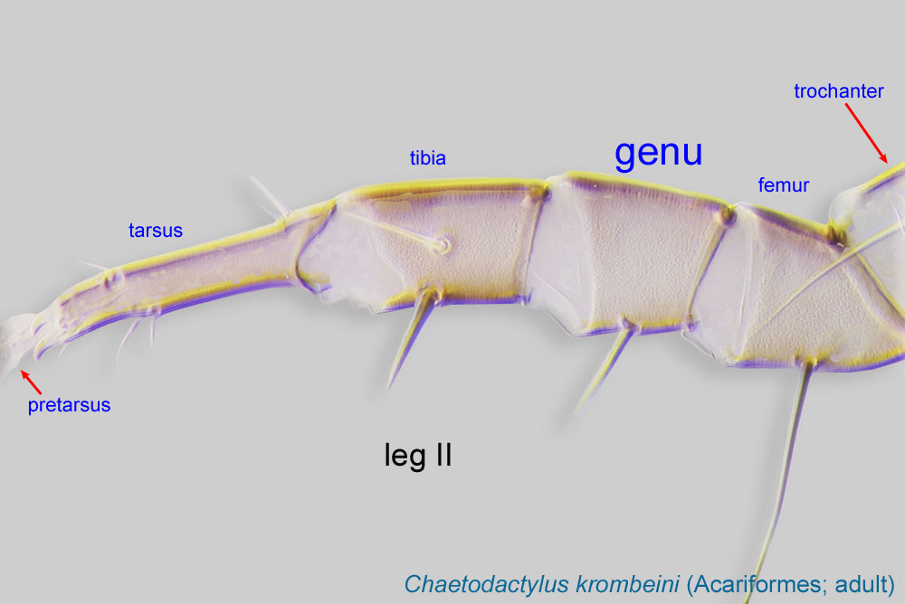
Leg or palp segment (also known as podomere or palpomere) between tibia and femur.
 I with solenidiasolenidion:
Thin-walled, terminally rounded or pointed filiform or peglike structure that is not birefringent in polarized light (unlike common setae in Acariformes). Often appears striated because of its internal structure. Found on the palpal tarsus on the gnathosoma and may also occur on the tarsus and tibia, less frequently on the genu, and occasionally on the femur of legs I-IV. In Acariformes, leg solenidia often arise from unsclerotized areas.
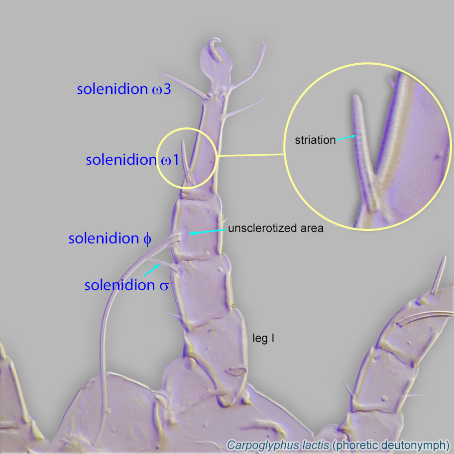 σ" and σ' subequal (Fig. 3). TarsiTarsus:
Terminal segment (also known as podomere or palpomere) of legs or palps. In Parasitoformes it can be subdivided into telotarsus and basitarsus.
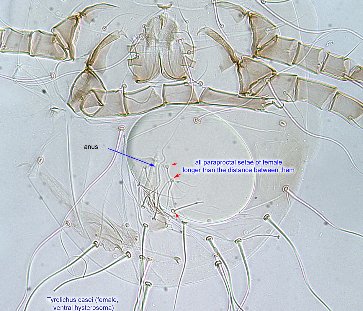
 without dorsal ridge (Fig. 6). All paraproctal setae (setae ad1-3 and ps1-3 surrounding anus) of female longer than the distance between them (Fig. 5).
This genus includes only one species, Tyrolichus casei.
The only species in this genus, Tyrolichus casei, is probably cosmopolitan. Records from beehives are from the Holarctic and Oriental regions (Canada, England, Germany, Poland, Ukraine, Azerbaijan, both western and eastern parts of Russia, and India).
honey bee (Apis) hives
temporarytemporary:
some life stages are associated with bees, while others are not
 are absent. Feeding stages disperse by active movements, air currents, or with a host.
This genus includes a single species, Tyrolichus casei. It has been found under bark, soil, decaying plant materials, bird and mammal nests, and stored food. Except for cheese (see below), infestation rate in stored food is relatively low. Tyrolichus does not form phoreticphoretic:
Pertaining to phoresy; using another organism (i.e., a host) for dispersal to new habitats. Phoresy can be distinguished from parasitism because feeding typically does not occur during phoresy.
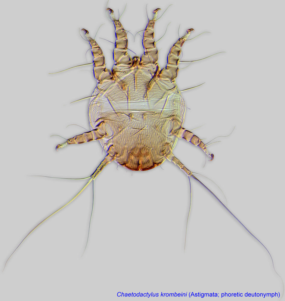
deutonymphsdeutonymph:
Ontogenetic stage between protonymph and tritonymph (or adult, if tritonymph is absent). See <a href="index.cfm?pageID=1720">Life stages page</a> for more details. and disperses as feeding stages.
There are multiple records of Tyrolichus casei from beehives (Apis mellifera and Apis cerana) throughout the world. The mites can reproduce in considerable numbers (Delfinado-Baker and Baker, 1987Delfinado-Baker and Baker, 1987:
Delfinado-Baker, M. amp; E. W. Baker. 1987. Notes on mites new to beehives in Puerto Rico and North America. American Bee Journal . 127 : 365-366.) in debris from old combs. Under laboratory conditions, they prefer bee bread, pollen, beehive debris and, to a lesser extent, honey, dead brood bees, and mold; they did not reproduce on royal jelly or propolispropolis:
A red or brown resinous substance collected by honey bees from tree buds that is used by them to fill crevices and to seal and varnish honeycombs.
(Chmielewski, 1991cChmielewski, 1991c:
Chmielewski, W. 1991c. Stored products mites (Acaroidea) in Polish bee hives. In Modern acarology. Volume I: proceedings of the 8 International Congress of Acarology, held in Ceske Budejovice, Czechoslovakia, 6-11 August 1990. , 615-619. The Hague, The Netherlands: SPB Academic Publishing.).
In New Zealand Tyrolichus casei has been recorded as a major pest of the cheese industry, responsible for severe infestations of cheese in curing rooms (Robertson, 1946Robertson, 1946:
Robertson, P. L. 1946. Tyroglyphid mites in stored products in New Zealand. Transactions of the Royal Society of New Zealand . 76 : 185-207, pl. 10-21.). In France and Germany this species has been historically used to produce "mite cheese:" Milbenkäse and Mimolette, for gourmet consumption. The mite is known to cause respiratory diseases to individuals handling infested cheese (Molina et al., 1974Molina et al., 1974:
Molina, C., J. M. Aiache, A. Tourreau amp; A. Jeanneret. 1974. Les troubles respiratoires des fromagers, rôle pathologique des acariens. La Nouvelle presse médicale . 3 : 1603-05.) as well as dermatitis (Hughes, 1976Hughes, 1976:
Hughes, A. M. 1976. The mites of stored food and houses. London: Her Majesty's Stationery Office. 400 pp.). In 2013 the US Food and Drug Administration (FDA) restricted import of Mimolette cheese to the USA due to mite infestation.
Authors: P. Klimov, B. OConnor, R. Ochoa, G. Bauchan, A. Redford, J. Scher
Last updated October 2016
tool images at ITP Node
idtools.org