

parasite and vector; infests brood cells and infects them with fungus, killing developing bees
Trochometridium Cross, 1965
Superorder Acariformes » Order Trombidiformes » Suborder Prostigmata » Infraorder Eleutherengona » Hyporder Heterostigmata » Family Trochometridiidae » Genus Trochometridium
Trochometridium tribulatum Cross, 1965
Female: First hysterosomal tergite C tripartite, with 2 lateral and 1 median lobes (Fig. 4). Femurfemur:
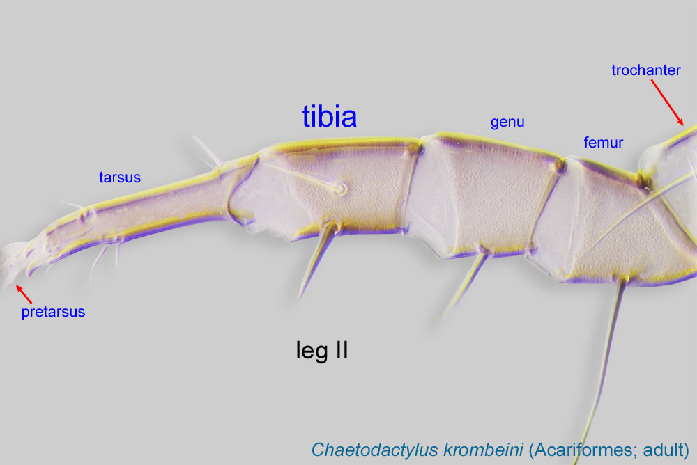
Leg or palp segment (also known as podomere or palpomere) between genu and trochanter. In ParasitIformes can be subdivided into telofemur and basifemur.
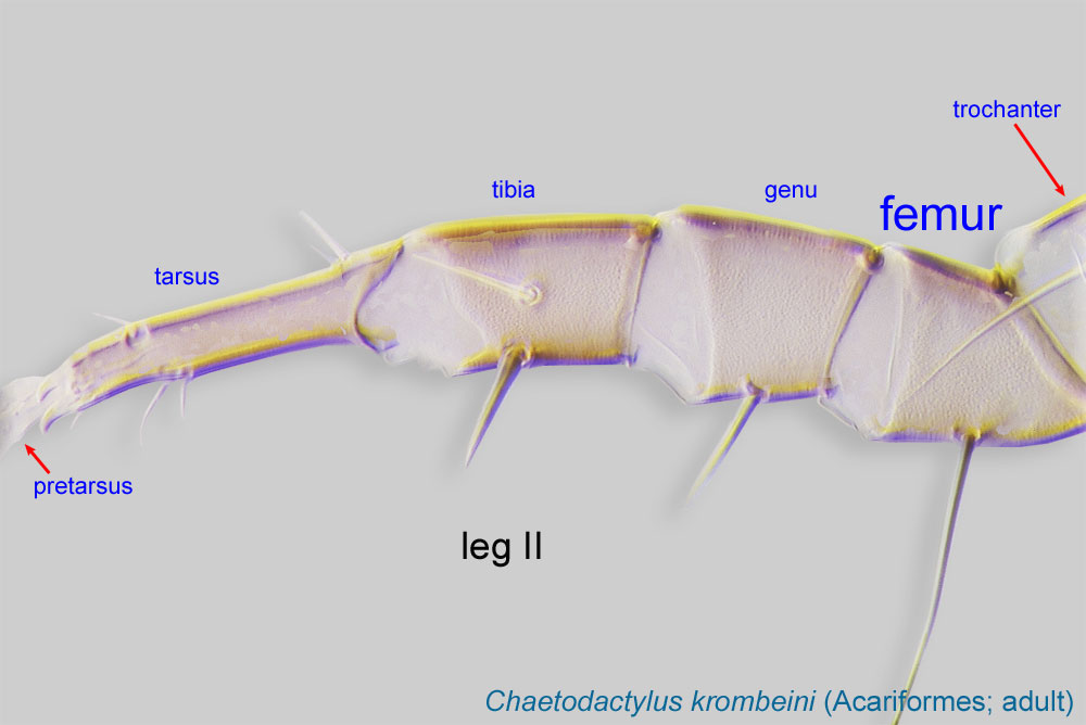 I and genugenu:
Leg or palp segment (also known as podomere or palpomere) between tibia and femur.
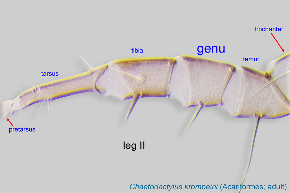 I each with 5 setae (Fig. 7).
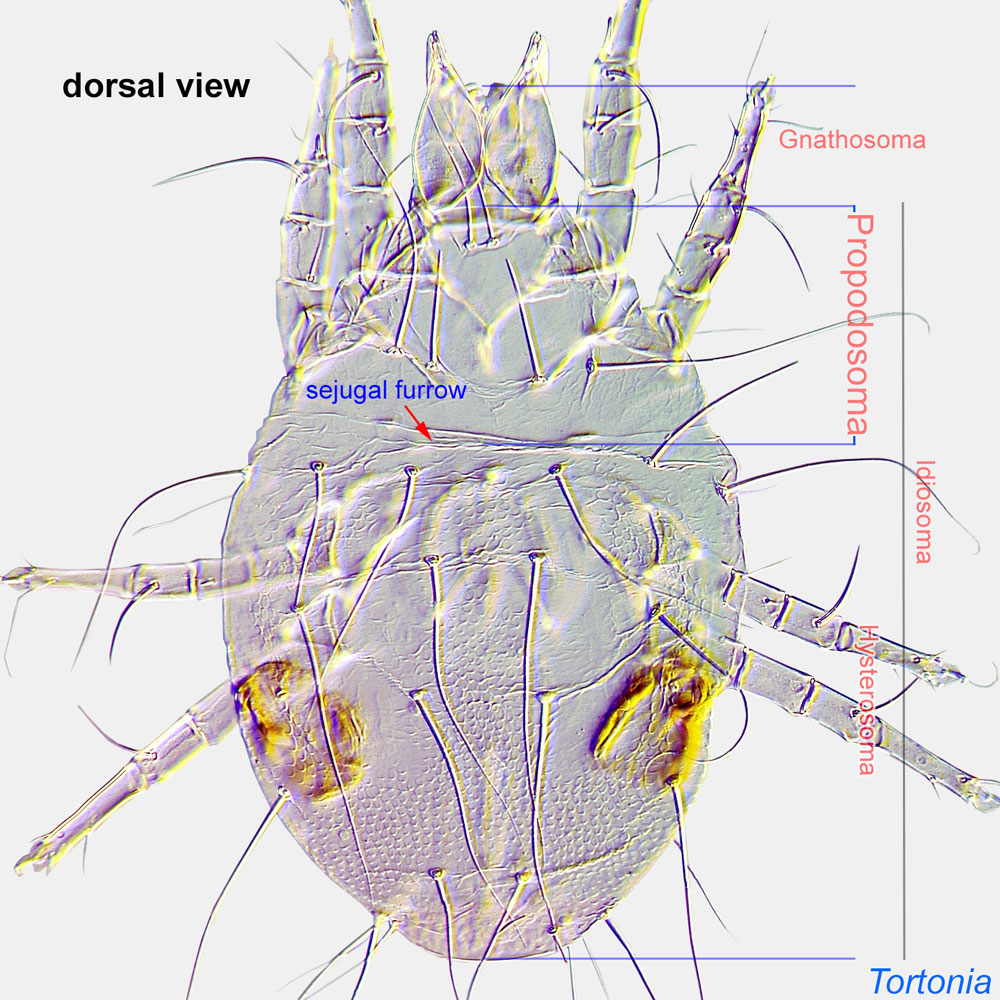
Female: With a pair of stigmata and associated tracheae anterolaterally on prodorsumprodorsum:
Dorsal surface of propodosoma.
 and with a pair of capitate bothridial setaebothridial seta:
A modified seta inserted in a cup-like base (bothridium); forms include filiform and capitate. Also known as bothridial sensillum and trichobothrium.
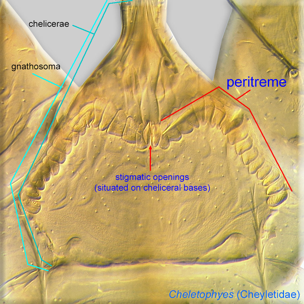
sc1 (Fig. 3). PeritremesPeritreme:
Paired, tubular, elaborated extensions of a tracheal system associated with stigmatic openings. Can be chambered, arch-like, and situated on the bases of chelicerae as in Cheyletidae (Prostigmata) or, in Mesostigmata, linear and situated on the lateral sides of the body.
 absent. Opisthosomaopisthosoma:
Body division posterior to legs IV; usually there is no distinct boundary delimiting this part of idiosoma.
usually with a longitudinal series of 4 dorsal overlapping plates (tergites). Tarsustarsus:
Terminal segment (also known as podomere or palpomere) of legs or palps. In Parasitoformes it can be subdivided into telotarsus and basitarsus.
 I with single claw (Fig. 7). TarsiTarsus:
Terminal segment (also known as podomere or palpomere) of legs or palps. In Parasitoformes it can be subdivided into telotarsus and basitarsus.
II-III with stalked, smooth empodiumempodium:
Here used only when empodial claw is not claw-like, i.e., it is pad-like or membranous.
, flanked by paired claws (Fig. 2). Legs IV with separate femurfemur:
Leg or palp segment (also known as podomere or palpomere) between genu and trochanter. In ParasitIformes can be subdivided into telofemur and basifemur.
and genugenu:
Leg or palp segment (also known as podomere or palpomere) between tibia and femur.
(Fig. 2), with total of 5-12 setae on tibiatibia:
Leg or palp segment (also known as podomere or palpomere) between tarsus and genu.
 and tarsustarsus:
Terminal segment (also known as podomere or palpomere) of legs or palps. In Parasitoformes it can be subdivided into telotarsus and basitarsus.
. TrochantersTrochanter:
Leg or palp segment (also known as podomere or palpomere) between femur and coxa. In Acariformes this is the most basal movable leg segment (or podomere) forming a joint with the body.
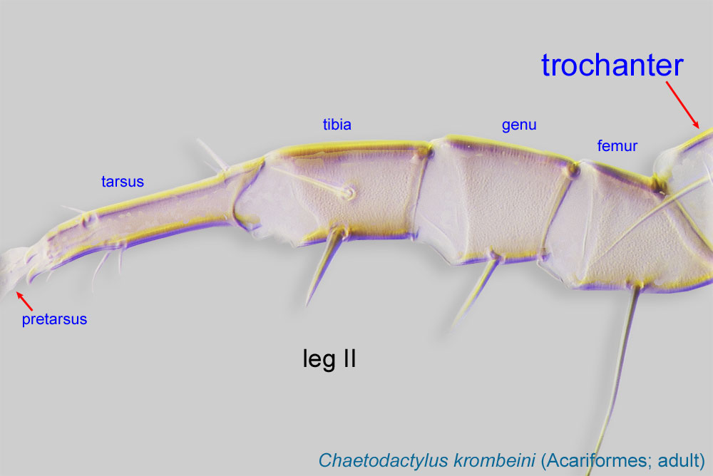 of legs I-IV each with a seta (Fig. 2). Trochantertrochanter:
Leg or palp segment (also known as podomere or palpomere) between femur and coxa. In Acariformes this is the most basal movable leg segment (or podomere) forming a joint with the body.
IV quadrangular, slightly longer than wide, different in form from subtriangular trochantertrochanter:
Leg or palp segment (also known as podomere or palpomere) between femur and coxa. In Acariformes this is the most basal movable leg segment (or podomere) forming a joint with the body.
III (Fig. 2). Female pharynxpharynx:
A muscular ectodermal structure that opens into a buccal cavity or mouth, and serves as a suction pump for ingesting food materials. In certain Heterostigmata pharyngal musculature is subdivided into 2-3 distinct parts.
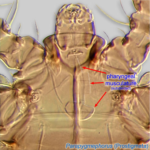 large, muscular, not divided into muscular sections. Prodorsumprodorsum:
Dorsal surface of propodosoma.
not covered posteriorly by tergite C (Fig. 4). Prodorsumprodorsum:
Dorsal surface of propodosoma.
with 3 pairs of filiform setae (v1, v2, sc2) (Fig. 3). Prodorsumprodorsum:
Dorsal surface of propodosoma.
with anterior pair of vertical setae (v1) inserted posterior to stigmata (Fig 3). Coxisternal (=coxal) plates I-II together with 6 pairs of setae (1a, b, c, 2a, b, c).
A dichotomous key is available in Hajiqanbar et al., 2009Hajiqanbar et al., 2009:
Hajiqanbar, H., A. Khaustov, K. Kamali, A. Saboori amp; H. Kamali. 2009. New taxa of the family Trochometridiidae (Acari: Heterostigmata) associated with insects from Iran. Journal of Natural History . 43 : 2701-2722.. It includes all described species, of which two have been found on bees.
Trochometridium tribulatum: Canada, USA, Mexico, Egypt, Sudan, and South Africa (Cross and Bohart, 1979Cross and Bohart, 1979:
Cross, E. A. amp; G. E. Bohart. 1979. Some observations of the habits and distribution of Trochometridium Cross, 1965 (Acarina: Pyemotidae). Acarologia . 20 : 286-293.; Lindquist, 1985Lindquist, 1985:
Lindquist, E. E. 1985. Discovery of sporothecae in adult female Trochometridium Cross, with notes on analogous structures in Siteroptes Amerling (Acari: Heterostigmata). Experimental and Applied Acarology . 1 : 73-85.; our data).
Trochometridium iranicum: Iran.
Species associated with non-bee hosts are distributed in the Palaearctic, Oriental and Australian regions.
Trochometridium iranicum (halictid bee Pseudapis nilotica) and Trochometridium tribulatum (Calliopsis, Pseudopanurgus (=Anthemurgus) (Andrenidae), Halictus, Nomia, Sphecodes (kleptoparasitic bee; phoreticphoretic:
Pertaining to phoresy; using another organism (i.e., a host) for dispersal to new habitats. Phoresy can be distinguished from parasitism because feeding typically does not occur during phoresy.
host only) (Halictidae), Megachile (Megachilidae), Diadasia, Exomalopsis, Melissodes, and Oreopasites (kleptoparasitic bee; phoreticphoretic:
Pertaining to phoresy; using another organism (i.e., a host) for dispersal to new habitats. Phoresy can be distinguished from parasitism because feeding typically does not occur during phoresy.
host only) (Apidae))
permanent permanent:
associated exclusively with bees or their close relative, wasps; cannot live without these hosts
(Trochometridium iranicum) or mostly permanentpermanent:
associated exclusively with bees or their close relative, wasps; cannot live without these hosts
(with a preference toward bees but host range includes non-bee hosts (Trochometridium tribulatum)
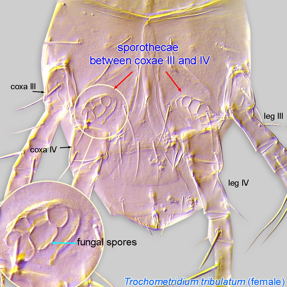 (pockets) on adult female body.
The dispersal stage is the adult female. The female has a pair of internal sacs called sporothecaesporotheca:
Internal paired sacks or external pouch serving for transferring fungal spores. In bee-associated mites, recorded in Trochometridium (paired sacks between coxae III and IV of females); certain Imparipes (Scutacaridae; paired sacks posterior to coxae IV of females); and Sennertia hipposideros-groups (hysterosomal pouch in phoretic deutonymphs).
between legs III and IV (Fig. 8). The sporothecaesporotheca:
Internal paired sacks or external pouch serving for transferring fungal spores. In bee-associated mites, recorded in Trochometridium (paired sacks between coxae III and IV of females); certain Imparipes (Scutacaridae; paired sacks posterior to coxae IV of females); and Sennertia hipposideros-groups (hysterosomal pouch in phoretic deutonymphs).
contain fungal spores that are transferred by the mite to provisioned cells of ground nesting bees such as Calliopsis, Nomia, Halictus, Anthemurgus, and Melissoides. The bee egg or young larva dies in the infested cell as a result of development of the fungus and mites. Mite eggs are laid instead of being retained within the physogastricphysogastric:
With entire body or only opisthosoma swollen to hold massive numbers of eggs or developing young.
mother. The eggs hatch into inactive larvae, which molt to males and females. The new generation of mites develops upon the fungal mycelium. The female is the only stage that feeds (Cross and Bohart, 1979Cross and Bohart, 1979:
Cross, E. A. amp; G. E. Bohart. 1979. Some observations of the habits and distribution of Trochometridium Cross, 1965 (Acarina: Pyemotidae). Acarologia . 20 : 286-293.; Lindquist, 1985Lindquist, 1985:
Lindquist, E. E. 1985. Discovery of sporothecae in adult female Trochometridium Cross, with notes on analogous structures in Siteroptes Amerling (Acari: Heterostigmata). Experimental and Applied Acarology . 1 : 73-85.; Neff and Rozen, 1995Neff and Rozen, 1995:
Neff, J. L. amp; J. G. Rozen. 1995. Foraging and nesting biology of the bee Anthemurgus passiflorae (Hymenoptera: Apoidea), descriptions of its immature stages, and observations on its floral host (Passifloraceae). American Museum Novitates . 3138 : 1-19.).
The host range for T. tribulatum includes wasps and beetles. Perhaps beetles become infested by mites dispersing from bees or wasps nesting in the same soil (i.e., the beetles are phoreticphoretic:
Pertaining to phoresy; using another organism (i.e., a host) for dispersal to new habitats. Phoresy can be distinguished from parasitism because feeding typically does not occur during phoresy.
hosts only).
Authors: P. Klimov, B. OConnor, R. Ochoa, G. Bauchan, A. Redford, J. Scher
Last updated October 2016
tool images at ITP Node
idtools.org


