kleptoparasitic; attempts to kill developing bees and feeds on pollen and waste
Tortonia Oudemans, 1911
Superorder Acariformes » Order Sarcoptiformes » Suborder Oribatida » Infraorder Desmonomata » Hyporder Astigmata » Family Suidasiidae » Genus Tortonia
Trichotarsus intermedius Oudemans, 1902
Ebertia Oudemans, 1924Oudemans, 1924:
Oudemans, A. C. 1924. Acarologische Aanteekehingen LXXVII. Entomologische Berichten (Amsterdam). 6: 317-336.; Neottiglyphus Volgin, 1974Volgin, 1974:
Volgin, V. I. 1974. New genera and species of acaroid mites (Acariformes, Acaroidea) from Kazakhstan. Entomologicheskoe Obozrenie . 53 : 218-225.; Hyohondania Sasa, 1952
Adult: Body with dense, equal, small, rounded protuberances (Fig. 4). Tarsal setae aa I absent (Fig. 6). Proral setae of tarsitarsus:
Terminal segment (also known as podomere or palpomere) of legs or palps. In Parasitoformes it can be subdivided into telotarsus and basitarsus.
 I-IV, enlarged, claw-like (Fig 6). Internal scapular setae (si) near posterior margin of prodorsalprodorsal:
Pertaining to the prodorsum.
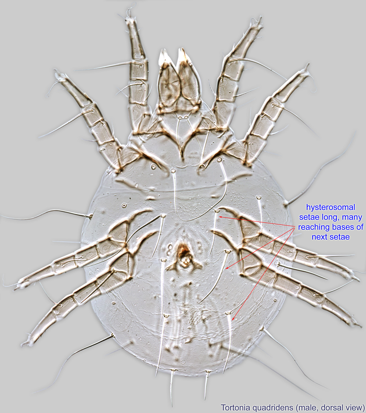
sclerite (Fig 7). Supracoxal setae filiform, barbed (Fig 7). Hysterosomal setae long, many reaching bases of next setae (Fig. 8).
Phoretic phoretic:
Pertaining to phoresy; using another organism (i.e., a host) for dispersal to new habitats. Phoresy can be distinguished from parasitism because feeding typically does not occur during phoresy.
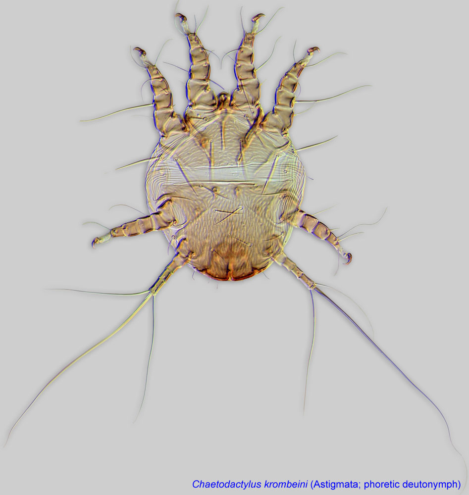
deutonymph: Setae ve present; setae ba I absent; ba II present (Fig. 3). Seta aa I absent (Fig. 3). Setae e I-II, short, filiform (no "saucer" at tip) (Fig. 3). Leg IV shorter than leg III (Figs. 2, 3a, and with three long apical setae (Fig. 2). Empodial clawsEmpodial claw:
Claw-like, membranous, or pad-like structure of setal origin. Present only on the pretarsus in Acariformes. In Astigmata, it is the only claw on the pretarsus and often referred to simply as the claw. In the remaining Acariformes, may be accomanied by two lateral claws. Also known as empodium, pretarsal empodium, or central claw.
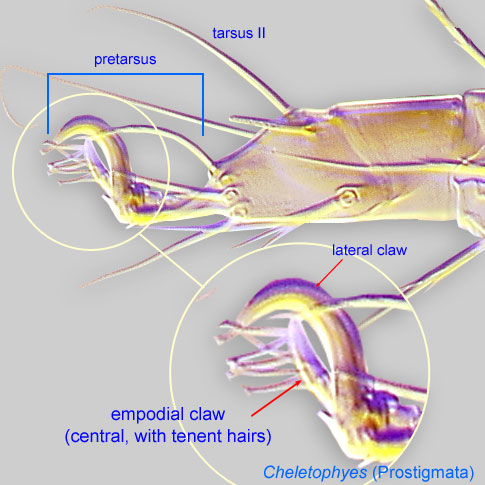 I-III arising directly from tarsal apices (no elongated membranous ambulacraambulacrum:
The claws and empodium of the apotele or pretarsus.
) (Fig. 2). Empodial clawempodial claw:
Claw-like, membranous, or pad-like structure of setal origin. Present only on the pretarsus in Acariformes. In Astigmata, it is the only claw on the pretarsus and often referred to simply as the claw. In the remaining Acariformes, may be accomanied by two lateral claws. Also known as empodium, pretarsal empodium, or central claw.
IV absent (Figs. 2, 3a).
Adult: Pretarsal ambulacrumambulacrum:
The claws and empodium of the apotele or pretarsus.
not greatly expanded (Fig. 6). Ventral subcapitulumsubcapitulum:
Ventral faces of the fused palpcoxae.
without external ridges (Fig. 9). Dorsal setae smooth (Fig. 4). Prodorsumprodorsum:
Dorsal surface of propodosoma.
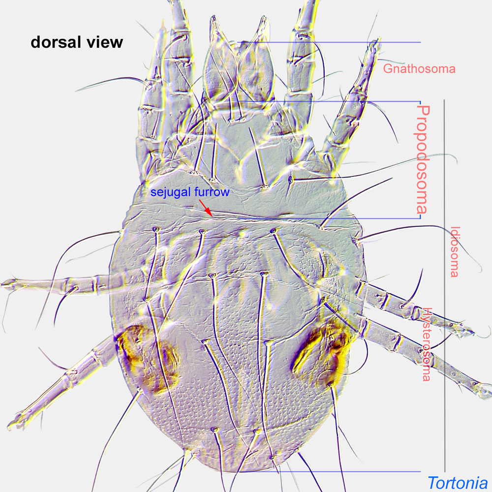 with external vertical setae ve present, situated at anterior level of prodorsalprodorsal:
Pertaining to the prodorsum.
sclerite (Fig. 7). Anus positioned near posterior margin of body (Fig. 5). Coxal apodemesapodeme:
Internal sclerite that serves as an attachment site for muscles. Most commonly used (as "coxal apodeme") to describe elements of coxae fused to the ventral body in Acariformes (coxae are free and not fused to the body in Parasitiformes), and may be variously referred to as ventral, sternal, anterior, or posterior.
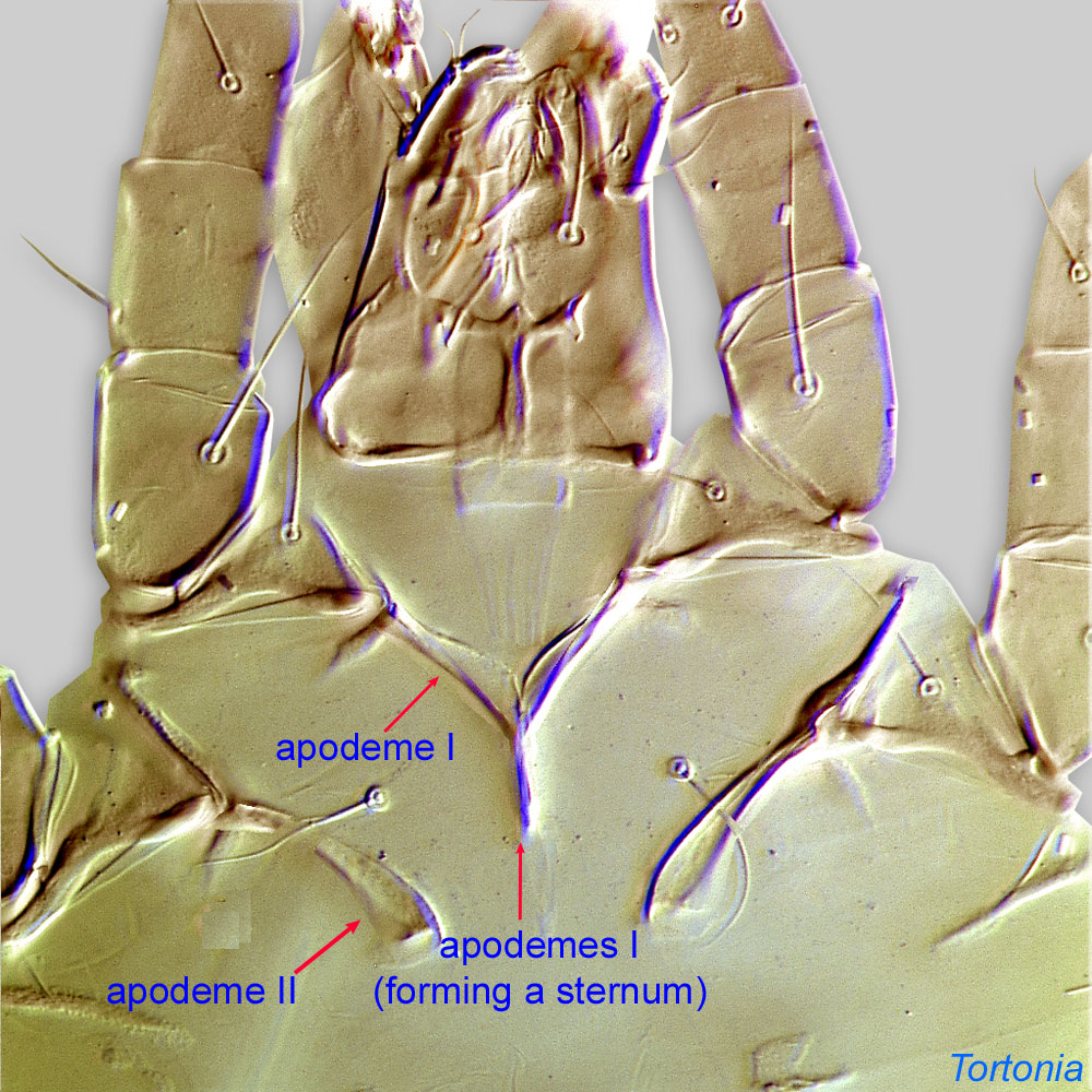 III-IV present (Fig. 5). Empodial clawsEmpodial claw:
Claw-like, membranous, or pad-like structure of setal origin. Present only on the pretarsus in Acariformes. In Astigmata, it is the only claw on the pretarsus and often referred to simply as the claw. In the remaining Acariformes, may be accomanied by two lateral claws. Also known as empodium, pretarsal empodium, or central claw.
simple (Fig 6). Solenidionsolenidion:
Thin-walled, terminally rounded or pointed filiform or peglike structure that is not birefringent in polarized light (unlike common setae in Acariformes). Often appears striated because of its internal structure. Found on the palpal tarsus on the gnathosoma and may also occur on the tarsus and tibia, less frequently on the genu, and occasionally on the femur of legs I-IV. In Acariformes, leg solenidia often arise from unsclerotized areas.
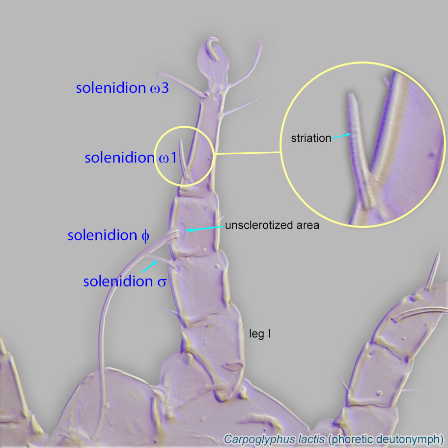 ω2 of tarsustarsus:
Terminal segment (also known as podomere or palpomere) of legs or palps. In Parasitoformes it can be subdivided into telotarsus and basitarsus.
I more distal than ω1, situated in distal half of tarsustarsus:
Terminal segment (also known as podomere or palpomere) of legs or palps. In Parasitoformes it can be subdivided into telotarsus and basitarsus.
(Fig 6). Setae e and f present on tarsitarsus:
Terminal segment (also known as podomere or palpomere) of legs or palps. In Parasitoformes it can be subdivided into telotarsus and basitarsus.
I-IV, both filiform (Fig 6).
This genus needs a revision. There is no key to species. Original species descriptions should be consulted.
Nearctic, Palaearctic (including Japan and North Africa), Neotropical, and Australian regions. Mites found on bees are known from the Nearctic and Palaearctic.
Phoretic phoretic:
Pertaining to phoresy; using another organism (i.e., a host) for dispersal to new habitats. Phoresy can be distinguished from parasitism because feeding typically does not occur during phoresy.
hosts include orchard mason bees (Osmia), leafcutting and resin bees (Megachile), bumble bees (Bombus), and large carpenter bees (Xylocopa), as well as kleptoparasitic bees of the genus Stelis (Megachilidae).
One lineage, including Tortonia dogaressa and Tortonia smitsvanburgsti, is exclusively associated with the megachilid bees of the genus Rhodanthidium.
permanent permanent:
associated exclusively with bees or their close relative, wasps; cannot live without these hosts
in nests
 (non-feeding stage) disperse on adult bees from one nest to another. Kleptoparasitic bees and wasps can also be used as transport.
In Japan, an undescribed species of Tortonia was found to be a kleptoparasitekleptoparasite:
An animal that takes prey or other food from another animal that has caught, collected, or otherwise prepared the food, including stored food. Both kleptoparasitic bees and kleptoparasitic mites feed on food provisioned in the host bee nest. Kleptoparasitic bees do not make their own nests; they stealthily deposit eggs in the nest of a bee host and can act as phoretic hosts for mites only because they deliver them to nests of actual bee hosts. Variant spelling: cleptoparasite.
of Osmia cornifrons, an important pollinator of apple trees (Qu et al., 2002Qu et al., 2002:
Qu, D., Y. Maeta, M. Goubara, K. J. Nakatsuka, J. Kozo amp; K. Kenji. 2002. Reproductive strategy in the two species of cleptoparasitic astigmatid mites, Chaetodactylus nipponicus and Tortonia sp. (Acari: Chaetodactylidae and Suidasiidae), infesting Osmia cornifrons (Hymenoptera: Megachilidae). I. Invasion/infestation patterns and partial use of the host food. Japanese Journal of Entomology (New Series). 5: 121-141.; Qu et al., 2003Qu et al., 2003:
Qu, D., Y. Maeta, K. J. Nakatsuka, K. Kenji amp; M. Goubara. 2003. Reproductive strategy in the two species of cleptoparasitic astigmatid mites, Chaetodactylus nipponicus and Tortonia sp. (Acari: Chaetodactylidae and Suidasiidae), infesting Osmia cornifrons (Hymenoptera: Megachilidae) II. Life history, phoretic positions, development and reproductivity. Japanese Journal of Entomology (New Series). 6: 55-73.): after infesting cells and prior to feeding on the stored pollen-mass, Tortonia sp. killed hosts, but only if the hosts were at stages egg to early third instar larva. The success of killing the host appears to positively depend on mite population sizes. Bees that survived formed normal cocoons.
Feeding stages and phoreticphoretic:
Pertaining to phoresy; using another organism (i.e., a host) for dispersal to new habitats. Phoresy can be distinguished from parasitism because feeding typically does not occur during phoresy.
deutonymphsdeutonymph:
Ontogenetic stage between protonymph and tritonymph (or adult, if tritonymph is absent). See <a href="index.cfm?pageID=1720">Life stages page</a> for more details. hibernate in bee nests. ProtonymphsProtonymph:
Ontogenetic stage between larva and deutonymph. See <a href="index.cfm?pageID=1720">Life stages page</a> for more details.
of Tortonia sp. that encountered shortages of food molted to phoreticphoretic:
Pertaining to phoresy; using another organism (i.e., a host) for dispersal to new habitats. Phoresy can be distinguished from parasitism because feeding typically does not occur during phoresy.
deutonymphsdeutonymph:
Ontogenetic stage between protonymph and tritonymph (or adult, if tritonymph is absent). See <a href="index.cfm?pageID=1720">Life stages page</a> for more details.. The percentage of phoreticphoretic:
Pertaining to phoresy; using another organism (i.e., a host) for dispersal to new habitats. Phoresy can be distinguished from parasitism because feeding typically does not occur during phoresy.
deutonymphsdeutonymph:
Ontogenetic stage between protonymph and tritonymph (or adult, if tritonymph is absent). See <a href="index.cfm?pageID=1720">Life stages page</a> for more details. phoretic on bees is lower (6.6% in total) as compared to Chaetodactylus.
Tortonia sp., when co-inhabiting with Chaetodactylus nipponicus in the same cell, shows lower population sizes, indicating its lower competitive ability. However, in cells with living hosts, the ratio of individual numbers of Tortonia sp. was higher than that of Ch. nipponicus, because the former can survive by feeding on host feces.
In the USA, mortality of developing wasps Monobia quadridens in cells infested with feeding stages of Tortonia quadridens has been observed, but the ultimate cause of death remains unknown. Mites have appeared to act as scavengers feeding on organic debris in the nests (Krombein, 1962Krombein, 1962:
Krombein, K. V. 1962. Natural history of Plummers Island, Maryland. XVI. Biological Notes on Chaetodactylus krombeini Baker, a parasitic mite of the megachilid bee, Osmia (Osmia) lignaria Say (Acarina, Chaetodactylidae). Proceedings of the Biological Society of Washington . 75 : 237-250.; Krombein, 1967Krombein, 1967:
Krombein, K. V. 1967. Trap-nesting wasps and bees: life histories, nests, and associates. Washington, D.C: Smithsonian press. 570 pp.). Similarly, all stages of Tortonia quadridens were found in a nest of Xylocopa virginica, on and in the pollen balls in empty (failed) cells. The ultimate cause of cell failure remains uncertain. These authors called Tortonia kleptoparasitic (Lombert et al., 1987Lombert et al., 1987:
Lombert, H. A. P. M., B. M. OConnor, F. S. Lukoschus amp; J. O. Whitaker, Jr. . 1987. Ontogeny, systematics and ecology of Sennertia ( Amsennertia ) americana Delfinado amp; Baker, 1976 (Acari: Chaetodactylidae) from the nest of the carpenter bee, Xylocopa virginica (Hymenoptera: Anthophoridae). International Journal of Acarology. 13: 113-129.).
Authors: P. Klimov, B. OConnor, R. Ochoa, G. Bauchan, A. Redford, J. Scher
Last updated October 2016
tool images at ITP Node
idtools.org