neutral; lives in inflorescences of palms and other monocots; uses bees as transport
Spadiseius Lindquist and Moraza, 2008Lindquist and Moraza, 2008:
Lindquist, E. E. amp; M. L. Moraza. 2008. A new genus of flower-dwelling melicharid mites (Acari : Mesostigmata : Ascoidea) phoretic on bats and insects in Costa Rica and Brazil. Zootaxa. 1-37.
Superorder Parasitiformes » Order Mesostigmata » Suborder Monogynaspida » Hyporder Dermanyssiae » Family Melicharidae » Genus Spadiseius
Spadiseius calyptrogynae Lindquist and Moraza, 2008Lindquist and Moraza, 2008:
Lindquist, E. E. amp; M. L. Moraza. 2008. A new genus of flower-dwelling melicharid mites (Acari : Mesostigmata : Ascoidea) phoretic on bats and insects in Costa Rica and Brazil. Zootaxa. 1-37.
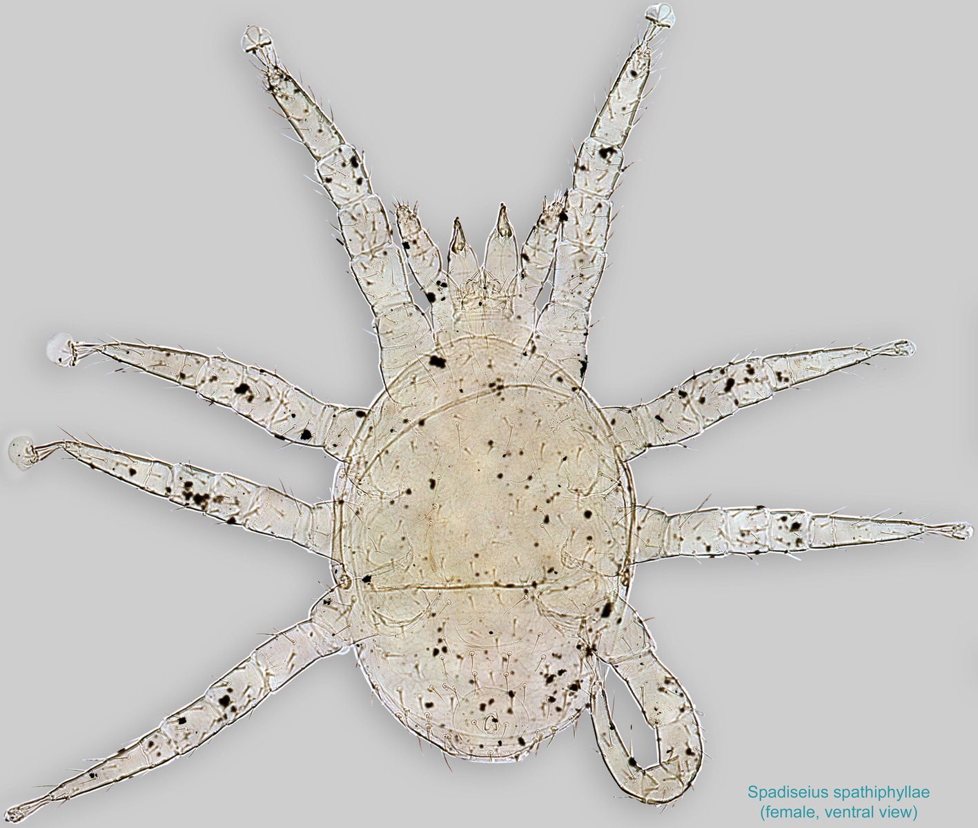
Adult: Dorsal shield entire, slightly to notably hypertrichoushypertrichous:
With many irregularly arranged setae.
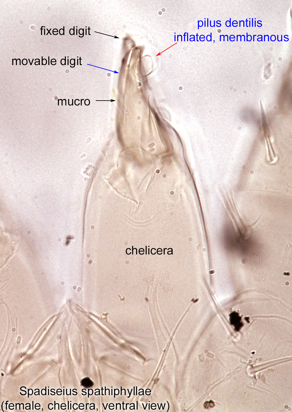
, with one to several pairs of supernumerary setae inserted near setae j6 and sometimes among setae z3–z5, s3–s5 on anterior region and sometimes among setae J1–J4 on posterior region (Fig. 3). Fixed cheliceral digit with pilus dentilispilus dentilis:
A seta-like or membranous sensory organ inserted ventrolaterally on the fixed digit of the chelicera of many Mesostigmata.
modified to an inflated, membranous lobe (Figs. 9, 11); movable cheliceral digit usually with a pointed process (mucro) on its mid-ventral face (Fig. 11). Peritrematic shield free posteriorly, not connected to exopodal plate IV (Fig. 7).
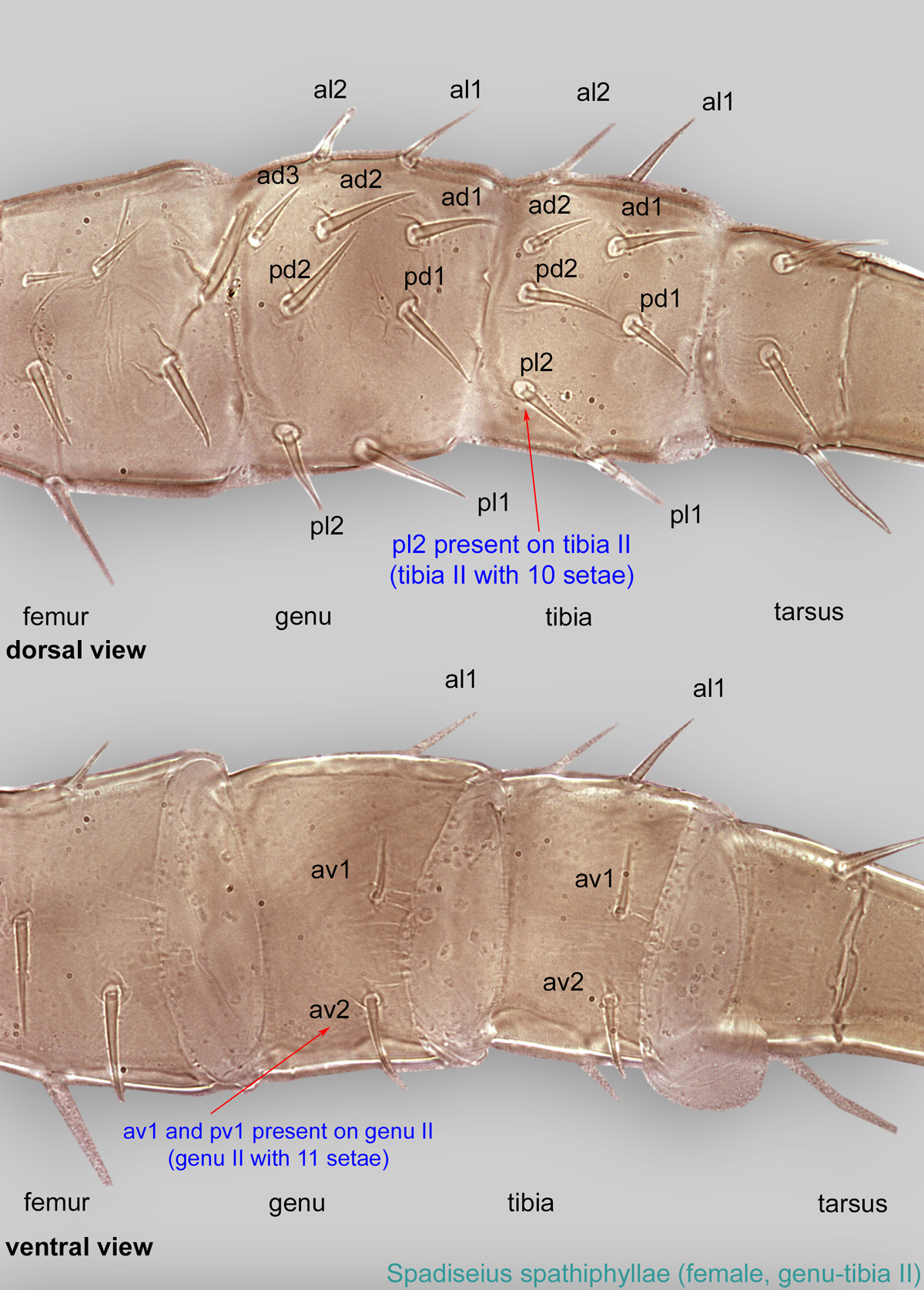
Female: Femurfemur:
Leg or palp segment (also known as podomere or palpomere) between genu and trochanter. In ParasitIformes can be subdivided into telofemur and basifemur.
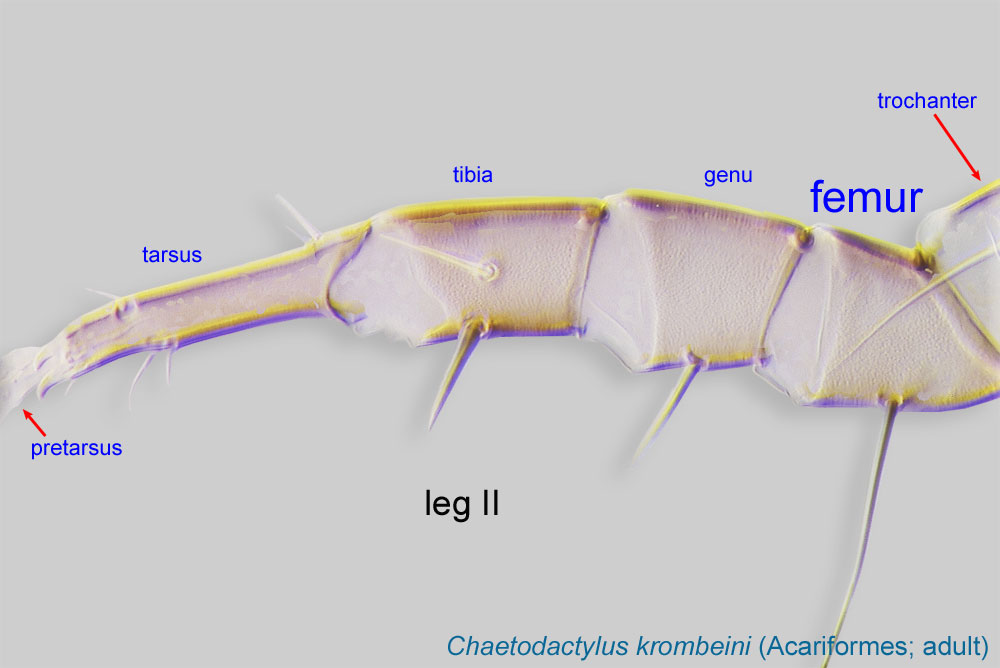 IV of female with dorsal seta ad1 untapered, rod-like, or oar-like, in contrast to adjacent simplesimple:
Of claws or setae; not modified or not bi- or trifurcate at tip.
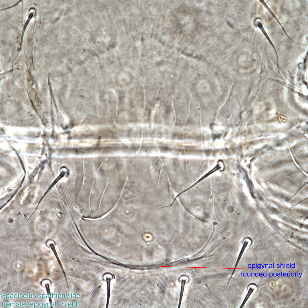
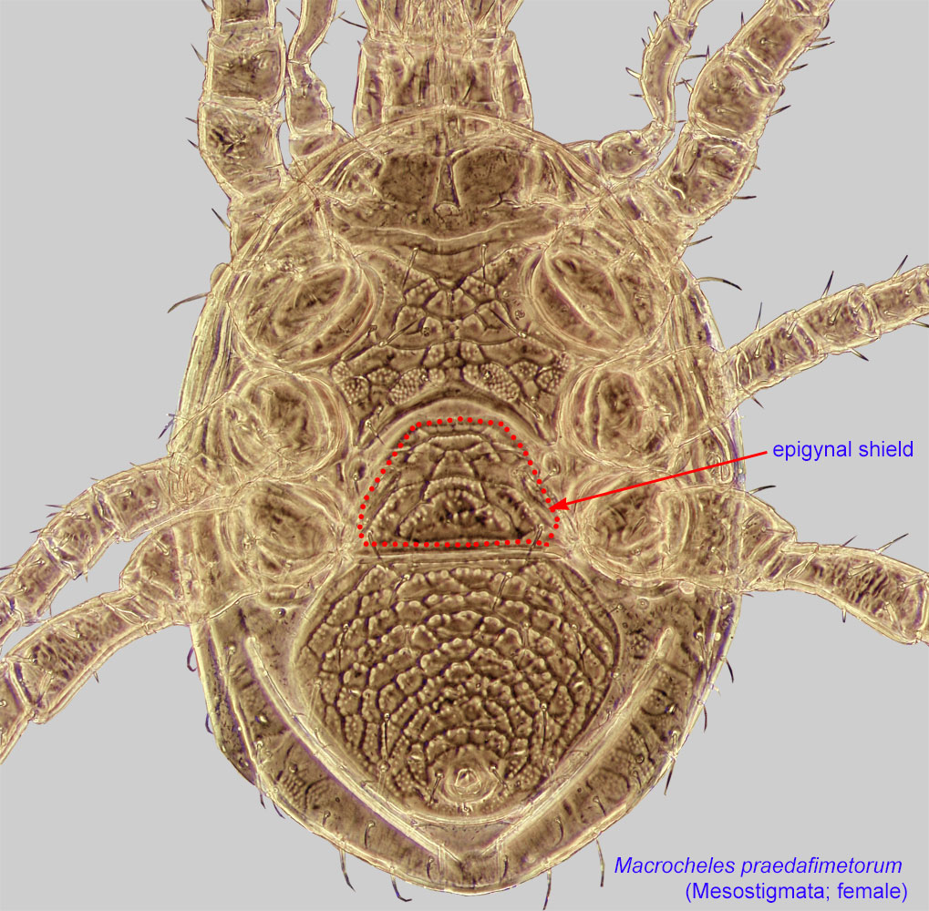
setae (Fig. 15). Epigynal shieldepigynal shield:
A shield protecting the female genital opening. Well-developed in Mesostigmata. Also known as epigynial shield.
 gently rounded posteriorly (Fig.5). Anal shieldanal shield:
In Mesostigmata, a ventral shield bearing the anal opening and circumanal setae (adanal or postanal setae), but without any ventral setae or pores (lyrifissures) on it. If ventral setae are present on shield than referred to as a ventrianal shield.
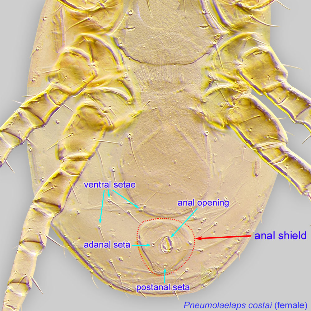 oval or elliptical, bearing only the 3 common circumanal setae (Fig. 6).
Male: Dorsal shield with peripheral setae enlarged to form a corona of basally thick and apically clavate, capitate, or attenuate setae, in contrast to their generally simplesimple:
Of claws or setae; not modified or not bi- or trifurcate at tip.
setal counterparts in females.
No dichotomous key is available, but the two known species can be identified based on their original descriptions in Lindquist and Moraza, 2008Lindquist and Moraza, 2008:
Lindquist, E. E. amp; M. L. Moraza. 2008. A new genus of flower-dwelling melicharid mites (Acari : Mesostigmata : Ascoidea) phoretic on bats and insects in Costa Rica and Brazil. Zootaxa. 1-37..
Neotropics
phoretic phoretic:
Pertaining to phoresy; using another organism (i.e., a host) for dispersal to new habitats. Phoresy can be distinguished from parasitism because feeding typically does not occur during phoresy.
on stingless bees (Meliponini)
facultativefacultative:
can complete entire life cycle without bees or their close relative, wasps
The entire life cycle of Spadiseius occurs in inflorescences of palms and aroids. Evidence for their feeding behavior is based on cheliceral morphology rather than direct observations. A single inflorescence of the aroid Spathiphyllum friedrichsthallii can harbor more than 300 mites. These mites disperse on various plant-visiting animals (e. g., scarab beetles, stingless bees, and bats).
Authors: P. Klimov, B. OConnor, R. Ochoa, G. Bauchan, A. Redford, J. Scher
Last updated October 2016
tool images at ITP Node
idtools.org