

probably not harmful; non bee-associated mites feed on decomposing organic materials and mold, but feeding habits of bee-associated species unknown
Saproglyphus Berlese, 1890
Superorder Acariformes » Order Sarcoptiformes » Suborder Oribatida » Infraorder Desmonomata » Hyporder Astigmata » Family Winterschmidtiidae » Genus Saproglyphus
Saproglyphus neglectus Berlese, 1890
Calvolia Oudemans, 1911 (in part, synonymy by B. OConnor, unpublished)
Adults: CondylophoresCondylophore:
In Acariformes, pretarsal paired sclerotized structures arising from the distal end of the tarsus and forming a joint with lateral claws (in Endeostigmata, Oribatida and Trombidiformes) and the empodial claw. Not to be confused with vertical sclerites of Parasitiformes.
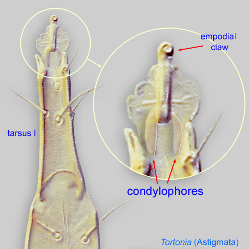 fused into a V-shaped scleritesclerite:
A component section of an exoskeleton; a plate forming the skeleton of an arthropod.
in base of ambulacral stalk (Fig. 11) and idiosomaidiosoma:
Body not including the gnathosoma.
 with 4-5 pairs of setae longer than the width of the body, these setae borne on angular protuberances (Fig. 8).
Phoretic phoretic:
Pertaining to phoresy; using another organism (i.e., a host) for dispersal to new habitats. Phoresy can be distinguished from parasitism because feeding typically does not occur during phoresy.
deutonymph: Setae ba I-II and aa I absent (Fig. 6). Pretarsal ambulacrumambulacrum:
The claws and empodium of the apotele or pretarsus.
and empodial clawempodial claw:
Claw-like, membranous, or pad-like structure of setal origin. Present only on the pretarsus in Acariformes. In Astigmata, it is the only claw on the pretarsus and often referred to simply as the claw. In the remaining Acariformes, may be accomanied by two lateral claws. Also known as empodium, pretarsal empodium, or central claw.
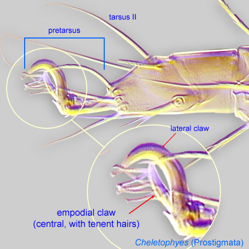 present on legs I-II (Fig. 6). Empodial clawsEmpodial claw:
Claw-like, membranous, or pad-like structure of setal origin. Present only on the pretarsus in Acariformes. In Astigmata, it is the only claw on the pretarsus and often referred to simply as the claw. In the remaining Acariformes, may be accomanied by two lateral claws. Also known as empodium, pretarsal empodium, or central claw.
I-III borne on long condylophorescondylophore:
In Acariformes, pretarsal paired sclerotized structures arising from the distal end of the tarsus and forming a joint with lateral claws (in Endeostigmata, Oribatida and Trombidiformes) and the empodial claw. Not to be confused with vertical sclerites of Parasitiformes.
(often poorly visible), encompassed by elongated membranous ambulacraambulacrum:
The claws and empodium of the apotele or pretarsus.
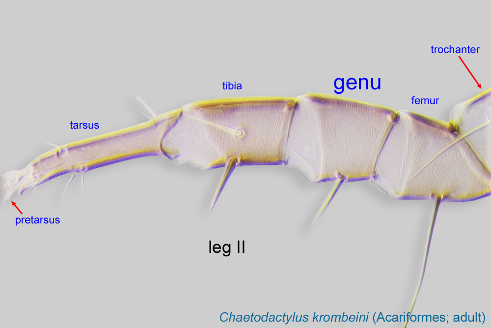
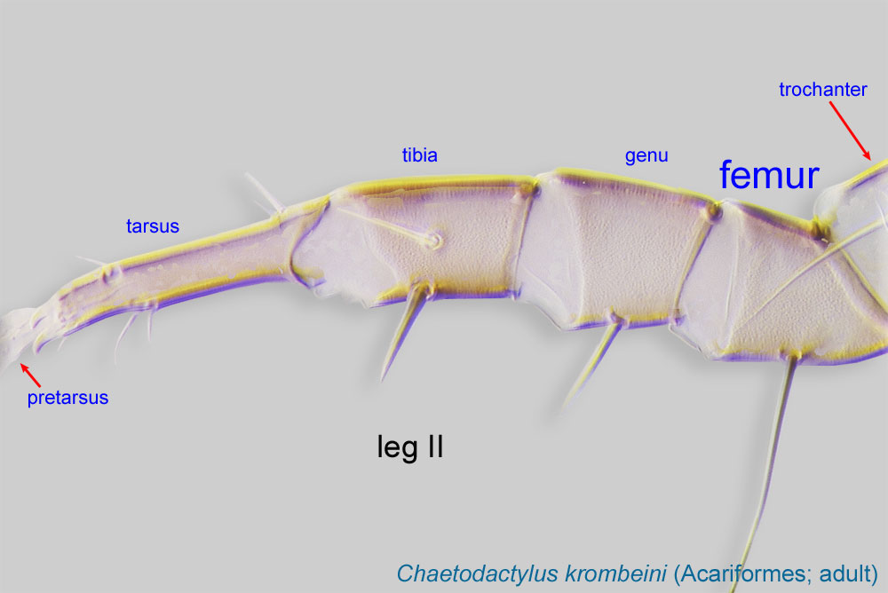
(Figs. 6, 7). Tarsustarsus:
Terminal segment (also known as podomere or palpomere) of legs or palps. In Parasitoformes it can be subdivided into telotarsus and basitarsus.
 I with solenidiasolenidion:
Thin-walled, terminally rounded or pointed filiform or peglike structure that is not birefringent in polarized light (unlike common setae in Acariformes). Often appears striated because of its internal structure. Found on the palpal tarsus on the gnathosoma and may also occur on the tarsus and tibia, less frequently on the genu, and occasionally on the femur of legs I-IV. In Acariformes, leg solenidia often arise from unsclerotized areas.
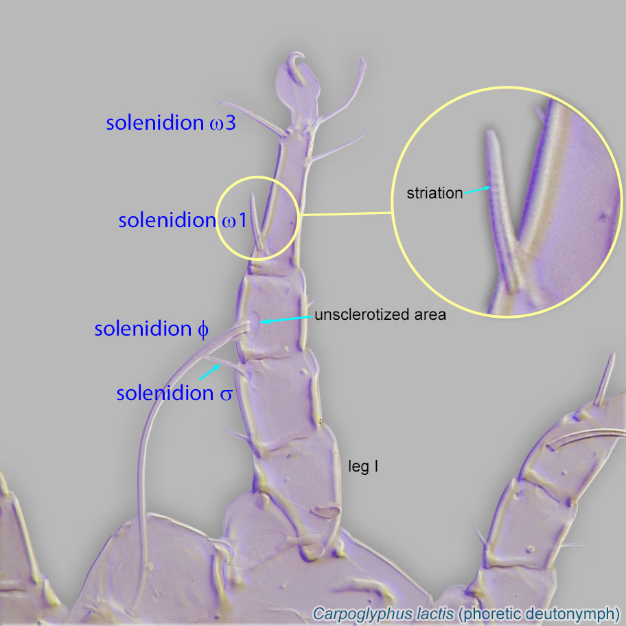 closely associated, usually in the basal half of the tarsustarsus:
Terminal segment (also known as podomere or palpomere) of legs or palps. In Parasitoformes it can be subdivided into telotarsus and basitarsus.
(Fig. 6). TarsiTarsus:
Terminal segment (also known as podomere or palpomere) of legs or palps. In Parasitoformes it can be subdivided into telotarsus and basitarsus.
I-II with 7 setae (e, f, d, p', la, ra, wa), of which 4 foliate (ra, la, f, p') (Fig. 6). TibiaeTibia:
Leg or palp segment (also known as podomere or palpomere) between tarsus and genu.
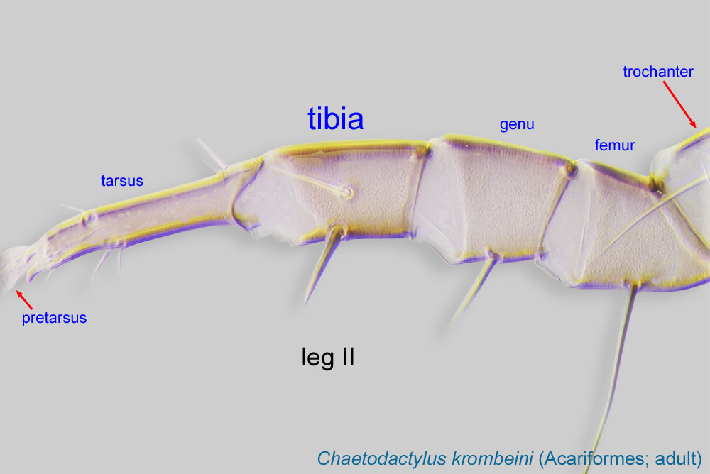 I-II with 1 ventral seta (gT I-II present, hT I-II absent) (Fig. 6). Empodial clawsEmpodial claw:
Claw-like, membranous, or pad-like structure of setal origin. Present only on the pretarsus in Acariformes. In Astigmata, it is the only claw on the pretarsus and often referred to simply as the claw. In the remaining Acariformes, may be accomanied by two lateral claws. Also known as empodium, pretarsal empodium, or central claw.
I-III small and simplesimple:
Of claws or setae; not modified or not bi- or trifurcate at tip.
(Figs. 6, 7). Empodial clawempodial claw:
Claw-like, membranous, or pad-like structure of setal origin. Present only on the pretarsus in Acariformes. In Astigmata, it is the only claw on the pretarsus and often referred to simply as the claw. In the remaining Acariformes, may be accomanied by two lateral claws. Also known as empodium, pretarsal empodium, or central claw.
IV absent (Fig. 7). Leg IV shorter than leg III (Fig. 7). Tarsustarsus:
Terminal segment (also known as podomere or palpomere) of legs or palps. In Parasitoformes it can be subdivided into telotarsus and basitarsus.
IV with 2-4 long apical setae (none foliate) (Fig. 7). Coxal apodemesapodeme:
Internal sclerite that serves as an attachment site for muscles. Most commonly used (as "coxal apodeme") to describe elements of coxae fused to the ventral body in Acariformes (coxae are free and not fused to the body in Parasitiformes), and may be variously referred to as ventral, sternal, anterior, or posterior.
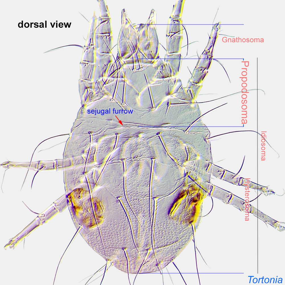
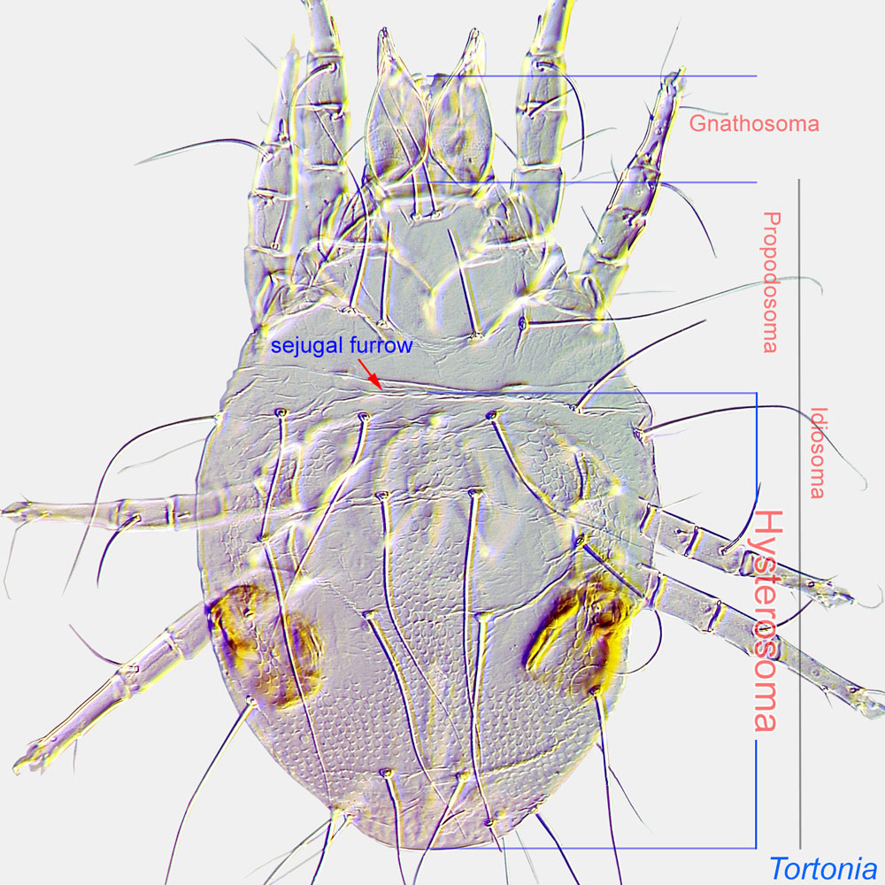
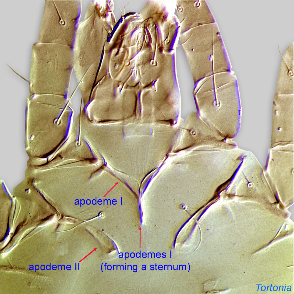 III fused together (Fig. 2). Ocelli present on anterior apex of propodosomapropodosoma:
Anterior part of idiosoma, in front of sejugal furrow.
 with separate lenses and pigment spots (Fig. 4).
Adults: Pretarsal ambulacrumambulacrum:
The claws and empodium of the apotele or pretarsus.
not greatly expanded, empodial clawsempodial claw:
Claw-like, membranous, or pad-like structure of setal origin. Present only on the pretarsus in Acariformes. In Astigmata, it is the only claw on the pretarsus and often referred to simply as the claw. In the remaining Acariformes, may be accomanied by two lateral claws. Also known as empodium, pretarsal empodium, or central claw.
present (Fig. 11). Prodorsumprodorsum:
Dorsal surface of propodosoma.
with external vertical setae (ve) absent (Fig. 10). Supracoxal glandsupracoxal gland:
Paired glands serving for maintenance of water balance in Astigmata. These glands are usually invisible on typical microscopic preparations. However, their presence can be detected by the well-sclerotized supracoxal gland openings, which are situated on supracoxal sclerites.
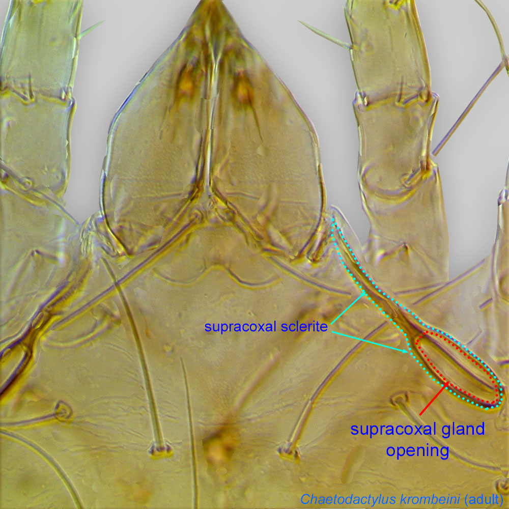 opening not associated with large scleritesclerite:
A component section of an exoskeleton; a plate forming the skeleton of an arthropod.
. Internal vertical setae (vi) at anterior edge of propodosomapropodosoma:
Anterior part of idiosoma, in front of sejugal furrow.
(Fig. 10). TibiaeTibia:
Leg or palp segment (also known as podomere or palpomere) between tarsus and genu.
I-II with 1 ventral seta (Fig. 11). Genugenu:
Leg or palp segment (also known as podomere or palpomere) between tibia and femur.
 III with a dorsal solenidionsolenidion:
Thin-walled, terminally rounded or pointed filiform or peglike structure that is not birefringent in polarized light (unlike common setae in Acariformes). Often appears striated because of its internal structure. Found on the palpal tarsus on the gnathosoma and may also occur on the tarsus and tibia, less frequently on the genu, and occasionally on the femur of legs I-IV. In Acariformes, leg solenidia often arise from unsclerotized areas.
(Fig. 12). Femurfemur:
Leg or palp segment (also known as podomere or palpomere) between genu and trochanter. In ParasitIformes can be subdivided into telofemur and basifemur.
 IV with 1 ventral seta (Fig. 9). TarsiTarsus:
Terminal segment (also known as podomere or palpomere) of legs or palps. In Parasitoformes it can be subdivided into telotarsus and basitarsus.
long, at least three times as long as wide (Figs. 8, 11, 12). Male without obvious striation pattern on hysterosomahysterosoma:
Division of body posterior to the sejugal furrow, bearing legs III and IV.
 .
.
This genus needs a revision. An older key, including the two described species found on bees (Saproglyphus hagensis and Saproglyphus reticulatus), is available in Zachvatkin, 1941Zachvatkin, 1941:
Zachvatkin, A. A. 1941. Tiroglifoidnue kleshchi Tyroglyphoidea [=Tyroglyphoid mites Tyroglyphoidea]. In Fauna SSSR: Paukoobraznuye, ed. S. A. Zernov, 475. Moscow-Leningrad: Akademiya Nauk SSSR (translated to English in a separate publication).. Notes: in this key (i) phoreticphoretic:
Pertaining to phoresy; using another organism (i.e., a host) for dispersal to new habitats. Phoresy can be distinguished from parasitism because feeding typically does not occur during phoresy.
deutonymphsdeutonymph:
Ontogenetic stage between protonymph and tritonymph (or adult, if tritonymph is absent). See <a href="index.cfm?pageID=1720">Life stages page</a> for more details.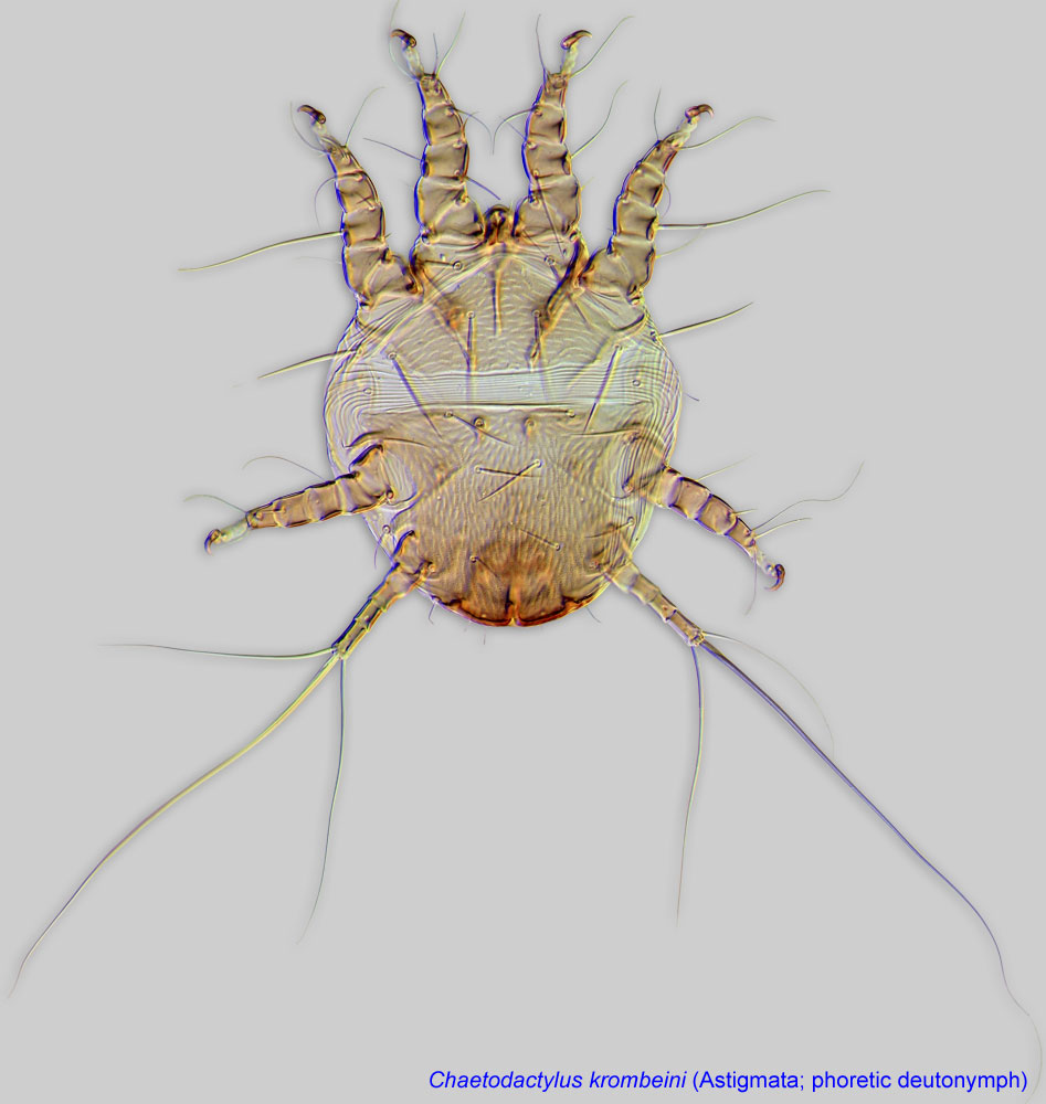 are given under the name Calvolia; (ii) membership of Calvolia reticulata in Saproglyphus needs to be verified; and (iii) Saproglyphus hagensis is included, but not illustrated; it has been illustrated and described based on type specimens in Fain, 1972Fain, 1972:
Fain, A. 1972. Notes sur les hypopes des Saproglyphidae (Acarina: Sarcoptiformes) 2. Redefinition des genres. Acarologia. 14: 225-249..
Palaearctic and Holarctic (only for species of Saproglyphus found in associations with bees).
Bee hosts include Hylaeus, Osmia, Hoplitis, and Apis.
facultative facultative:
can complete entire life cycle without bees or their close relative, wasps
(Saproglyphus hagensis); may be permanentpermanent:
associated exclusively with bees or their close relative, wasps; cannot live without these hosts
but uncertain (Saproglyphus reticulatus).
disperse on different animals, including bees.Phoretic phoretic:
Pertaining to phoresy; using another organism (i.e., a host) for dispersal to new habitats. Phoresy can be distinguished from parasitism because feeding typically does not occur during phoresy.
deutonymphsdeutonymph:
Ontogenetic stage between protonymph and tritonymph (or adult, if tritonymph is absent). See <a href="index.cfm?pageID=1720">Life stages page</a> for more details. are most common on bark beetles, but they have also been found on dung beetles, longhorn beetles, leaf beetles (Aphthona), earwigs, flies, springtails, and rodents. Feeding stages of Saproglyphus are found in decomposing organic materials, including fungi, or under bark, feeding primarily on fungi. Generalist species live in a variety of habitats and use different animals for dispersal. Saproglyphus hagensis is an example of a generalist species, a fewphoretic deutonymphsdeutonymph:
Ontogenetic stage between protonymph and tritonymph (or adult, if tritonymph is absent). See <a href="index.cfm?pageID=1720">Life stages page</a> for more details. of which have been found dispersing on the bee Hylaeus nivalis (Kuhlmann, 1998Kuhlmann, 1998:
Kuhlmann, M. 1998. Nachweise mit Bienen und Wespen (Hymenoptera Aculeata) assoziierter Milben (Acari) und Fächerflügler (Strepsiptera). Linzer biologische Beiträge. 30: 69-80.).
Several species of Saproglyphus show some degree of specificity (e.g., mites associated with bark beetles). A few species (Saproglyphus reticulatus from Hoplitis, and an undescribed species from the USA from Osmia) may be specific to bees, although it is unknown whether their feeding stages live only in bee nests or in other habitats as well. It is possible that these deutonymphsdeutonymph:
Ontogenetic stage between protonymph and tritonymph (or adult, if tritonymph is absent). See <a href="index.cfm?pageID=1720">Life stages page</a> for more details. opportunistically attach to bees that come in close proximity to their principal habitat.
Finally, a single deutonymphdeutonymph:
Ontogenetic stage between protonymph and tritonymph (or adult, if tritonymph is absent). See <a href="index.cfm?pageID=1720">Life stages page</a> for more details. of an unidentified species of Saproglyphus was found in a beehive of Apis mellifera (Haragsim et al., 1987Haragsim et al., 1987:
Haragsim, O., K. Samšiňák amp; E. Vobrázková. 1978. The mites inhabiting the bee hives in ČSR. Zeitschrift für Angewandte Entomologie. 87: 52-67.). This is probably an accidental record. It is impossible to evaluate the degree of specificity of this species.
Authors: P. Klimov, B. OConnor, R. Ochoa, G. Bauchan, A. Redford, J. Scher
Last updated October 2016
tool images at ITP Node
idtools.org


