mutualists or commensals; feed on fatty acids from floral oils and most likely on fungi
Roubikia OConnor, 1993
Superorder Acariformes » Order Sarcoptiformes » Suborder Oribatida » Infraorder Desmonomata » Hyporder Astigmata » Family Chaetodactylidae » Genus Roubikia
Chaetodactylus panamensis Baker, Roubik and Delfinado-Baker, 1987
Phoretic phoretic:
Pertaining to phoresy; using another organism (i.e., a host) for dispersal to new habitats. Phoresy can be distinguished from parasitism because feeding typically does not occur during phoresy.
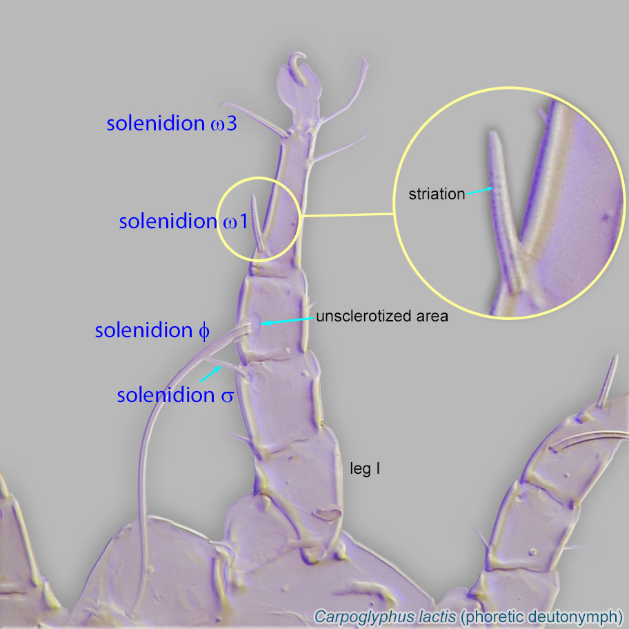
deutonymph: Gnathosomal solenidionsolenidion:
Thin-walled, terminally rounded or pointed filiform or peglike structure that is not birefringent in polarized light (unlike common setae in Acariformes). Often appears striated because of its internal structure. Found on the palpal tarsus on the gnathosoma and may also occur on the tarsus and tibia, less frequently on the genu, and occasionally on the femur of legs I-IV. In Acariformes, leg solenidia often arise from unsclerotized areas.
 and palppalp:
Second (after chelicera) paired appendage of the gnathosoma. Has a sensory function, but may be variously modified for other functions (e.g., raptorial, attachment to host, or filtering).
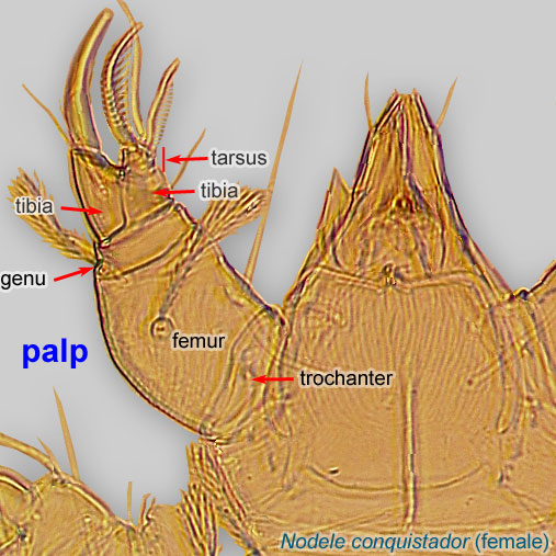 setae present and free palpspalp:
Second (after chelicera) paired appendage of the gnathosoma. Has a sensory function, but may be variously modified for other functions (e.g., raptorial, attachment to host, or filtering).
absent (Fig. 9). Coxal fields IV closed (Fig. 10). ApodemesApodeme:
Internal sclerite that serves as an attachment site for muscles. Most commonly used (as "coxal apodeme") to describe elements of coxae fused to the ventral body in Acariformes (coxae are free and not fused to the body in Parasitiformes), and may be variously referred to as ventral, sternal, anterior, or posterior.
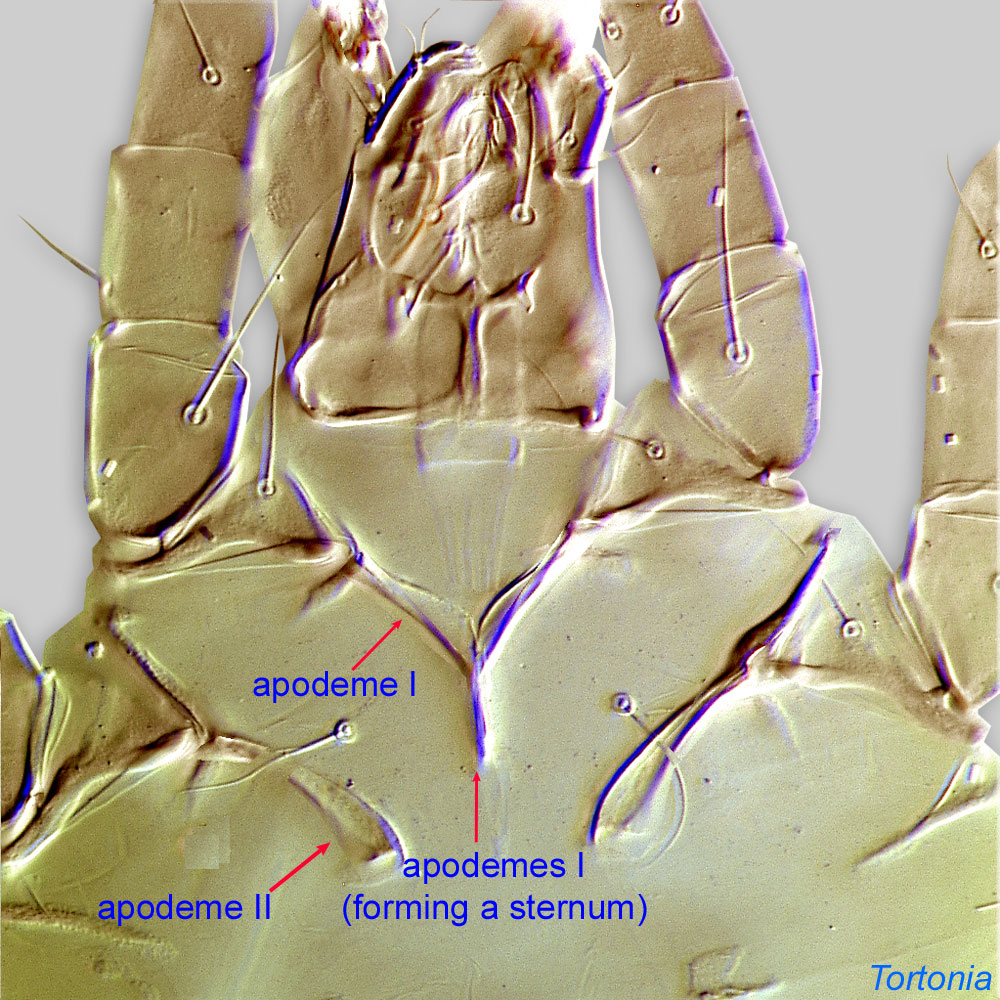 of ps1 partially fused anteriorly (Fig. 11). Dorsal cuticular folds of ambulacraambulacrum:
The claws and empodium of the apotele or pretarsus.
I-III weakly developed, with distal part smaller than proximal.
Adult: Supracoxal setae scx spiniform, with rounded tip (Fig. 12).
Female: SpermatophoresSpermatophore:
Any structure that carries a packet of sperm.
present. Inseminatory canal cylindrical, well sclerotized, protruding into spermathecaspermatheca:
A structure in the female for storing sperm, typically sac-like.
(Fig. 13). CondylophoresCondylophore:
In Acariformes, pretarsal paired sclerotized structures arising from the distal end of the tarsus and forming a joint with lateral claws (in Endeostigmata, Oribatida and Trombidiformes) and the empodial claw. Not to be confused with vertical sclerites of Parasitiformes.
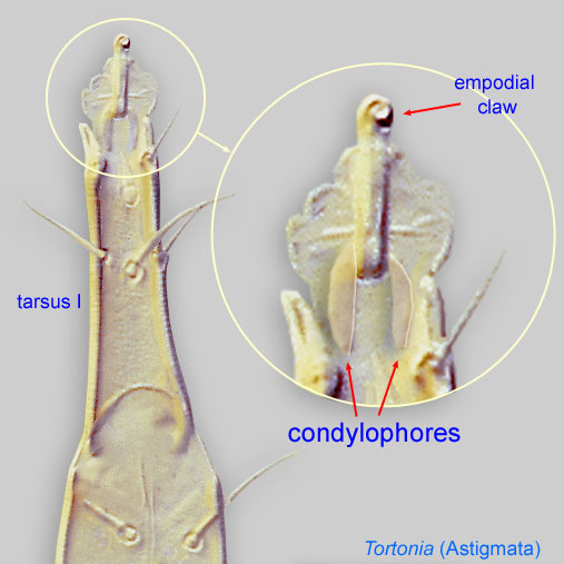 with short sclerotized portion and distinct proximal unsclerotized portion connected to the tarsustarsus:
Terminal segment (also known as podomere or palpomere) of legs or palps. In Parasitoformes it can be subdivided into telotarsus and basitarsus.
 .
.
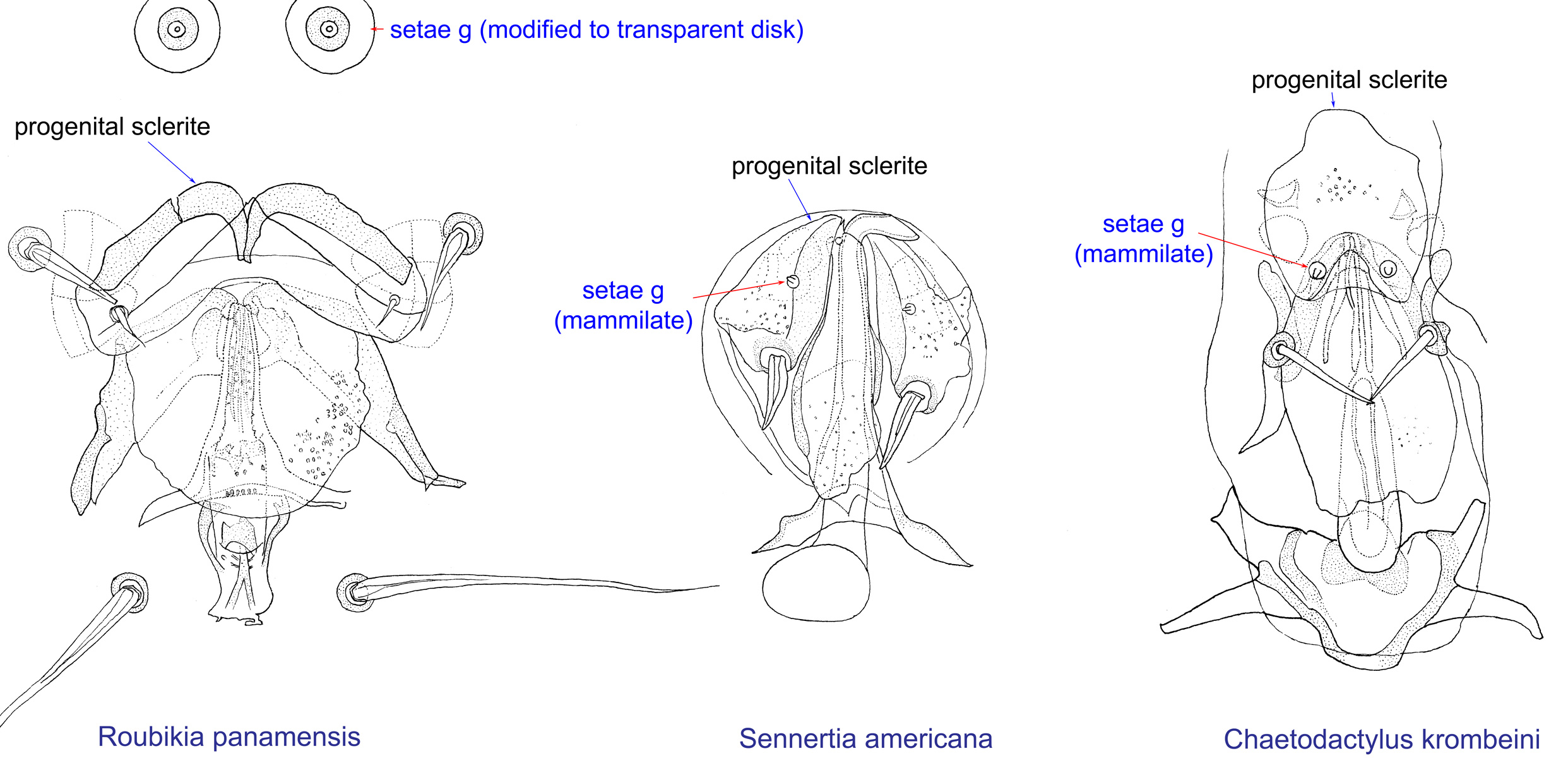
Male: Genital setae (g) represented by transparent disk (Fig. 14). Genital setae distinctly (more than their diameter at base) anterior to progenital scleritesprogenital sclerite:
A paired or unpaired sclerite situated anterior to the oviporus (female) or genital apparatus (male). In some astigmatid females it is a single, enlarged sclerite, which is often called an epigynum in descriprive works.
(Fig. 14). Tarsal setae e III-IV absent. Setae s and w IV separated: w submedial, s subapical. Sclerotized portions of condylophorescondylophore:
In Acariformes, pretarsal paired sclerotized structures arising from the distal end of the tarsus and forming a joint with lateral claws (in Endeostigmata, Oribatida and Trombidiformes) and the empodial claw. Not to be confused with vertical sclerites of Parasitiformes.
fused and incorporated into disto-ventral sclerotized tarsal wall (Fig. 15). Pretarsal suckers not developed (Fig. 15). Heteromorphic male present.
A dichotomous key to phoreticphoretic:
Pertaining to phoresy; using another organism (i.e., a host) for dispersal to new habitats. Phoresy can be distinguished from parasitism because feeding typically does not occur during phoresy.
deutonymphsdeutonymph:
Ontogenetic stage between protonymph and tritonymph (or adult, if tritonymph is absent). See <a href="index.cfm?pageID=1720">Life stages page</a> for more details.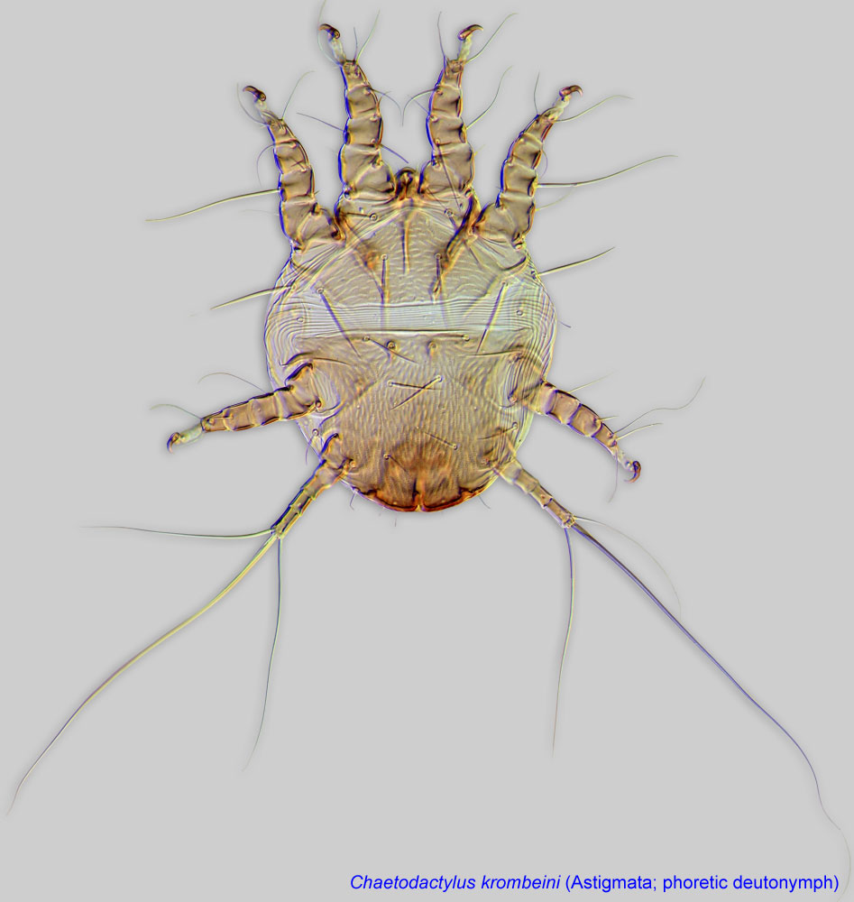 is available from the bee-associated mites website and Klimov and OConnor, 2008Klimov and OConnor, 2008:
Klimov, P. B. amp; B. M. OConnor. 2008. Morphology, evolution, and host associations of bee-associated mites of the family Chaetodactylidae (Acari: Astigmata), with a monographic revision of North American taxa. Miscellaneous Publications Museum of Zoology University of Michigan.199: 1-243.. Adults are known only from one species, Roubikia panamensis, and are described in Baker et al., 1987Baker et al., 1987:
Baker, E. W., D. W. Roubik, and M. Delfinado-Baker. 1987. The developmental stages and dimorphic males of Chaetodactylus panamensis , n. sp. (Acari: Chaetodactylidae) associated with solitary bee (Apoidea: Anthophoridae). International Journal of Acarology 13:65-73. and Klimov and OConnor, 2008Klimov and OConnor, 2008:
Klimov, P. B. amp; B. M. OConnor. 2008. Morphology, evolution, and host associations of bee-associated mites of the family Chaetodactylidae (Acari: Astigmata), with a monographic revision of North American taxa. Miscellaneous Publications Museum of Zoology University of Michigan.199: 1-243..
Neotropical region
apid bees of the genus Tetrapedia; can be phoreticphoretic:
Pertaining to phoresy; using another organism (i.e., a host) for dispersal to new habitats. Phoresy can be distinguished from parasitism because feeding typically does not occur during phoresy.
on Coelioxoides spp. (kleptoparasites of Tetrapedia)
permanentpermanent:
associated exclusively with bees or their close relative, wasps; cannot live without these hosts
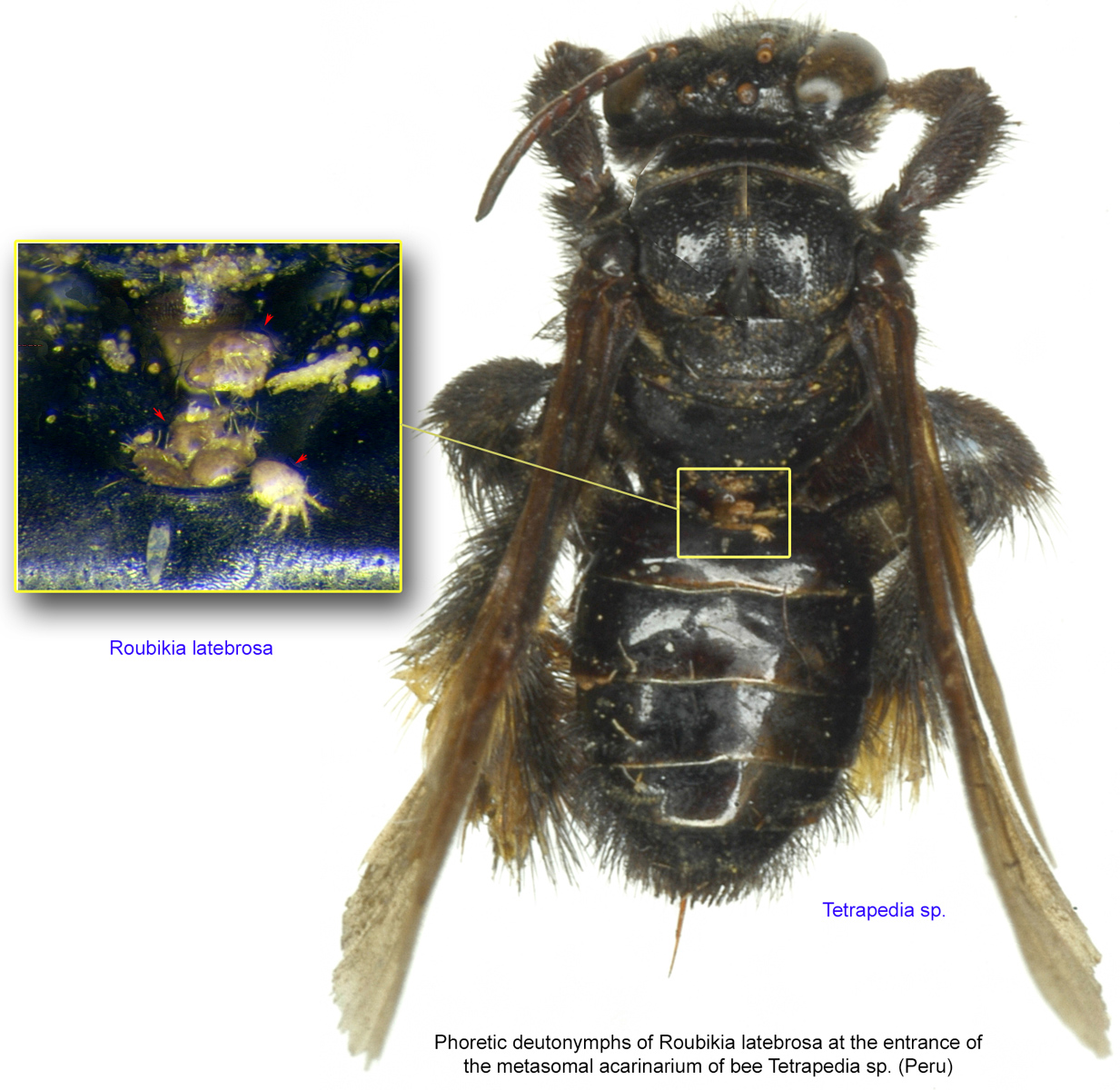
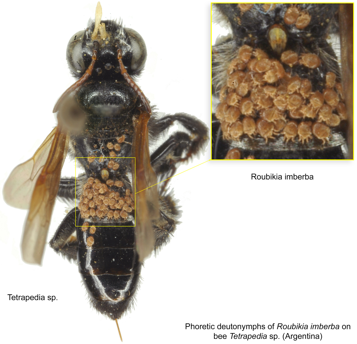
disperse on adult bees from one nest to another (Figs. 3-8).This genus is exclusively associated with Tetrapedia (Apidae: Tetrapediini). Adults live in the bee nests, and deutonymphsdeutonymph:
Ontogenetic stage between protonymph and tritonymph (or adult, if tritonymph is absent). See <a href="index.cfm?pageID=1720">Life stages page</a> for more details. disperse on the adult bees (Figs. 6, 7). DeutonymphsDeutonymph:
Ontogenetic stage between protonymph and tritonymph (or adult, if tritonymph is absent). See <a href="index.cfm?pageID=1720">Life stages page</a> for more details. of Roubikia panamensis and R. imberba can also be found dispersing on kleptoparasitic bees of the genus Coelioxoides (Apidae: Tetrapediini) (Figs. 5, 8). This kleptoparasitic 'cuckoo' bee does not build its own nests but uses nests of Tetrapedia as a resource for its brood (Alves-Dos-Santos et al., 2002Alves-Dos-Santos et al., 2002:
Alves-Dos-Santos, I., G. A. R. Melo amp; J. G. Rozen. 2002. Biology and immature stages of the bee tribe Tetrapediini (Hymenoptera: Apidae). American Museum Novitates. 3377: 1-45.). For Roubikia, Coelioxoides bees may serve as transport, dispersing the mites to different nests of Tetrapedia.
In Brazil, mites associated with Tetrapedia diversipes have been shown to be beneficial to the bee as the bee mortality rate in nests was inversely correlated to the level of mite infestation (Cordeiro et al., 2011Cordeiro et al., 2011:
Cordeiro, G. D., M. Taniguchi, C. H. W. Flechtmann amp; I. Alves-dos-Santos. 2011. Phoretic mites (Acari: Chaetodactylidae) associated with the solitary bee Tetrapedia diversipes (Apidae: Tetrapediini). Apidologie.42: 128-139.). The mites presumably feed on fungi harmful to the bee larvae inside the bee nests (Cordeiro et al., 2011Cordeiro et al., 2011:
Cordeiro, G. D., M. Taniguchi, C. H. W. Flechtmann amp; I. Alves-dos-Santos. 2011. Phoretic mites (Acari: Chaetodactylidae) associated with the solitary bee Tetrapedia diversipes (Apidae: Tetrapediini). Apidologie.42: 128-139.).
A single species, Roubikia latebrosa, is phoreticphoretic:
Pertaining to phoresy; using another organism (i.e., a host) for dispersal to new habitats. Phoresy can be distinguished from parasitism because feeding typically does not occur during phoresy.
in the metasomal acarinariumacarinarium:
A specialized morphological structure that facilitates retention of mites on the body of an organism, typically a bee or wasp.
of the host (Figs. 3, 4). Dispersal to unrelated host bees can be accomplished via phoresyphoresy:
Attaching to or boarding another organism (i.e., a host) for dispersal to new habitats. Can be distinguished from parasitism because feeding typically does not occur.
on kleptoparasitic bees attacking the mite's principal hosts (Alves-Dos-Santos et al., 2002Alves-Dos-Santos et al., 2002:
Alves-Dos-Santos, I., G. A. R. Melo amp; J. G. Rozen. 2002. Biology and immature stages of the bee tribe Tetrapediini (Hymenoptera: Apidae). American Museum Novitates. 3377: 1-45.). Mites may disembark from the bees and attach to new host females at places where the bees collect loose dirt for nest material or forage (Roubik, 1987Roubik, 1987:
Roubik, D. W. 1987. Notes on the biology of anthophorid bee Tetrapedia and the mite Chaetodactylus panamensis Baker, Roubik and Delfinado-Baker (Acari: Chaetodactylidae). International Journal of Acarology . 13 : 75-76.).
|
Fig. 9. Gnathosomal morphology is a key character separating genera of the family Chaetodactylidae (Centriacarus, Roubikia, Achaetodactylus, Chaetodactylus, and Sennertia) based on phoretic deutonymphs. In particular, Roubikia can be distinguished by its gnathosomal solenidion and palp setae present and free palps absent; drawing of Achaetodactylus leleupi courtesy of Belgian GTI Focal Point 2009, http://www.taxonomy.be. |
|
|
Fig. 12. Supracoxal sclerite of homeomorphic males of the family Chaetodactylidae (female supracoxal sclerite of Ch. krombeini given for comparison), showing the shape of supracoxal setae (scx). Roubikia is unique in having a spiniform scx with rounded tip, while in all other genera scx is filiform with a pointed tip. |
|
|
Fig. 13. Spermatheca and inseminatory canal of Chaetodactylidae. Roubikia is unique in having a cylindrical inseminatory canal that protrudes into the spermatheca and distinct, filariform spermatophores. In other chaetodactylid genera, the inseminatory canal is distinctly widened at its entrance to the spermatheca and does not protrude into the spermatheca; spermatophores are absent. |
|
|
Fig. 15. Leg tarsi of males of Chaetodactylidae. Roubikia can be distinguished from other chaetodactylid genera with known males (Sennertia and Chaetodactylus) by the absence of pretarsal suckers. In the other two genera, male pretarsal suckers are present. For Roubikia heteromorphic males are shown, for the others homeomorphic males. |
Authors: P. Klimov, B. OConnor, R. Ochoa, G. Bauchan, A. Redford, J. Scher
Last updated October 2016
tool images at ITP Node
idtools.org