larvae are parasites that feed on hemolymph, while deutonymphsdeutonymph:
Ontogenetic stage between protonymph and tritonymph (or adult, if tritonymph is absent). See <a href="index.cfm?pageID=1720">Life stages page</a> for more details. and adults are largely neutral predators of small invertebrates in bee nests
Neotrombidium Leonardi, 1902
Superorder Acariformes » Order Trombidiformes » Suborder Prostigmata » Infraorder Anystina » Hyporder Parasitengona » Family Neotrombidiidae » Genus Neotrombidium
Trombidium (Neotrombidium) furcigerum Leonardi, in Berlese and Leonardi, 1902
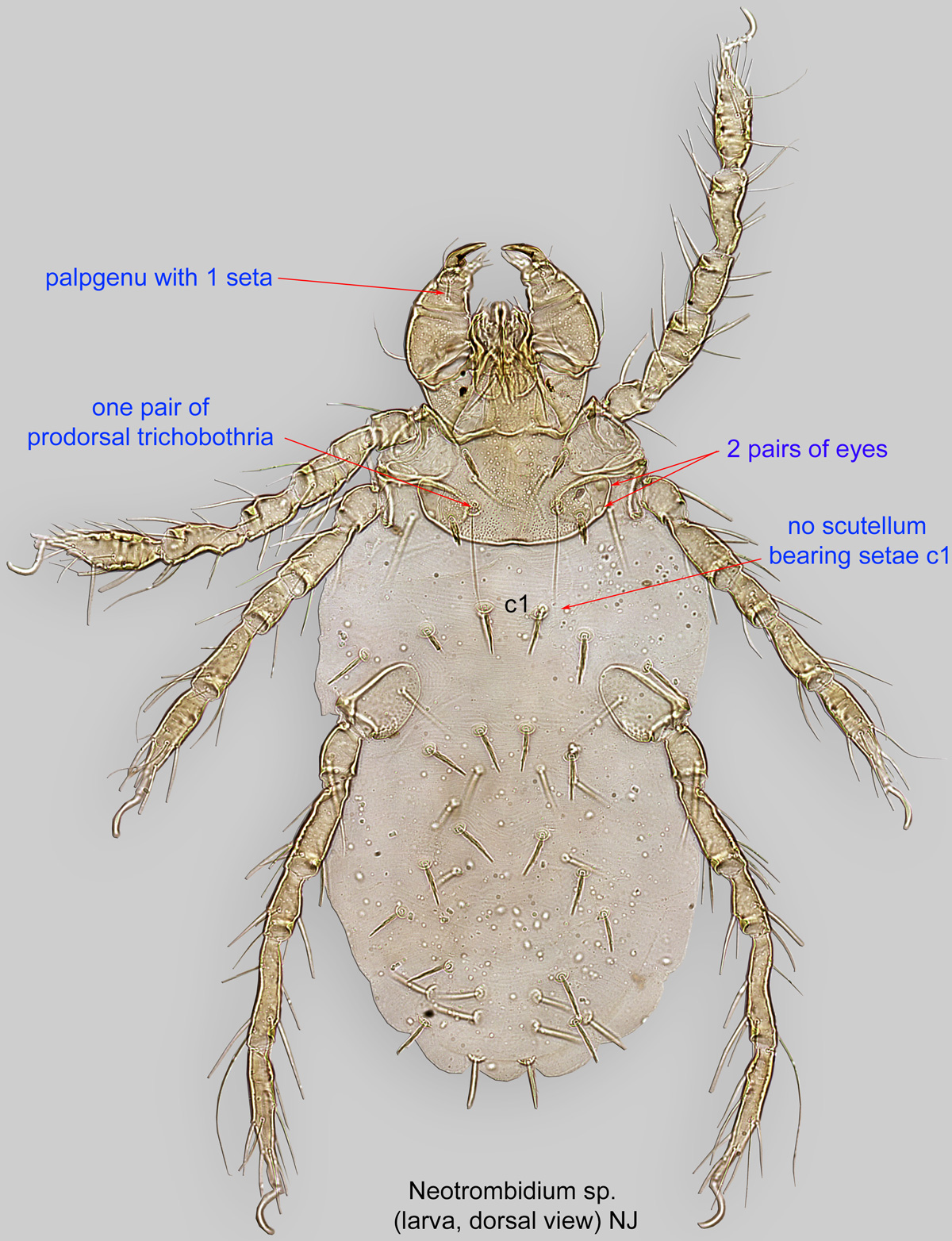
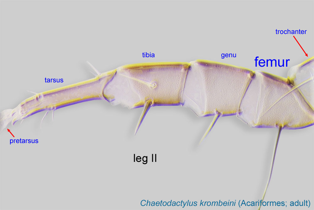
Larva: Palpal genugenu:
Leg or palp segment (also known as podomere or palpomere) between tibia and femur.
 with one seta (Fig. 1). One pair of dorsal trichobothriatrichobothrium:
Modified seta that can be distinguished from true seta by its distinct large socket (complex cup-like cavity), and often, shape (which may be filiform, ciliate, pectinate, or thickened or clubbed). Present in many Acariformes (except Astigmata) and Opilioacarida (Parasitiformes). When paired and situated on the propodosoma, termed "prodorsal."
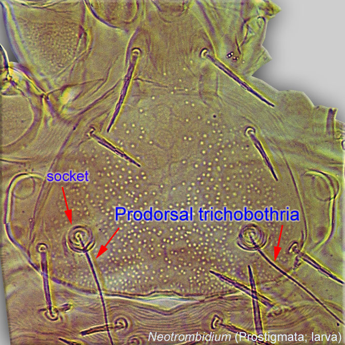 on prodorsalprodorsal:
Pertaining to the prodorsum.
sclerite (Fig. 1). Scutellum bearing setae c1 absent (Fig. 1). Claparède’s organs (urstigmata) present (Fig. 2). One claw at the apex of leg tarsitarsus:
Terminal segment (also known as podomere or palpomere) of legs or palps. In Parasitoformes it can be subdivided into telotarsus and basitarsus.
 I-III (Fig. 2). Posteroapical corner of at least the posterior leg coxacoxa:
In Parasitiformes, most basal leg segment (or podomere) forming a joint with the body. Areas delimited by coxal apodemes are called coxal fields in Astigmata or coxisternal plates in Prostigmata.
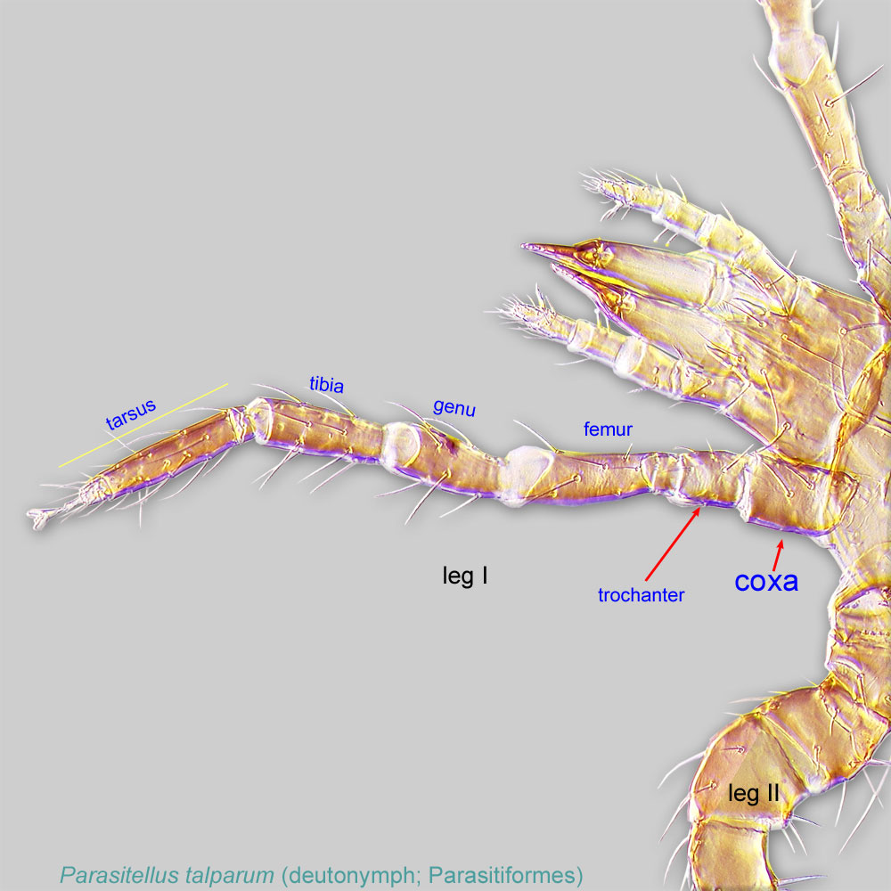 ornamented with characteristic areas of porosity in adults and larvae (Fig. 2). The number of barbed setae on the femorafemur:
Leg or palp segment (also known as podomere or palpomere) between genu and trochanter. In ParasitIformes can be subdivided into telofemur and basifemur.
 of the anterior, mid, and posterior legs is 7-6-6, a combination unique to this genus. Femurfemur:
Leg or palp segment (also known as podomere or palpomere) between genu and trochanter. In ParasitIformes can be subdivided into telofemur and basifemur.
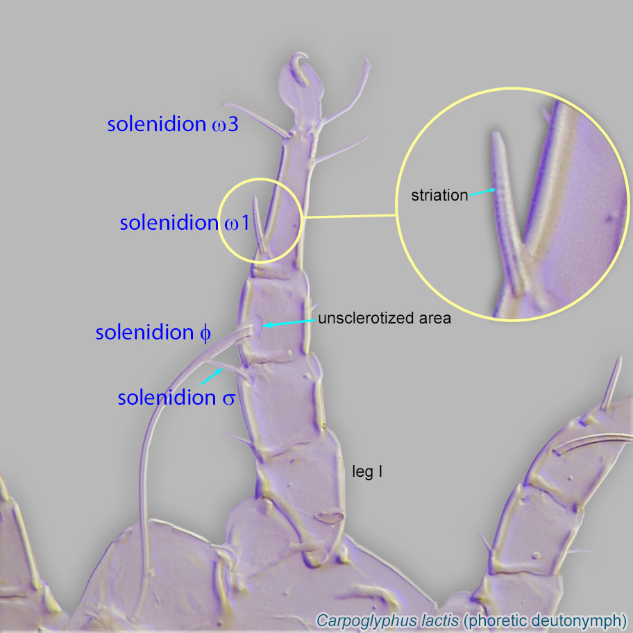
I with at least one solenidionsolenidion:
Thin-walled, terminally rounded or pointed filiform or peglike structure that is not birefringent in polarized light (unlike common setae in Acariformes). Often appears striated because of its internal structure. Found on the palpal tarsus on the gnathosoma and may also occur on the tarsus and tibia, less frequently on the genu, and occasionally on the femur of legs I-IV. In Acariformes, leg solenidia often arise from unsclerotized areas.
 , femorafemur:
Leg or palp segment (also known as podomere or palpomere) between genu and trochanter. In ParasitIformes can be subdivided into telofemur and basifemur.
II-III with at least two solenidiasolenidion:
Thin-walled, terminally rounded or pointed filiform or peglike structure that is not birefringent in polarized light (unlike common setae in Acariformes). Often appears striated because of its internal structure. Found on the palpal tarsus on the gnathosoma and may also occur on the tarsus and tibia, less frequently on the genu, and occasionally on the femur of legs I-IV. In Acariformes, leg solenidia often arise from unsclerotized areas.
. Lateral walls of base of gnathosomagnathosoma:
Division of body anterior to the propodosoma bearing two pairs of appendages (palps and chelicerae).
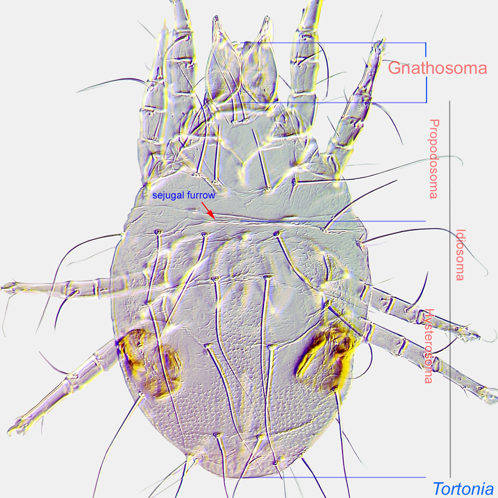 (gnathobase) densely punctate (Fig. 2).
A dichotomous key is available in Lindquist and Vercammen-Grandjean, 1971Lindquist and Vercammen-Grandjean, 1971:
Lindquist, E. E. amp; P. H. Vercammen-Grandjean. 1971. Revision of the chigger-like larvae of the genera Neotrombidium Leonardi and Monunguis Wharton, with a redefinition of the subfamily Neotrombidiinae Feider in the Trombidiidae (Acarina: Prostigmata). Canadian Entomologist.103: 1557-1590..
This genus is worldwide in distribution except for the polar regions. A single species associated with bees has been found in the USA (Florida).
large carpenter bee Xylocopa micans
temporarytemporary:
some life stages are associated with bees, while others are not
This genus includes 16 species (Beron, 1995Beron, 1995:
Beron, P. 1995. A new larval species of Neotrombidium (Acariformes, Actinedida: Neotrombidiidae) from Cuba. Historia Naturalis Bulgarica . 5 : 13-18.; Lindquist and Vercammen-Grandjean, 1971Lindquist and Vercammen-Grandjean, 1971:
Lindquist, E. E. amp; P. H. Vercammen-Grandjean. 1971. Revision of the chigger-like larvae of the genera Neotrombidium Leonardi and Monunguis Wharton, with a redefinition of the subfamily Neotrombidiinae Feider in the Trombidiidae (Acarina: Prostigmata). Canadian Entomologist.103: 1557-1590.; Singer, 1971Singer, 1971:
Singer, G. 1971. Neotrombidium Leonardi (Acarina: Trombidioidea) Part I: Revision of the genus, with description of two new species. Acarologia . 13 : 119-142.) with adults found in subcorticalsubcortical:
Under tree bark.
habitats and forest litter. Larvae are parasitic on subcorticalsubcortical:
Under tree bark.
beetles and wood-boring carpenter bees (Xylocopa).
Authors: P. Klimov, B. OConnor, R. Ochoa, G. Bauchan, A. Redford, J. Scher
Last updated October 2016
tool images at ITP Node
idtools.org