primarily kleptoparasitic when feed on pollen but can be neutral to beneficial when feed on fungus in bee nests
Horstiella Turk, 1948Turk, 1948:
Turk, F. A. 1948. Insecticolous Acari from Trinidad, B.W.I. Proceedings of the Zoological Society of London.118: 82-125.
Superorder Acariformes » Order Sarcoptiformes » Suborder Oribatida » Infraorder Desmonomata » Hyporder Astigmata » Family Acaridae » Genus Horstiella
Horstiella armata Turk, 1948Turk, 1948:
Turk, F. A. 1948. Insecticolous Acari from Trinidad, B.W.I. Proceedings of the Zoological Society of London.118: 82-125.
Adult: Only one solendion σ on genugenu:
Leg or palp segment (also known as podomere or palpomere) between tibia and femur.
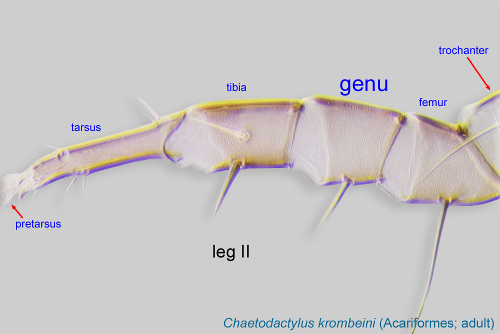 I (Fig. 9).
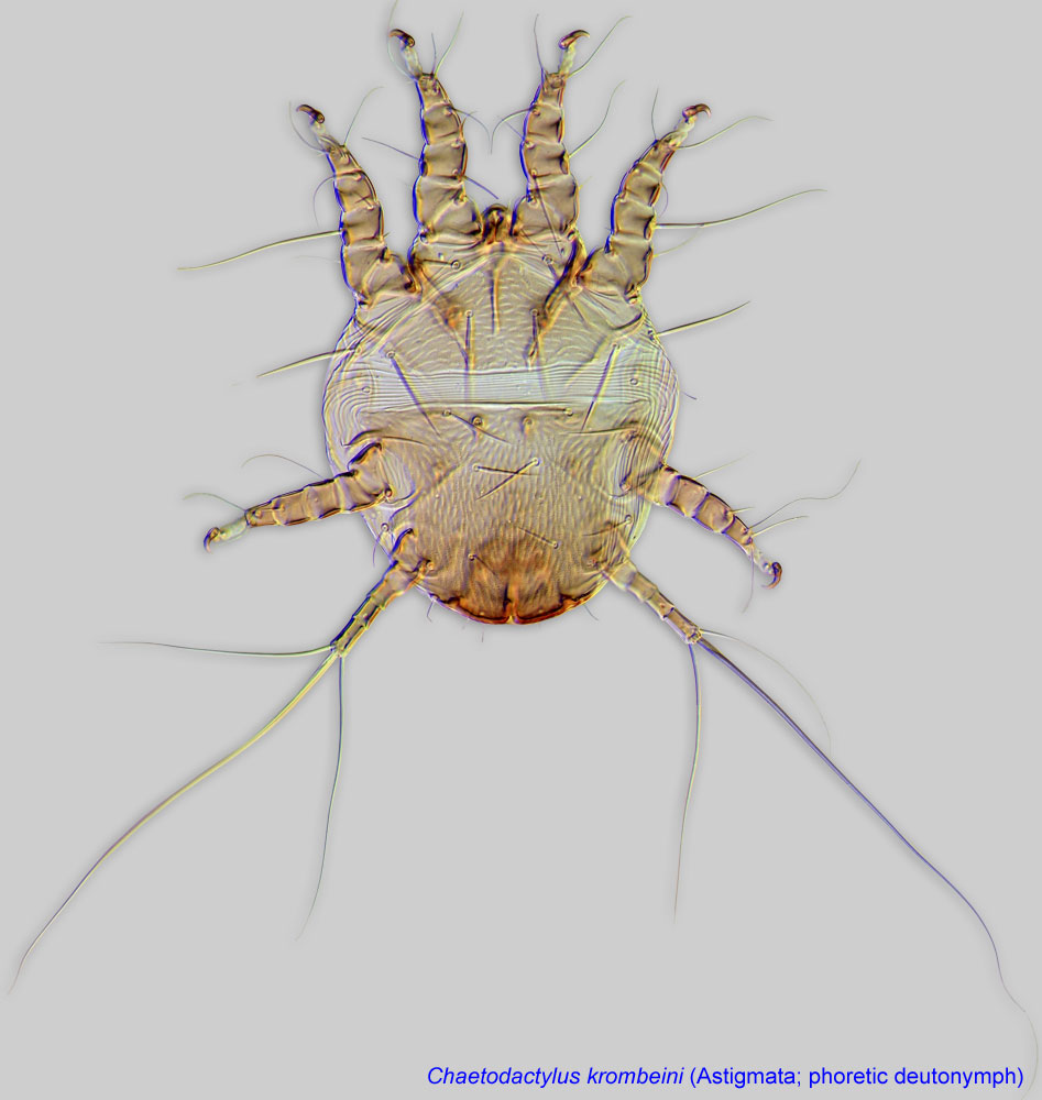
Phoretic phoretic:
Pertaining to phoresy; using another organism (i.e., a host) for dispersal to new habitats. Phoresy can be distinguished from parasitism because feeding typically does not occur during phoresy.
deutonymph: Propodosomal shieldpropodosomal shield:
Unpaired shield situated on the dorsal propodosoma.
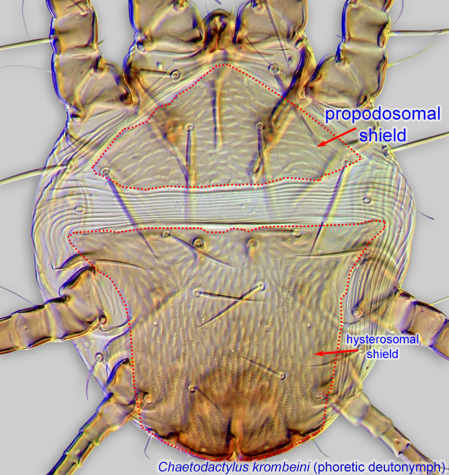 transversely striated, hysterosomal shieldhysterosomal shield:
Unpaired shield situated on the dorsal hysterosoma.
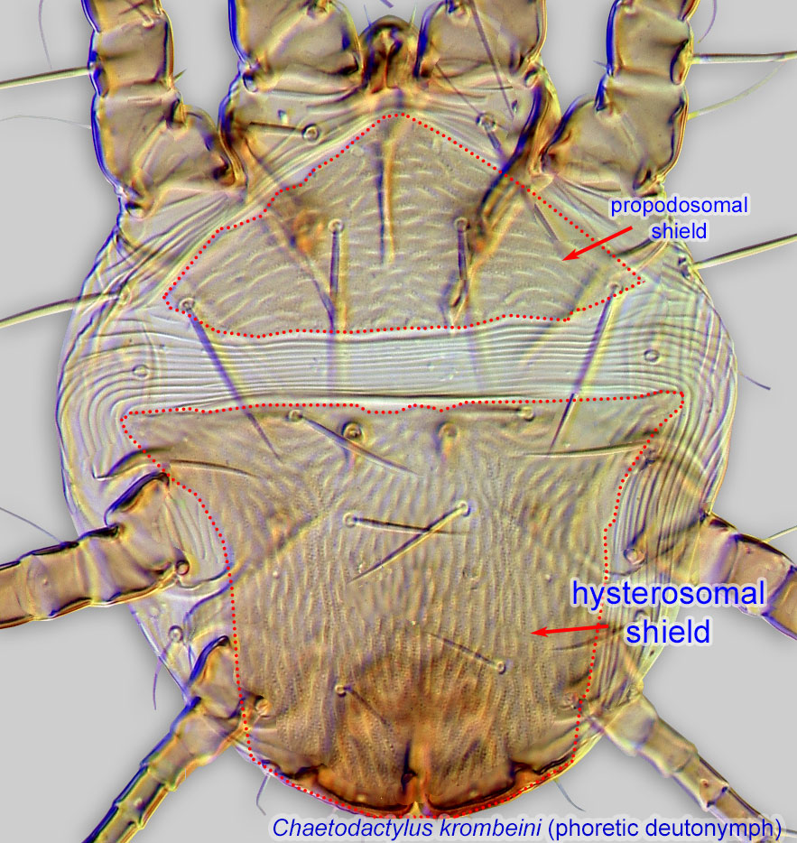 with longitudinal and transverse striations (striate pattern may be faint) (Fig. 1). External vertical setae ve represented by vestigial alveoli (Fig. 3). Supracoxal setae scx absent (Fig. 3). Coxal setae 1a, 3a, 4b alveolar; in contrast, coxal setae 4a in the form of large, striate conoids (Figs. 2, 4). Setae aa I, and ba I-II absent (Figs. 3). Solenidionsolenidion:
Thin-walled, terminally rounded or pointed filiform or peglike structure that is not birefringent in polarized light (unlike common setae in Acariformes). Often appears striated because of its internal structure. Found on the palpal tarsus on the gnathosoma and may also occur on the tarsus and tibia, less frequently on the genu, and occasionally on the femur of legs I-IV. In Acariformes, leg solenidia often arise from unsclerotized areas.
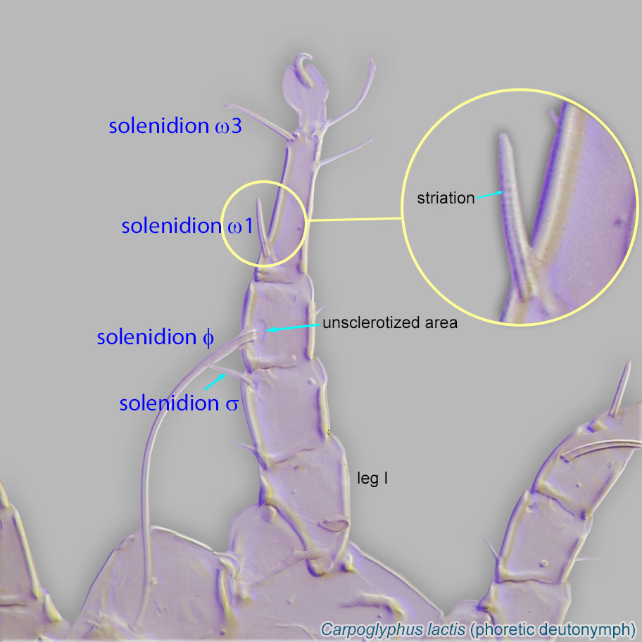 ω2 absent from tarsustarsus:
Terminal segment (also known as podomere or palpomere) of legs or palps. In Parasitoformes it can be subdivided into telotarsus and basitarsus.
 I, represented by vestigial alveolus (Fig. 3). Solenidionsolenidion:
Thin-walled, terminally rounded or pointed filiform or peglike structure that is not birefringent in polarized light (unlike common setae in Acariformes). Often appears striated because of its internal structure. Found on the palpal tarsus on the gnathosoma and may also occur on the tarsus and tibia, less frequently on the genu, and occasionally on the femur of legs I-IV. In Acariformes, leg solenidia often arise from unsclerotized areas.
σ III and seta nG III absent from genugenu:
Leg or palp segment (also known as podomere or palpomere) between tibia and femur.
III (Fig. 4). Tarsustarsus:
Terminal segment (also known as podomere or palpomere) of legs or palps. In Parasitoformes it can be subdivided into telotarsus and basitarsus.
IV with 1 seta (d IV) longer than leg IV (Fig. 4).
Adult: External vertical setae (ve) represented by alveoli, located on lateral sides of prodorsalprodorsal:
Pertaining to the prodorsum.
shield, approximately midway between vi and scapular setae se (Fig. 9). Hysterosomal setae c1, c2, d1, and f2 present (Figs. 5, 7, 8; notice that f2 is very short and may not be easily seen in female). Grandjean's organGrandjean's organ:
Paired, finger-shaped, lobe-shaped, or otherwise elaborated structure situated on the lateral sides of the propodosoma, typically in association with the podocephalic canal. Its free edges may be strongly fimbriate.
biramous (Fig. 8) and setae ba I-II absent (Fig. 9). Male with sclerotized projection extending from posterior opisthosomaopisthosoma:
Body division posterior to legs IV; usually there is no distinct boundary delimiting this part of idiosoma.
(Figs. 7, 8). Projection is broad, with setae h2 and h3 dorsally and setae ps1, ps2, and f2 ventrally (Figs. 7, 8).
A dichotomous key is available in Ochoa and OConnor, 2000Ochoa and OConnor, 2000:
Ochoa, R. amp; B. M. OConnor. 2000. Revision of the genus Horstiella (Acari: Acaridae): mites associated with Neotropical Epicharis bees (Hymenoptera: Apidae). Annals of the Entomological Society of America . 93 : 713-737..
Neotropical region
Ground-nesting apid bees of the genus Epicharis, subgenera Triepicharis, Parepicharis, Hoplepicharis, Epicharis (s.str.), and Epicharana. Epicharis subgenera Epicharoides and Epicharitides lack these mites.
permanentpermanent:
associated exclusively with bees or their close relative, wasps; cannot live without these hosts
 disperse on bee hosts.
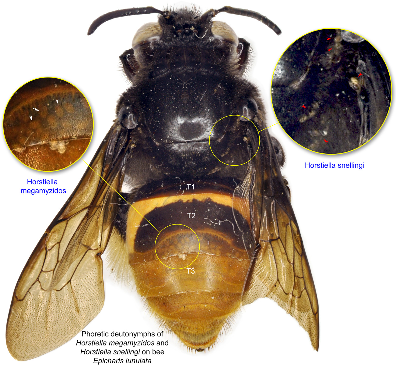
Species of Horstiella are exclusively associated with the ground-nesting bee genus Epicharis in the Neotropics (Figs. 13-19). This association is quite unusual in that most species of Epicharis harbor two species of Horstiella, a condition known as synhospitality (Ochoa and OConnor, 2000Ochoa and OConnor, 2000:
Ochoa, R. amp; B. M. OConnor. 2000. Revision of the genus Horstiella (Acari: Acaridae): mites associated with Neotropical Epicharis bees (Hymenoptera: Apidae). Annals of the Entomological Society of America . 93 : 713-737.) (Figs. 14, 16); the mite species pairs were almost always spatially segregated on an individual host (Fig. 14).
Most Horstiella species are commonly found on the bee's mesosoma and 1st metasoma tergite, though Horstiella megamyzidos specifically attaches under the lateral edges of the metasomal tergites (Fig. 14) and under the sternites. In male bees, the more frequent mite attachment site is the ventral region of the mesosoma and metasoma (Fig. 18), which correlates with the mating position of the host bees. Because only the female bee makes nests, mites developing in a cell with a male bee would normally have no opportunity to found new colonies. Migrating to the ventral surface of male bees would, therefore, give the mites a selective advantage because this can facilitate transfer onto the body of a female bee during copulation, which occurs with the male above the female (Ochoa and OConnor, 2000Ochoa and OConnor, 2000:
Ochoa, R. amp; B. M. OConnor. 2000. Revision of the genus Horstiella (Acari: Acaridae): mites associated with Neotropical Epicharis bees (Hymenoptera: Apidae). Annals of the Entomological Society of America . 93 : 713-737.).
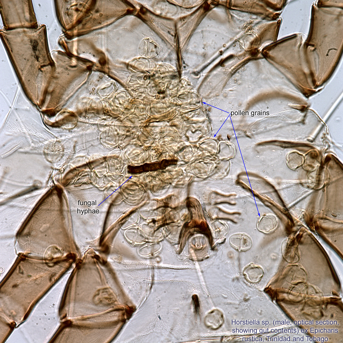
Large quantities of pollen grains as well as fungal conidia were found in guts of adult mites found in nests of Epicharis rustica (Figs. 11, 12; our data, unpublished). These observations suggest that the mites are primarily kleptoparasitic pollen-feeders but can also be fungivorousfungivorous:
Feeding on fungi.
in bee nests.
Authors: P. Klimov, B. OConnor, R. Ochoa, G. Bauchan, A. Redford, J. Scher
Last updated October 2016
tool images at ITP Node
idtools.org