neutral; live on bodies of developing bee larvae; probably feed on microorganisms
Glyphanoetus Oudemans, 1929
Superorder Acariformes » Order Sarcoptiformes » Suborder Oribatida » Infraorder Desmonomata » Hyporder Astigmata » Family Histiostomatidae » Genus Glyphanoetus
Glyphanoetus fulmeki Oudemans, 1929
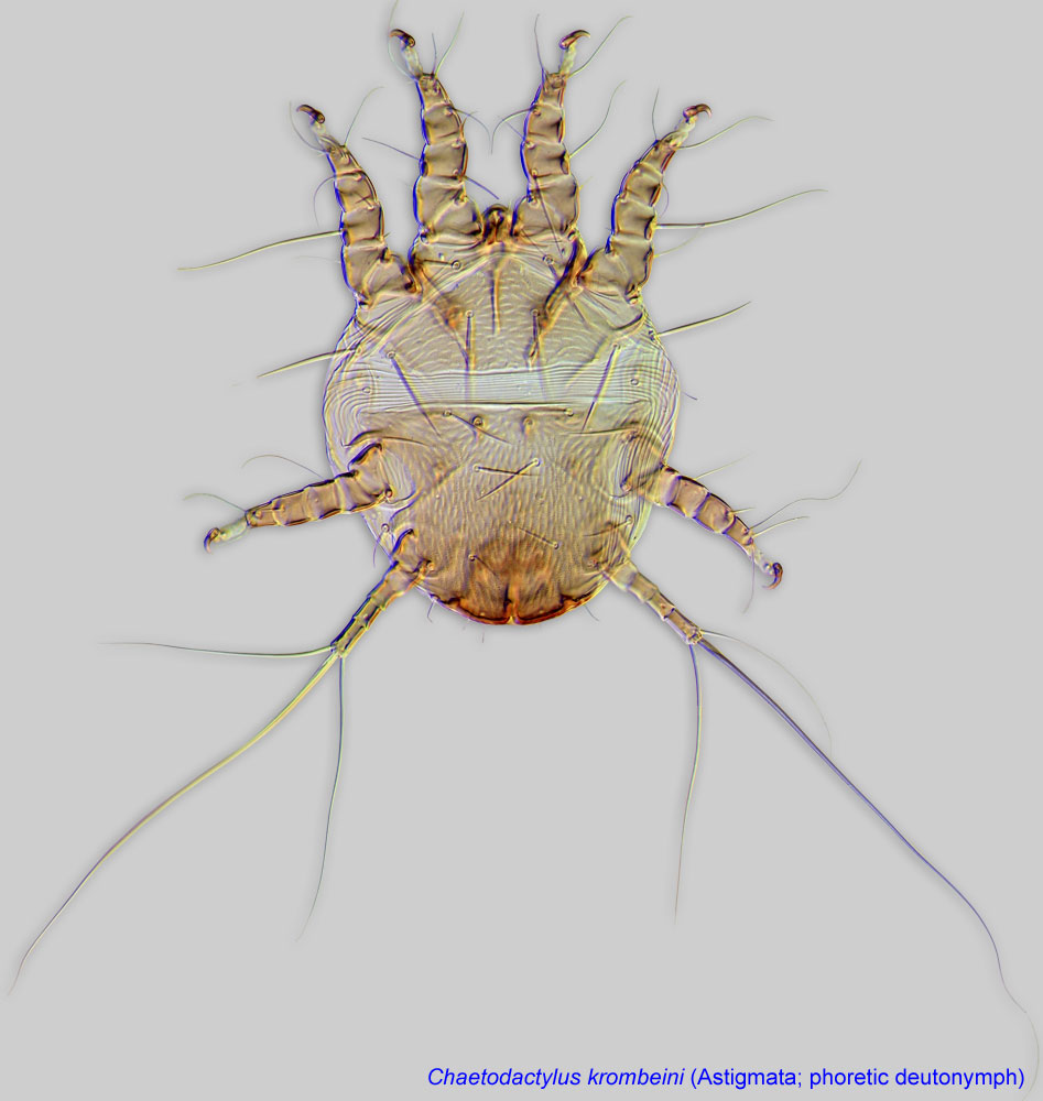
Phoretic phoretic:
Pertaining to phoresy; using another organism (i.e., a host) for dispersal to new habitats. Phoresy can be distinguished from parasitism because feeding typically does not occur during phoresy.
deutonymph: Solenidionsolenidion:
Thin-walled, terminally rounded or pointed filiform or peglike structure that is not birefringent in polarized light (unlike common setae in Acariformes). Often appears striated because of its internal structure. Found on the palpal tarsus on the gnathosoma and may also occur on the tarsus and tibia, less frequently on the genu, and occasionally on the femur of legs I-IV. In Acariformes, leg solenidia often arise from unsclerotized areas.
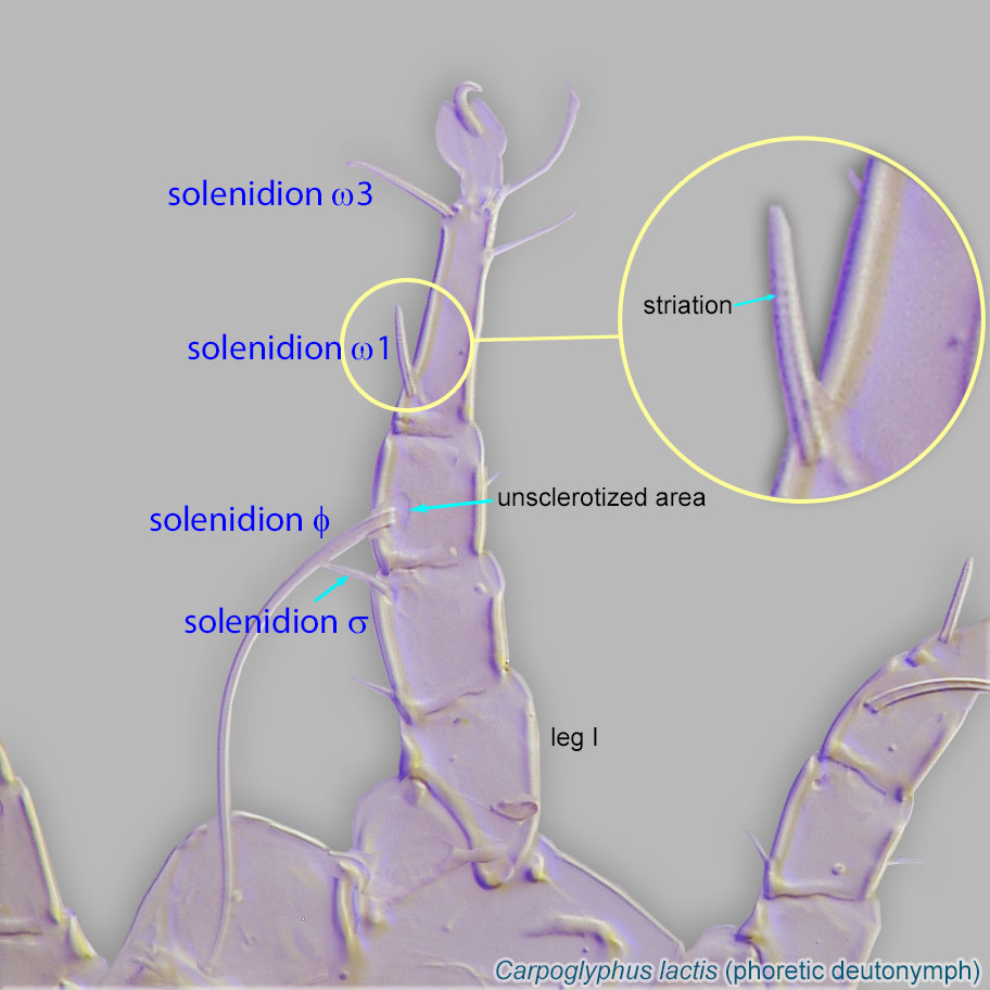 ω1 of leg I positioned directly on tibiatibia:
Leg or palp segment (also known as podomere or palpomere) between tarsus and genu.
 , associated with tibial solenidionsolenidion:
Thin-walled, terminally rounded or pointed filiform or peglike structure that is not birefringent in polarized light (unlike common setae in Acariformes). Often appears striated because of its internal structure. Found on the palpal tarsus on the gnathosoma and may also occur on the tarsus and tibia, less frequently on the genu, and occasionally on the femur of legs I-IV. In Acariformes, leg solenidia often arise from unsclerotized areas.
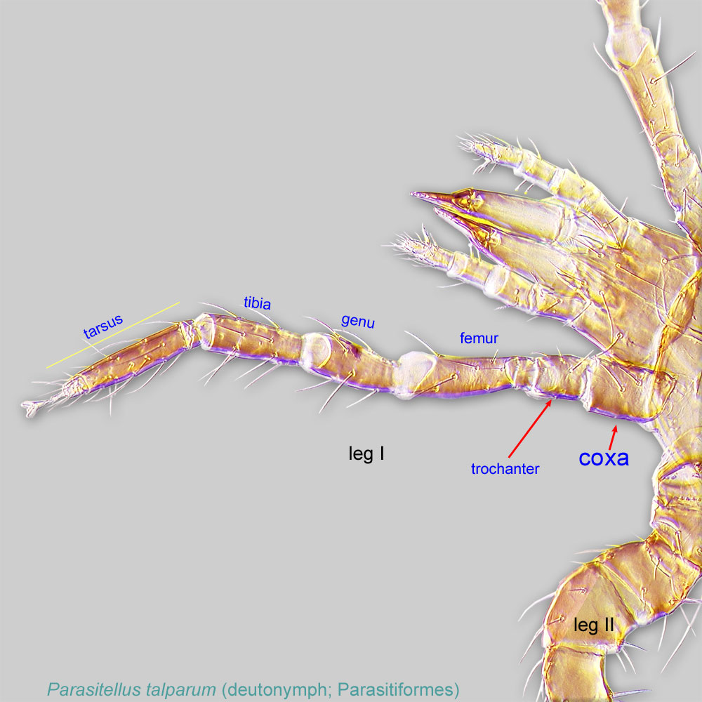
phi (φ) (Fig. 6). Setae of coxacoxa:
In Parasitiformes, most basal leg segment (or podomere) forming a joint with the body. Areas delimited by coxal apodemes are called coxal fields in Astigmata or coxisternal plates in Prostigmata.
 I (1a) filiform (dung mites) or vestigial (bee-associated mites). Setae of coxaecoxa:
In Parasitiformes, most basal leg segment (or podomere) forming a joint with the body. Areas delimited by coxal apodemes are called coxal fields in Astigmata or coxisternal plates in Prostigmata.
III (3a) conoidal (Fig.4).
Phoretic phoretic:
Pertaining to phoresy; using another organism (i.e., a host) for dispersal to new habitats. Phoresy can be distinguished from parasitism because feeding typically does not occur during phoresy.
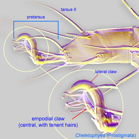
deutonymph: Empodial clawsEmpodial claw:
Claw-like, membranous, or pad-like structure of setal origin. Present only on the pretarsus in Acariformes. In Astigmata, it is the only claw on the pretarsus and often referred to simply as the claw. In the remaining Acariformes, may be accomanied by two lateral claws. Also known as empodium, pretarsal empodium, or central claw.
 I-IV simplesimple:
Of claws or setae; not modified or not bi- or trifurcate at tip.
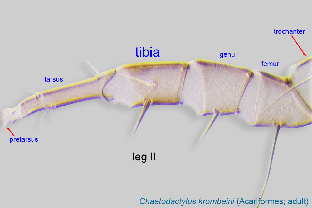
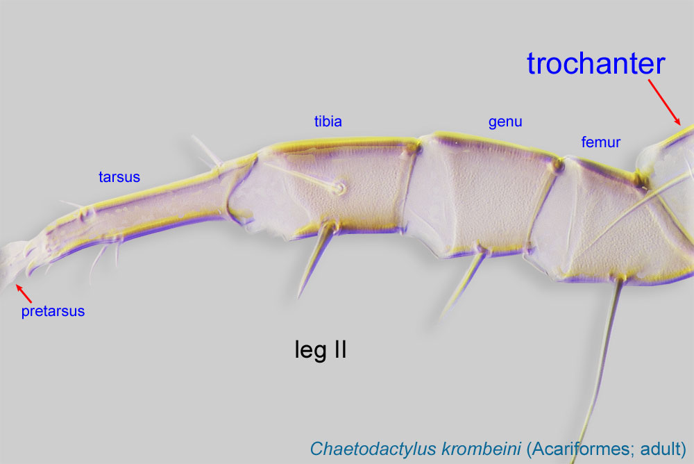
(not bifurcated) (Fig. 6). Tarsustarsus:
Terminal segment (also known as podomere or palpomere) of legs or palps. In Parasitoformes it can be subdivided into telotarsus and basitarsus.
 III and IV with a weak, flexible region in middle of segment (Fig. 7). Pretarsuspretarsus:
Terminal leg or palpal segment distal to tarsus.
III with empodial clawempodial claw:
Claw-like, membranous, or pad-like structure of setal origin. Present only on the pretarsus in Acariformes. In Astigmata, it is the only claw on the pretarsus and often referred to simply as the claw. In the remaining Acariformes, may be accomanied by two lateral claws. Also known as empodium, pretarsal empodium, or central claw.
, pretarsuspretarsus:
Terminal leg or palpal segment distal to tarsus.
IV with a claw that is distinctly thinner than claw III (Fig. 7). TrochantersTrochanter:
Leg or palp segment (also known as podomere or palpomere) between femur and coxa. In Acariformes this is the most basal movable leg segment (or podomere) forming a joint with the body.
 I-II without setae. Anterior prodorsumprodorsum:
Dorsal surface of propodosoma.
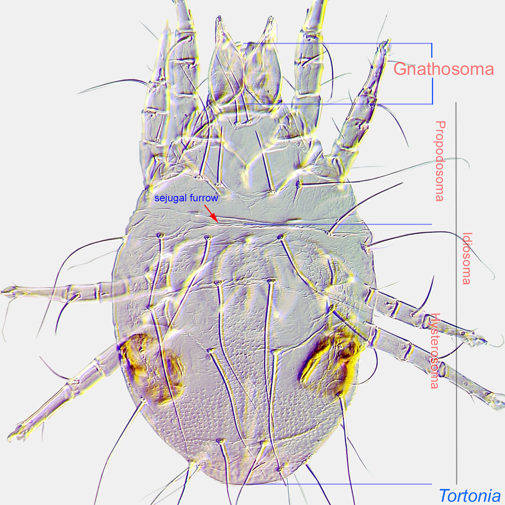
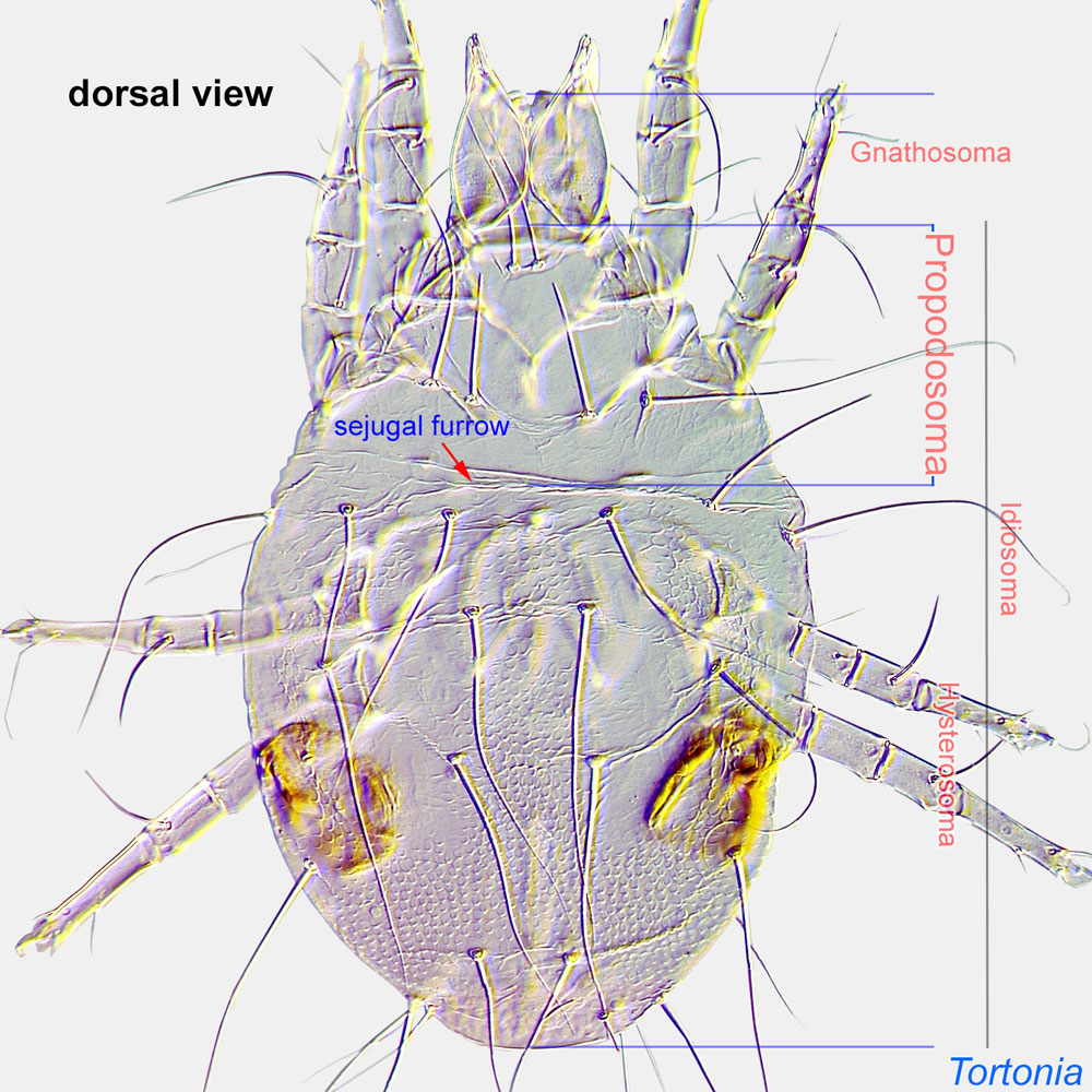 without a pair of brown pigmented areas (Fig. 6). Anterior edge of dorsal hysterosomahysterosoma:
Division of body posterior to the sejugal furrow, bearing legs III and IV.
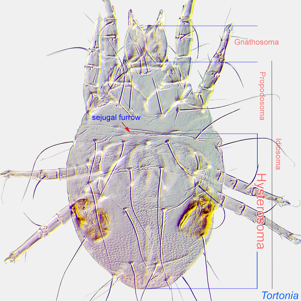 entire, smooth (not scalloped) (Fig. 1). Anterolateral region of hysterosomahysterosoma:
Division of body posterior to the sejugal furrow, bearing legs III and IV.
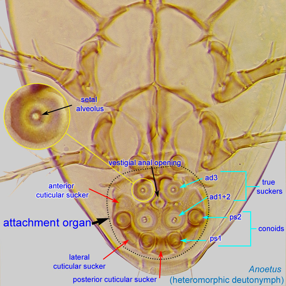
without lens-like organs (Fig. 1). Hysterosomal setae c1, d1 and e1 filiform (not lanceolate) (Fig. 1). Attachment organattachment organ:
Complex unpaired structure in phoretic deutonymphs of Astigmata situated on the posteroventral end of the body that serves for attachment to the host during phoresy. In deutonymphs phoretic on insects, attachment organ consists of a vestigial anal opening and two types of attachment elements: true suckers that create negative pressure and conoids that create adhesive forces. Not to be confused with pedicel.
 wider than long (Fig. 5). Coxal fields IV closed (Fig. 4).
Female: PretarsiPretarsus:
Terminal leg or palpal segment distal to tarsus.
with a bilobed membranous ambulacrumambulacrum:
The claws and empodium of the apotele or pretarsus.
and empodial clawempodial claw:
Claw-like, membranous, or pad-like structure of setal origin. Present only on the pretarsus in Acariformes. In Astigmata, it is the only claw on the pretarsus and often referred to simply as the claw. In the remaining Acariformes, may be accomanied by two lateral claws. Also known as empodium, pretarsal empodium, or central claw.
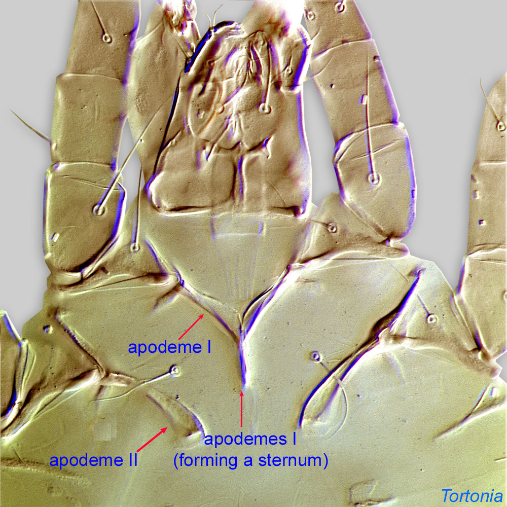
. Posterior rings adjacent to or directly anterior to the anus. Anterior apodemesapodeme:
Internal sclerite that serves as an attachment site for muscles. Most commonly used (as "coxal apodeme") to describe elements of coxae fused to the ventral body in Acariformes (coxae are free and not fused to the body in Parasitiformes), and may be variously referred to as ventral, sternal, anterior, or posterior.
 of coxaecoxa:
In Parasitiformes, most basal leg segment (or podomere) forming a joint with the body. Areas delimited by coxal apodemes are called coxal fields in Astigmata or coxisternal plates in Prostigmata.
I fused medially. Most dorsal setae elongate (c1 always elongate); setae spine-like, foliate or heavily barbed. Dorsal setae borne on individual tubercles.
Male: Paranal suckers absent. Aedeagusaedeagus:
An external organ of a male arthropod that is specialized to deliver sperm during copulation.
thin, usually short and often posteriorly directed. Without large spine-like setae at base of aedeagusaedeagus:
An external organ of a male arthropod that is specialized to deliver sperm during copulation.
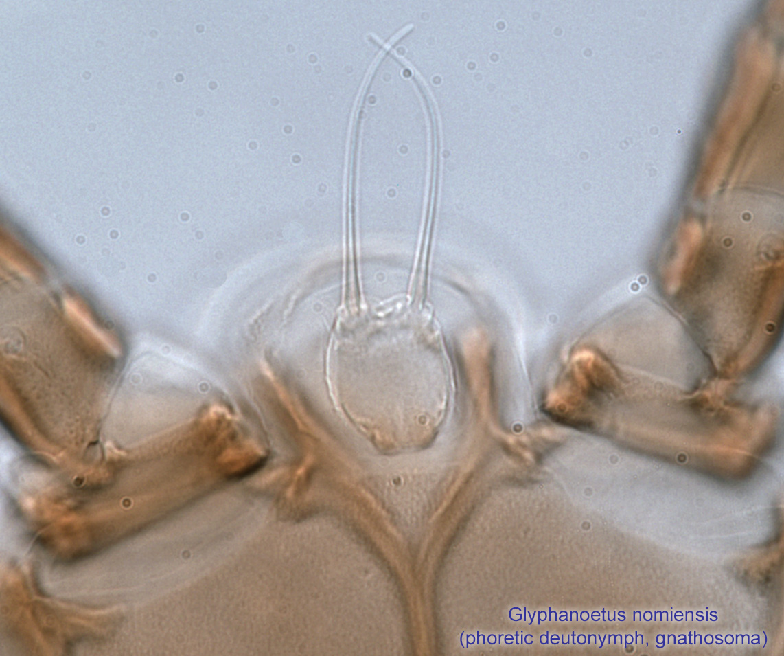
. With two pairs of genital "rings." Mouthparts and setation normal. External vertical setae long and spine-like, extending over the gnathosomagnathosoma:
Division of body anterior to the propodosoma bearing two pairs of appendages (palps and chelicerae).
 . Many dorsal setae long, often heavily barbed or blade-like.
A dichotomous key is not available. Most species of Glyphanoetus have been described from dung or dung beetles. The only species of Glyphanoetus described from bees can be identified using Cross, 1968Cross, 1968:
Cross, E. A. 1968. Descriptions of three new species of mites phoretic upon native bees. Southwestern Naturalist. 13: 325-334..
Palaearctic, Nearctic, Neotropical, and Afrotropical regions. Species associated with bees have been recorded in the Nearctic region.
Glyphanoetus nomiensis and undescribed species have been found in association with halictid bees of the genus Nomia.
permanent permanent:
associated exclusively with bees or their close relative, wasps; cannot live without these hosts
(Glyphanoetus nomiensis and related undescribed species)
 disperse on adult bees (males and females). They generally attach to the wings, but in male bees, they prefer "pockets" under sternite 5 (ST5).
Most species of Glyphanoetus are phoreticphoretic:
Pertaining to phoresy; using another organism (i.e., a host) for dispersal to new habitats. Phoresy can be distinguished from parasitism because feeding typically does not occur during phoresy.
on beetles (Coleoptera) and flies (Sphaeroceridae) that visit dung and related habitats. Observations on phoresyphoresy:
Attaching to or boarding another organism (i.e., a host) for dispersal to new habitats. Can be distinguished from parasitism because feeding typically does not occur.
of Glyphanoetus nomiensis associated with Nomia melanderi were reported in Cross and Bohart, 1969Cross and Bohart, 1969:
Cross, E. A. amp; G. E. Bohart. 1969. Phoretic behavior of four species of alkali bee mites as influenced by season and host sex. Journal of the Kansas Entomological Society, . 42 : 195-219.. The bees probably obtain their mites largely or entirely from the natal cell (i.e., mites do not migrate through the burrows or soil to find a phoreticphoretic:
Pertaining to phoresy; using another organism (i.e., a host) for dispersal to new habitats. Phoresy can be distinguished from parasitism because feeding typically does not occur during phoresy.
host). In general, the mites attach themselves to the upper surfaces of all four wings of both sexes, but they are often found in even larger numbers in the lateral depressions of the modified fifth metasomal sternite of males (lateral "pockets"). The largest mite load was detected in a male (62 deutonymphsdeutonymph:
Ontogenetic stage between protonymph and tritonymph (or adult, if tritonymph is absent). See <a href="index.cfm?pageID=1720">Life stages page</a> for more details., of which 32 were under sternite 5). Newly emerged female bees were found to have a maximum of 50 mites.
Immature and adult mites have been found on the mucous-covered body of the developing bee larvae. Their food source is not known, and they appear to cause little or no direct larval mortality by feeding (Cross, 1968Cross, 1968:
Cross, E. A. 1968. Descriptions of three new species of mites phoretic upon native bees. Southwestern Naturalist. 13: 325-334.).
Authors: P. Klimov, B. OConnor, R. Ochoa, G. Bauchan, A. Redford, J. Scher
Last updated October 2016
tool images at ITP Node
idtools.org