
Phytophthora kwongonina
|
Phytophthora spp. in subclade 6a: portion of the seven-loci ML phylogeny featuring the type cultures of 212 described species (by T. Bourret). Notice the position of P. kwongonina Ex-type CBS 143060 = S&T BL187. Gloria Abad, USDA S&T.
|
|
Phytophthora spp. in subclade 6a: Morphological Tabular key (PDF) and Tabular key legends (PDF) in IDphy2 KEY SECTION. Notice the data of P. kwongonina Ex-type CBS 143060 = S&T BL187. Gloria Abad, USDA S&T.
|
|
colony morphology after 5 d growth at 20ºC on carrot agar, V8 agar, malt extract agar, and potato-dextrose agar (from left to right) |
|
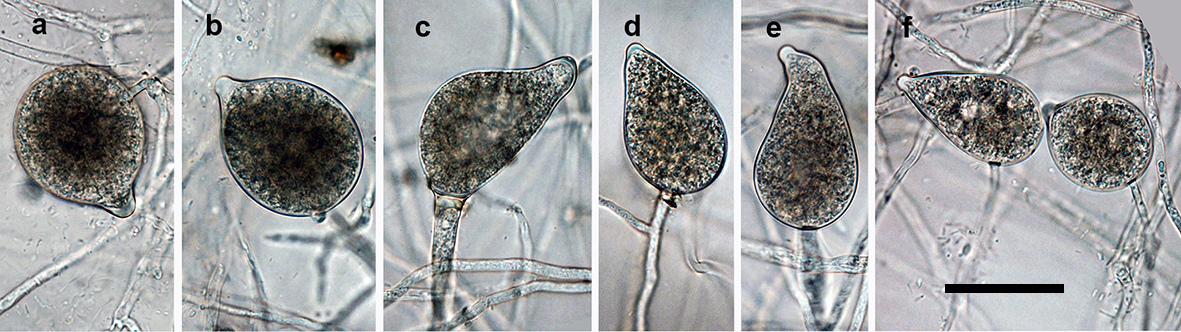
persistent, nonpapillate, predominantly ovoid to elongated ovoid sporangia with nested and extended internal proliferation; scale bar = 25µm |
|
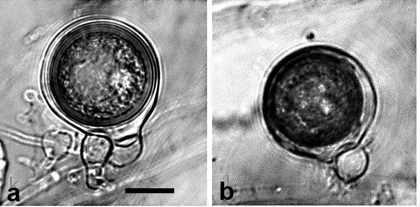
oogonia with wavy walls containing aplerotic oospores, with large ooplasts and thick walls which were pale on maturity; antheridia exclusively paragynous generally situated adjacent to the oogonial stalk; scale bar = 25µm |
|
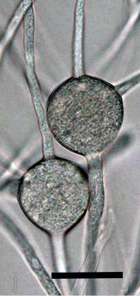
spherical hyphal swellings with radiating hyphae which appear like small chlamydospores except that the wall doesn’t form; scale bar = 25µm |
.JPG)
.JPG)

Name and publication
Phytophthora kwongonina T.I. Burgess (2018)
Burgess TI, Simamora AV, White D, Wiliams B, Schwager M, Stukely MJC, and Hardy GE StJ. 2018. New species from Phytophthora Clade clade:
a taxonomic group of organisms classified together on the basis of homologous features traced to a common ancestor
6a: evidence for recent radiation. Persoonia 41: 1–17.
Corresponding author: tburgess@murdoch.edu.au
Nomenclature
from Burgess et al. (2018)
Mycobank
Etymology
refers to association with the kwongon vegetation in the southwest of Western Australia
Typification
Type: AUSTRALIA, Western Australia, Bunbury, from rhizosphere soil of dying Banksia grandis, isolated by the VHS, 2010 (holotype MURU 477, dried culture on V8A, Herbarium of Murdoch University, Western Australia)
Ex-type: CBS 143060 and VHS 23298
Sequences for ex-type in original manuscript: Phytophthora kwongonina strain VHS 23298 = ITS JN547636, TUB MF326824, HSP MF326876, COX MF326847 and NADH MF326914
Ex-type in other collections
(ET) CBS 143060, VHS23298, S&T BL 187 (Abad)
Molecular identification
Voucher sequences for barcoding genes (ITS rDNA and COI) of the ex-type (see Molecular protocols page)
Phytophthora kwongonina isolate VHS 23298 ITS rDNA JN547636
Phytophthora kwongonina isolate CPHST BL 187 COI MH477747
Voucher sequences for Molecular Toolbox with seven genes (ITS, β-tub, COI, EF1α, HSP90, L10, and YPT1
(see Molecular protocols page) (In Progress)
Voucher sequences for Metabarcoding High-throughput Sequencing (HTS) Technologies [Molecular Operational Taxonomic Unit (MOTU)]
(see Molecular protocols page) (In Progress)
Sequences with multiple genes for ex-type in other sources
- NCBI: Phytophthora kwongonina CPHST BL 187
- NCBI: Phytophthora kwongonina VHS 23298
- EPPO-Q-bank: Phytophthora kwongonina
- BOLDSYSTEMS: Phytophthora kwongonina
Position in multigenic phylogeny with 7 genes (ITS, β-tub, COI, EF1α, HSP90, L10, and YPT1)
Clade clade:
a taxonomic group of organisms classified together on the basis of homologous features traced to a common ancestor
6a
Morphological identification
Colonies and cardinal temperatures
Colony colony:
assemblage of hyphae which usually develops form a single source and grows in a coordinated way
morphology on V8 agar, CA, and PDA was cottony with a slight petaloid pattern, growth was appressed with striations on MEA. Minimum growth temperature 4°C, optimum 25°C, and maximum 35°C.
Conditions for growth and sporulation
SporangiaSporangia:
sac within which zoospores form, especially when water is cooled to about 10°C below ambient temperature; in solid substrates, sporangia usually germinate by germ tubes
are produced in water cultures (soil extract or river water) and not observed in solid media. OogoniaOogonia:
the female gametangium in which the oospore forms after fertilization by the antheridium
are formed readily in single-strain culture on CA and V8 after about 14 d.
Asexual phase
SporangiaSporangia:
sac within which zoospores form, especially when water is cooled to about 10°C below ambient temperature; in solid substrates, sporangia usually germinate by germ tubes
were nonpapillatenonpapillate:
pertaining to the production of a non-distinct, or inconspicuous, papilla at the distal end of the sporangium (cf. papillate and semipapillate)
, persistentpersistent:
pertaining to sporangia that remain attached to the sporangiophore and do not separate or detach easily (cf. caducous)
, and predominantly ovoidovoid:
egg-shaped, with the widest part at the base of the sporangium and the narrow part at the apex
to elongated ovoidovoid:
egg-shaped, with the widest part at the base of the sporangium and the narrow part at the apex
in shape. SporangiaSporangia:
sac within which zoospores form, especially when water is cooled to about 10°C below ambient temperature; in solid substrates, sporangia usually germinate by germ tubes
averaged 57.5 ± 11.2 x 36.0 ± 6.9 µm (overall range 34.6–87.0 x 23.2–56.5 µm). Sporangiophores were simple with internal proliferationinternal proliferation:
internal proliferation occurs when the sporangiophore continues to grow through an empty sporangium
both nested and extended. Hyphal swellings were predominantly spherical, 12.2–46.4 µm in diameter. ChlamydosporesChlamydospores:
an asexual spore with a thickened inner wall that is delimited from the mycelium by a septum; may be terminal or intercalary, and survives for long periods in soil
absent.
Sexual phase
Homothallic. OogoniaOogonia:
the female gametangium in which the oospore forms after fertilization by the antheridium
had wavy walls and averaged 45.4 ± 3.4 mm (36.7–52.4 µm). OosporesOospores:
zygote or thick-walled spore that forms within the oogonium after fertilization by the antheridium; may be long-lived
were highly apleroticaplerotic:
pertaining to a mature oospore that does not fill the oogonium; i.e. there is room left between the oospore wall and oogonium wall (cf. plerotic)
, globoseglobose:
having a rounded form resembling that of a sphere
with very think walls, and pale on maturity, average size 37.1 ± 2.9 µm (31.9–44.1). AntheridiaAntheridia:
the male gametangium; a multinucleate, swollen hyphal tip affixed firmly to the wall of the female gametangium (the oogonium)
are paragynousparagynous:
pertaining to the sexual stage in which the antheridium is attached to the side of the oogonium (cf. amphigynous)
.
Most typical characters
Phytophthora kwongonina closely resembles P. rosacearum, P. pseudorosacerum, and P. cooljarloo. Its most distinguishing characteristic are the spherical hyphal swellings with radiating hyphaehyphae:
single, tubular filament of a fungal or oomycete thallus; the basic structural unit of a fungus or oomycete
which appear like small chlamydosporeschlamydospores:
an asexual spore with a thickened inner wall that is delimited from the mycelium by a septum; may be terminal or intercalary, and survives for long periods in soil
except that the wall doesn’t form.
Specimen(s) evaluated
Australia, Western Australia, Bunbury, from rhizosphere soil of dying Banksia grandis, isolated by the VHS, 2010, CBS 143060 = VHS 23298; Cervantes, from rhizosphere soil of dying Banksia prionotes, TC Hill, 1986, TCH009; Fitzgerald River National Park, from rhizosphere soil of dying Xanthorrhoea platyphylla, isolated by the VHS, 1993, DDS 3599
Hosts and distribution
NOT FOUND as of June 22, 2018 in U.S. National Fungus Collections Nomenclature Database.
Additional references and links
- SMML USDA-ARS: Phytophthora kwongonina
- EPPO Global Database: Phytophthora kwongonina
- Forest Phytophthoras of the world: Phytophthora kwongonina
- CABI Digital Library: Phytophthora kwongonina
- Encyclopedia of Life (EOL): Phytophthora kwongonina
- Index Fungorum (IF): Phytophthora kwongonina
- Google All Phytophthora kwongonina
- Google Images Phytophthora kwongonina
- Google Scholar Phytophthora kwongonina
Fact sheet authors
Treena Burgess, Ph.D., Phytophthora Science and Management, Harry Butler Institute, Murdoch University, Australia
Z. Gloria Abad, Ph.D., USDA-APHIS-PPQ-S&T Plant Pathogen Confirmatory Diagnostics Laboratory (PPCDL), United States of America.
