
Phytophthora kernoviae
|
Phytophthora spp. in subclade 10a: portion of the seven-loci ML phylogeny featuring the type cultures of 212 described species (by T. Bourret). Notice the position of P. kernoviae Ex-type NRRL 64375 = S&T BL 91. Gloria Abad, USDA S&T.
|
|
Phytophthora spp. in subclade 10a: Morphological Tabular key (PDF) and Tabular key legends (PDF) in IDphy2 KEY SECTION. Notice the data of P. kernoviae Ex-type NRRL 64375 = S&T BL 91. Gloria Abad, USDA S&T.
|
|
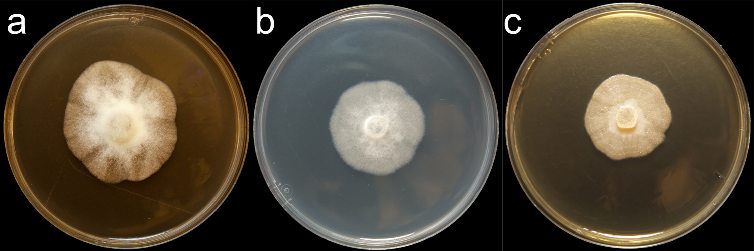
Phytophthora kernoviae (CPHST BL 91) colonies of the ex-type grown for 7 days on (a) V8® Agar, (b) potato dextrose agar, and (c) malt extract agar; photo by Krysta Jennings and Leandra Knight, USDA-APHIS-PPQ |
|
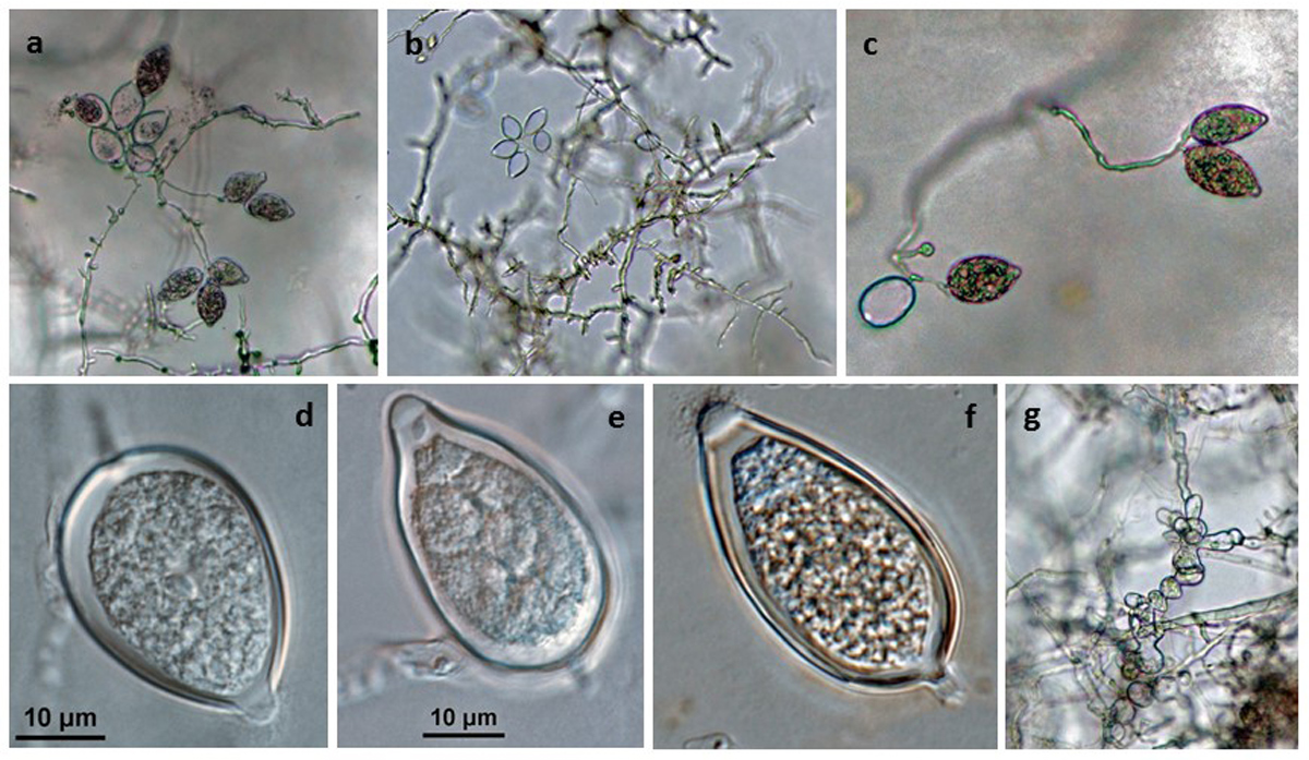
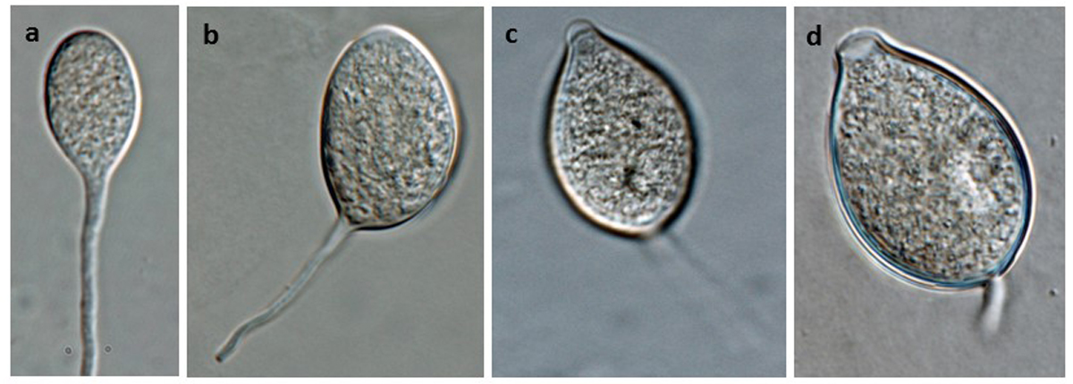
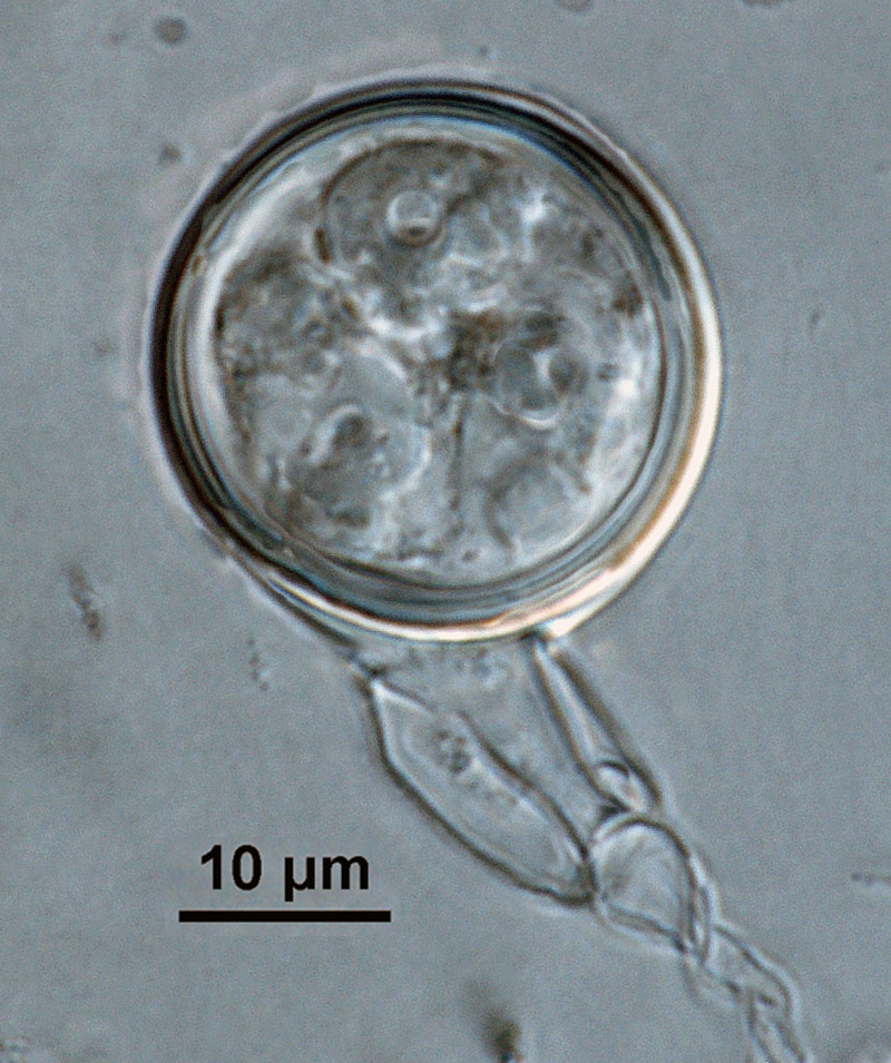
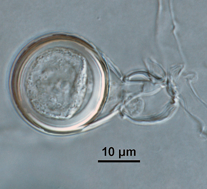
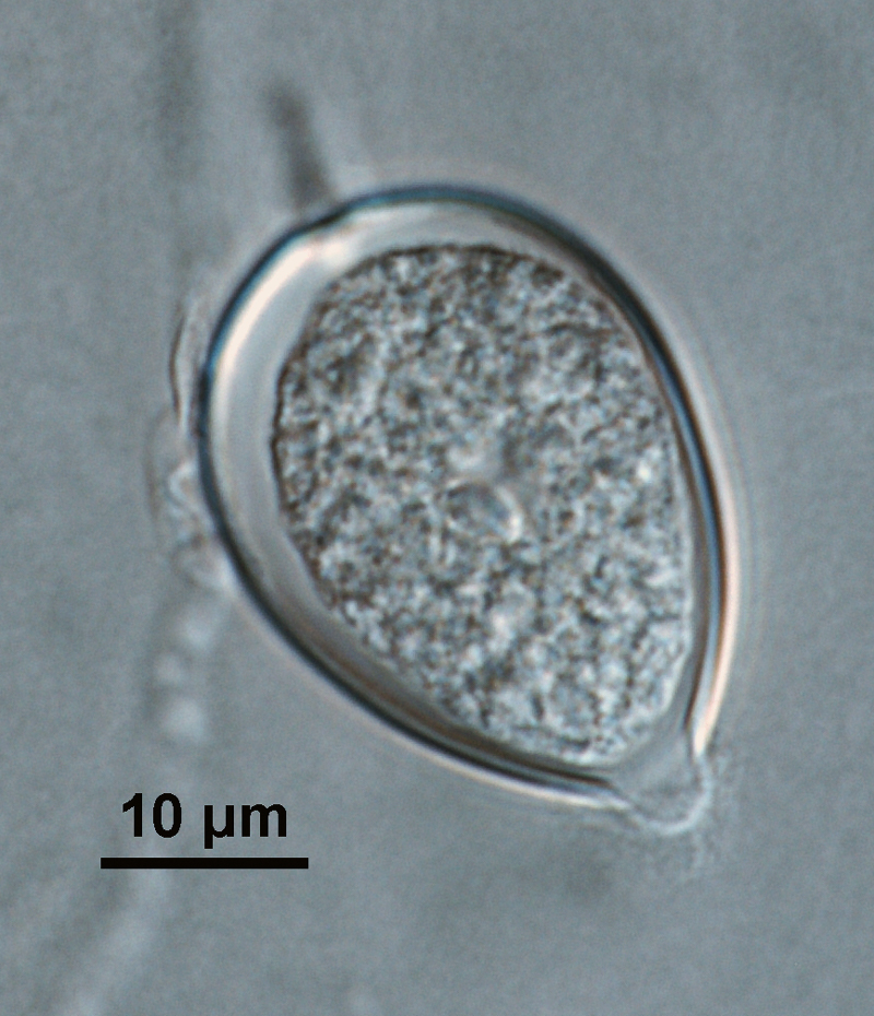
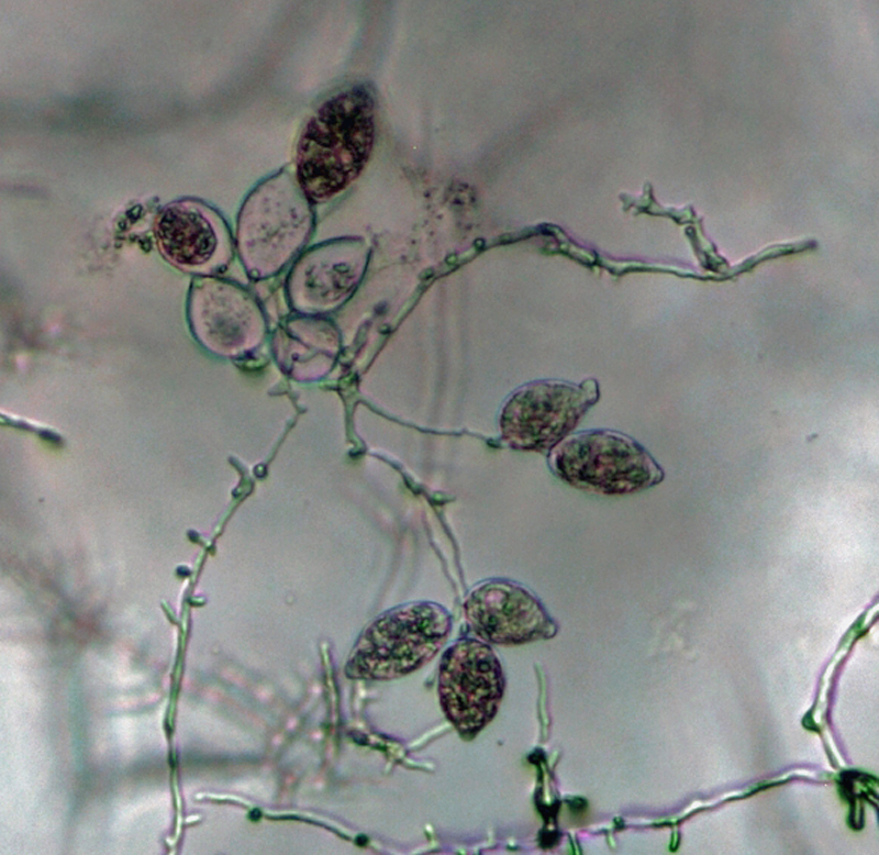
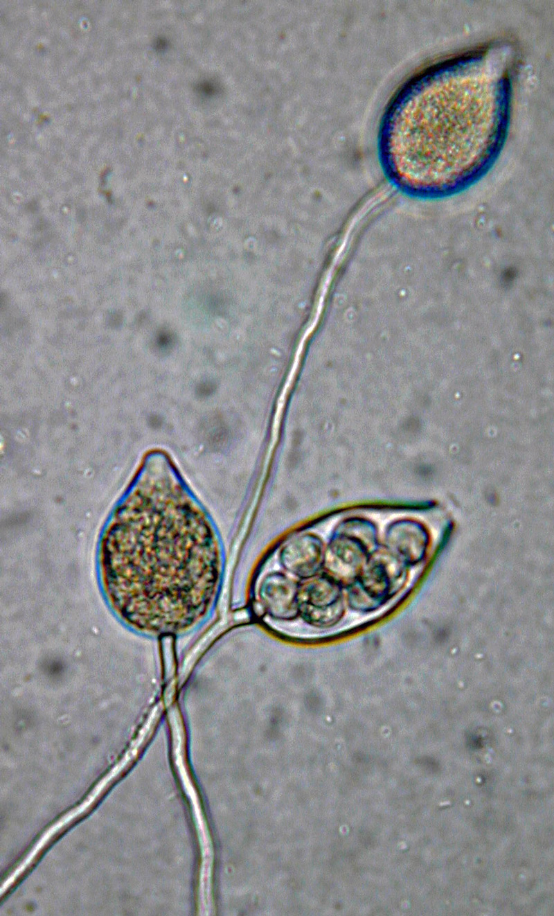
Phytophthora kernoviae (CPHST BL 91 ex-type) asexual phase: (a–c) sporangia produced in simple sympodial sporangiophores, (d–f) papillate sporangia with medium-length pedicels formed on V8 agar flooded with soil extract, (d) ovoid, (e) obpyriform, and (f) ellipsoid shapes, (g) coralloid/toruloid hyphal swellings; photos by Gloria Abad, USDA-APHIS-PPQ. |
|
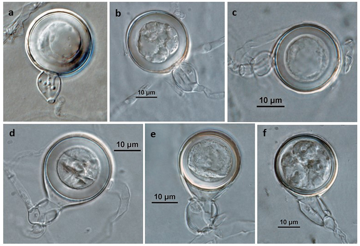
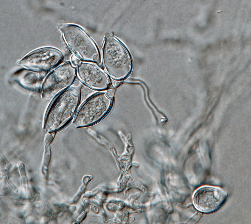
Phytophthora kernoviae (CPHST BL 91, ex-type) sexual phase: (a–f) oogonia smooth-walled, (d–f) with tapered base; (a–f) antheridia amphigynous (different shapes); oospores plerotic with thick walls, except (f); photos by Gloria Abad, USDA-APHIS-PPQ. |
|
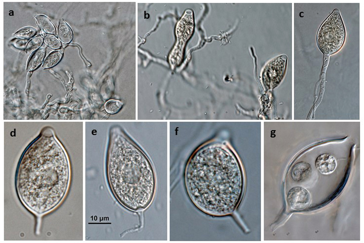
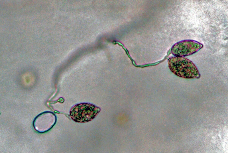
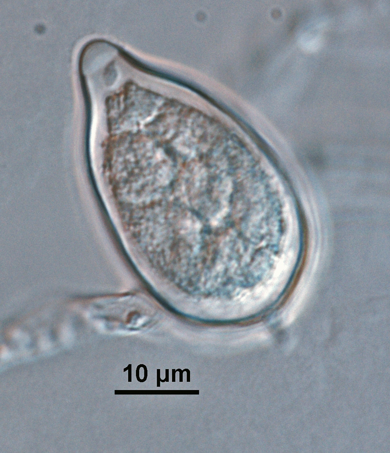
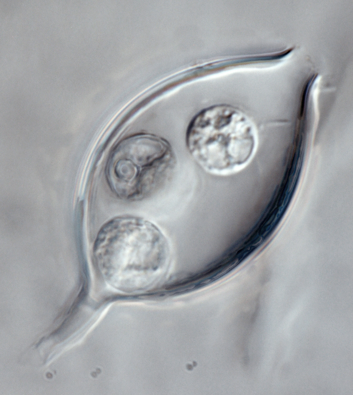
Phytophthora kernoviae (selected specimen P10681) asexual phase: (a–c) sporangia produced in (a) close simple sympodial and (b, c) unbranched sporangiophores; (d–f) papillate sporangia with medium-length pedicels formed on V8 agar flooded with soil extract; (d) ovoid, (e) ellipsoid with tapered top, (f) irregular shape; (g) zoospores in sporangium; photos by Gloria Abad, USDA-APHIS-PPQ. |
|
Phytophthora kernoviae (selected specimen P10956): colony shape on (a) V8 agar, (b) potato dextrose agar; (c, d) sporangia born in sporangiophores; (e–f) papillated sporangia with pedicels (e) short, (f) medium-length; (g–e) sexual phase with smooth-walled oogonia with amphigynous antheridia, (g) thick-walled plerotic oospore, (e) medium thick-walled plerotic oospore; photos by Gloria Abad, USDA-APHIS-PPQ |
|
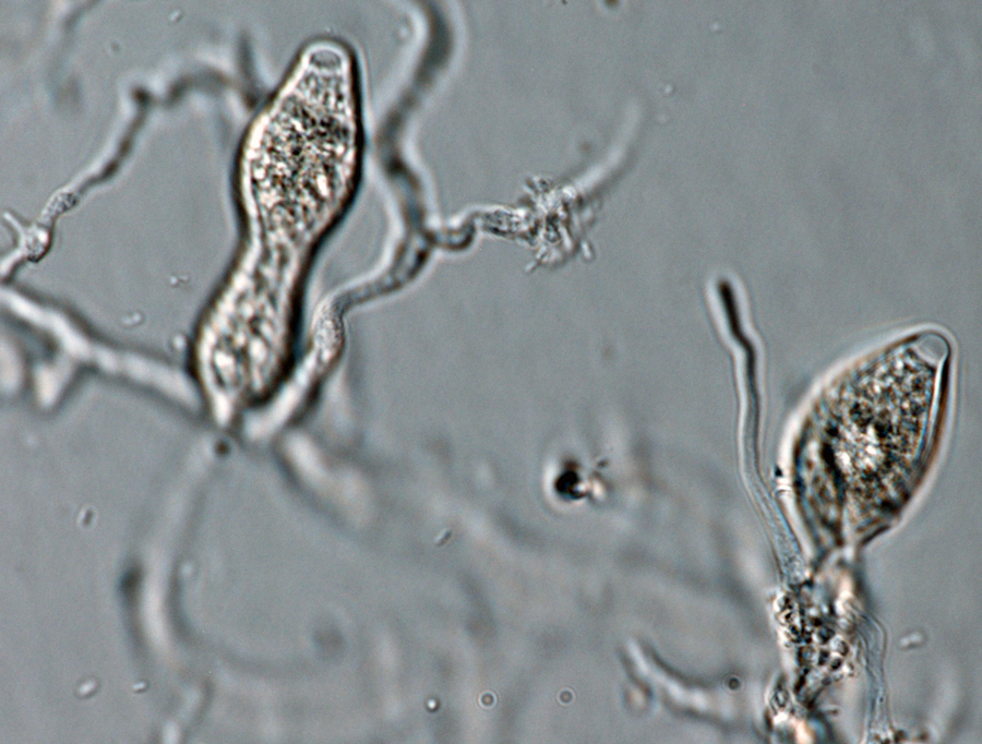
Phytophthora kernoviae (selected specimen P10957) asexual phase: (a) sporangia produced in unbranched sporangiophore; (b, c) sporangia with long pedicels formed on V8 agar flooded with soil extract; (d) sporangium with medium pedicel; photos by Gloria Abad, USDA-APHIS-PPQ. |
|
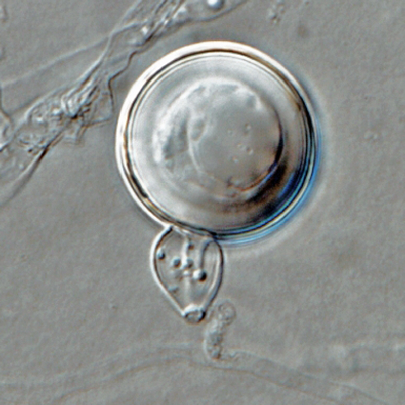
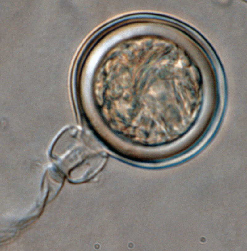
Phytophthora kernoviae (CPHST BL 91, ex-type) sexual phase: smooth-walled oogonium, antheridium amphigynous,and plerotic oospore with thick walls; photo by Gloria Abad, USDA-APHIS-PPQ. |
|
Phytophthora kernoviae (CPHST BL 91 ex-type) asexual phase: coralloid/toruloid hyphal swellings; photo by Gloria Abad, USDA-APHIS-PPQ. |
|
Phytophthora kernoviae (CPHST BL 91, ex-type) sexual phase: smooth-walled oogonium, antheridium amphigynous,and plerotic oospore with thick walls; photo by Gloria Abad, USDA-APHIS-PPQ. |
|
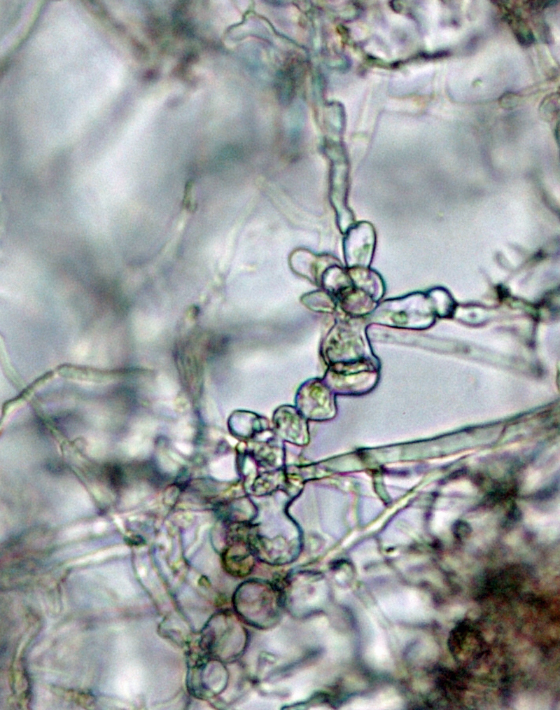
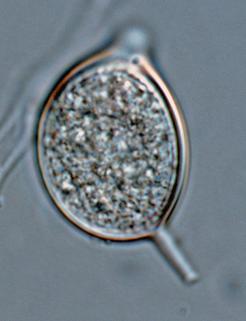
Phytophthora kernoviae (CPHST BL 91 ex-type) asexual phase: sporangia produced in simple sympodial sporangiophores; photo by Gloria Abad, USDA-APHIS-PPQ. |
|
Phytophthora kernoviae (CPHST BL 91, ex-type) sexual phase: smooth-walled oogonium, antheridium amphigynous,and plerotic oospore with thick walls; photo by Gloria Abad, USDA-APHIS-PPQ. |
|
Phytophthora kernoviae (CPHST BL 91, ex-type) sexual phase: smooth-walled oogonium with tapered bases, antheridium amphigynous,and plerotic oospore with thick walls; photo by Gloria Abad, USDA-APHIS-PPQ. |
|
Phytophthora kernoviae (CPHST BL 91, ex-type) sexual phase: smooth-walled oogonium with tapered bases, antheridium amphigynous,and plerotic oospore with thick walls; photo by Gloria Abad, USDA-APHIS-PPQ. |
|
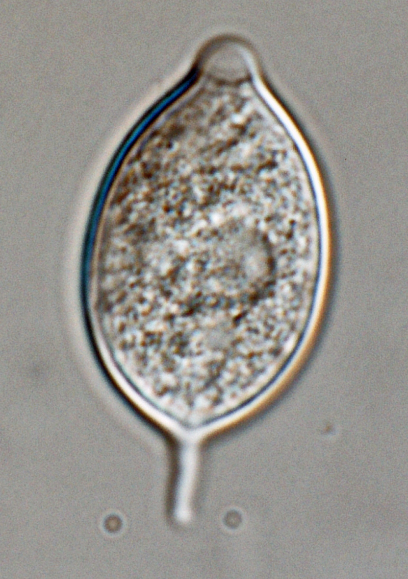
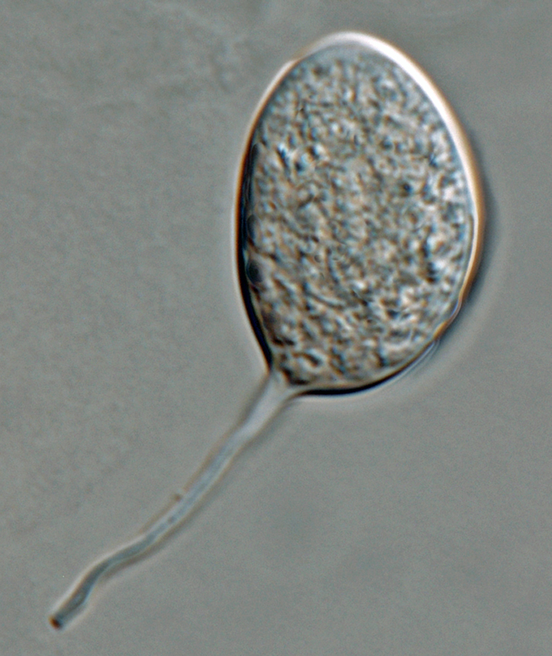
Phytophthora kernoviae (CPHST BL 91 ex-type) asexual phase: papillate ovoid sporangium with medium-length pedicel formed on V8 agar flooded with soil extract; photo by Gloria Abad, USDA-APHIS-PPQ. |
|
Phytophthora kernoviae (CPHST BL 91 ex-type) asexual phase: papillate obpyriform sporangium with medium-length pedicel formed on V8 agar flooded with soil extract; photo by Gloria Abad, USDA-APHIS-PPQ. |
|
Phytophthora kernoviae (CPHST BL 91 ex-type) asexual phase: papillate ellipsoid sporangium with medium-length pedicel formed on V8 agar flooded with soil extract; photo by Gloria Abad, USDA-APHIS-PPQ. |
|
Phytophthora kernoviae (CPHST BL 91 ex-type) asexual phase: sporangia produced in simple sympodial sporangiophores; photo by Gloria Abad, USDA-APHIS-PPQ. |
|
Phytophthora kernoviae (CPHST BL 91 ex-type) asexual phase: sporangia produced in simple sympodial sporangiophores; photo by Gloria Abad, USDA-APHIS-PPQ. |
|
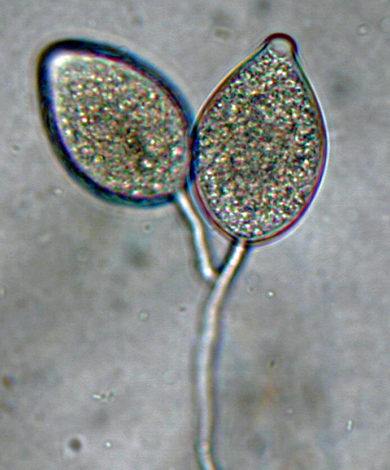
Phytophthora kernoviae (selected specimen P10681) asexual phase: sporangia produced in close simple sympodial sporangiophores; photo by Gloria Abad, USDA-APHIS-PPQ. |
|
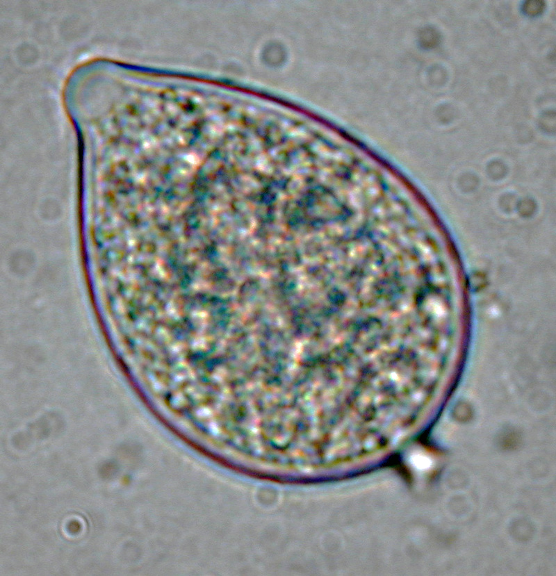
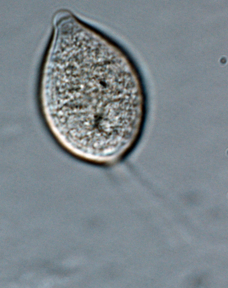
Phytophthora kernoviae (selected specimen P10681) asexual phase: papillate ellipsoid sporangium with tapered top and medium-length pedicel, formed on V8 agar flooded with soil extract; photo by Gloria Abad, USDA-APHIS-PPQ. |
|
Phytophthora kernoviae (selected specimen P10681) asexual phase: zoospores in sporangium; photo by Gloria Abad, USDA-APHIS-PPQ. |
|
Phytophthora kernoviae (selected specimen P10681) asexual phase: papillate sporangia produced in unbranched sporangiophores; photo by Gloria Abad, USDA-APHIS-PPQ. |
|
Phytophthora kernoviae (selected specimen P10681) asexual phase: papillate ovoid sporangium with medium-length pedicel, formed on V8 agar flooded with soil extract; photo by Gloria Abad, USDA-APHIS-PPQ. |
|
Phytophthora kernoviae (selected specimen P10681) asexual phase: sporangium borne in simple sympodial sporangiophore; photo by Gloria Abad, USDA-APHIS-PPQ. |
|
Phytophthora kernoviae (selected specimen P10681) asexual phase: papillate irregular shaped sporangium with medium-length pedicel, formed on V8 agar flooded with soil extract; photo by Gloria Abad, USDA-APHIS-PPQ. |
|
Phytophthora kernoviae (selected specimen P10956) asexual phase: papillate sporangium with short pedicel; photo by Gloria Abad, USDA-APHIS-PPQ. |
|
Phytophthora kernoviae (selected specimen P10956) asexual phase: papillate sporangia borne in simple sympodial sporangiophore; photo by Gloria Abad, USDA-APHIS-PPQ. |
|
Phytophthora kernoviae (selected specimen P10956) sexual phase: medium thick-walled plerotic oospore smooth-walled oogonium and amphigynous antheridium; photo by Gloria Abad, USDA-APHIS-PPQ. |
|
Phytophthora kernoviae (selected specimen P10956) sexual phase: smooth-walled oogonium with amphigynous antheridium; photo by Gloria Abad, USDA-APHIS-PPQ. |
|
Phytophthora kernoviae (selected specimen P10956) asexual phase: papillate sporangium with short pedicel; photo by Gloria Abad, USDA-APHIS-PPQ. |
|
Phytophthora kernoviae (selected specimen P10956) asexual phase: papillate sporangia borne in simple sympodial sporangiophore; photo by Gloria Abad, USDA-APHIS-PPQ. |
|
Phytophthora kernoviae (selected specimen P10957) asexual phase: sporangium produced in unbranched sporangiophore; photo by Gloria Abad, USDA-APHIS-PPQ. |
|
Phytophthora kernoviae (selected specimen P10957) asexual phase: sporangium with long pedicel formed on V8 agar flooded with soil extract; photo by Gloria Abad, USDA-APHIS-PPQ. |
|
Phytophthora kernoviae (selected specimen P10957) asexual phase: sporangium with long pedicel formed on V8 agar flooded with soil extract; photo by Gloria Abad, USDA-APHIS-PPQ. |
|
Phytophthora kernoviae (selected specimen P10957) asexual phase: sporangium with long pedicel formed on V8 agar flooded with soil extract; photo by Gloria Abad, USDA-APHIS-PPQ. |
.JPG)
.JPG)


Name and publication
Phytophthora kernoviae Brasier, Beales & S.A. Kirk (2005)
Brasier CM, Beales PA, Kirk SA, Denman S, and Rose J. 2005. Phytophthora kernoviae sp. nov., an invasive pathogen causing bleeding stem lesions on forest trees and foliar necrosis of ornamentals in the UK. Mycol. Res. 109: 853–859.
Corresponding author: clive.brasier@forestry.gsi.gov.uk
previously known informally as Phytophthora taxon C (PtC)
Nomenclature
from Brasier et al. (2005)
Mycobank
Etymology
from ‘Kernow’, the Cornish noun for Cornwall
Typification
Type: UNITED KINGDOM, Cornwall, Caerhays, isolate from bark of Fagus sylvatica, Jan. 2004, C. M. Brasier, IMI 393170 – holotypus, dried culture on carrot agar; previously Forestry Commission Phytophthora culture collection isolate P1571
Ex-type: IMI 393170
NOTE: IMI 393170 indicates that a culture is available from CABI Collection, however a culture of IMI 393170 is not available from CABI.
Sequences for ex-type in original manuscript: Phytophthora kernoviae isolate P1571 (FCPCC) = ITS AY940661
Ex-type in other collections
(ET) NRRL 64375, CABI IMI393170 (AVA), P1571 (FCPCC), WPC P19827, S&T BL 91 (Abad), CAE4
Molecular identification
Voucher sequences for barcoding genes (ITS rDNA and COI) of the ex-type (see Molecular protocols page)
Phytophthora kernoviae isolate CPHST BL 91 (= P19827 WPC) = ITS rDNA MG865521, COI MH136915
Voucher sequences for Molecular Toolbox with seven genes (ITS, β-tub, COI, EF1α, HSP90, L10, and YPT1
(see Molecular protocols page) (In Progress)
Voucher sequences for Metabarcoding High-throughput Sequencing (HTS) Technologies [Molecular Operational Taxonomic Unit (MOTU)]
(see Molecular protocols page) (In Progress)
Sequences with multiple genes for ex-type in other sources
- NCBI: Phytophthora kernoviae CPHST BL 91
- NCBI: Phytophthora kernoviae P19827
- NCBI: Phytophthora kernoviae P1571 (FCPCC)
- EPPO-Q-bank: Phytophthora kernoviae
- BOLDSYSTEMS: Phytophthora kernoviae (barcoding COI & ITS)
Position in multigenic phylogeny with 7 genes (ITS, β-tub, COI, EF1α, HSP90, L10, and YPT1)
Clade 10a
Genome sequence
Phytophthora kernoviae strain ex-type P19827. Accession genome CPHST-BL-91 reference, BioProject PRJNA554119, USDA-APHIS-PPQ-S&T (2019), Srivastava et al. 2020
Morphological identification
adapted from Brasier et al. (2005)
Colonies and cardinal temperatures
Colony colony:
assemblage of hyphae which usually develops form a single source and grows in a coordinated way
morphology after 7 days of growth on potato dextrose agar, malt extract agar, and V8 agar with light rosette pattern. Minimum temperature for growth is 3°C, optimum 21°C, and maximum 24°C.
Conditions for growth and sporulation
Sporangia and oosporesoospores:
zygote or thick-walled spore that forms within the oogonium after fertilization by the antheridium; may be long-lived
formed in V8 cultures flooded in 10% soil solution.
Asexual phase
SporangiaSporangia:
sac within which zoospores form, especially when water is cooled to about 10°C below ambient temperature; in solid substrates, sporangia usually germinate by germ tubes
papillatepapillate:
pertaining to the production of a distinct papilla at the distal end of the sporangium (cf. nonpapillate and semipapillate)
; caducouscaducous:
pertaining to sporangia that become dislodged readily (i.e. deciduous) and separate from the sporangiophore (cf. persistent)
with medium to long pedicels (3–28 µm long); ellipsoidellipsoid:
refers to a solid body that forms an ellipse in the longitudinal plane and a circle in cross section; many fungal spores are ellipsoidal or elliptic
, ovoidovoid:
egg-shaped, with the widest part at the base of the sporangium and the narrow part at the apex
, obpyriformobpyriform:
inversely pear-shaped, i.e. with the widest part at the point of attachment (cf. pyriform)
, limoniform (19–62 L x 12–31 W µm), mouse-shaped, some with irregular shapes and some with tapered bases; most with conspicuous vacuole; originated in simple sympodial or unbranched sporangiophores. Hyphal swellings coralloid/toruloid occasionally produced. ChlamydosporesChlamydospores:
an asexual spore with a thickened inner wall that is delimited from the mycelium by a septum; may be terminal or intercalary, and survives for long periods in soil
absent.
Sexual phase
Homothallic. OogoniaOogonia:
the female gametangium in which the oospore forms after fertilization by the antheridium
smooth-walled, often with tapered basetapered base:
pertaining to the base of a sporangium or oogonium; funnel-shaped
(22–30 µm diam); antheridia amphigynous; oosporesoospores:
zygote or thick-walled spore that forms within the oogonium after fertilization by the antheridium; may be long-lived
spherical (18–28 µm diam) pleroticplerotic:
pertaining to an oospore that fills the oogonium (cf. aplerotic)
to slightly apleroticaplerotic:
pertaining to a mature oospore that does not fill the oogonium; i.e. there is room left between the oospore wall and oogonium wall (cf. plerotic)
.
Most typical characters
Phytophthora kernoviae is characterized by the presence of sporangiasporangia:
sac within which zoospores form, especially when water is cooled to about 10°C below ambient temperature; in solid substrates, sporangia usually germinate by germ tubes
with caducouscaducous:
pertaining to sporangia that become dislodged readily (i.e. deciduous) and separate from the sporangiophore (cf. persistent)
medium to long pedicelpedicel:
the hyphal base of a sporangium that remains attached after the sporangium separates, or is shed, from the sporangiophore; the pedicel may be short (< 5 µm), medium (5–20 µm), or long (> 20 µm)
and by the presence of young oosporesoospores:
zygote or thick-walled spore that forms within the oogonium after fertilization by the antheridium; may be long-lived
with thick walls.
Additional specimen(s) evaluated
Phytophthora kernoviae, ex-type CPHST BL 91, duplicate of P19827 (World Phytophthora Collection), which is a duplicate of ex-type IMI 393170
Hosts and distribution
Notes: previously reported as Phytophthora taxon C
Distribution: Europe (UK); New Zealand, South America (Chile)
Substrate: bark, leaves, shoots
Disease note: on beech, associated with bark necrosis and canker and bleeding stem lesions, especially on aerial stems; pathogenicity confirmed by inoculation studies; also associated with shoot dieback, foliar necroses, and wilting of rhododendron
Host: Fagus sylvatica (beech), Quercus robur (oak), (Fagaceae); also Liriodendron tulipifera (Magnoliaceae), Rhododendron ponticum (Ericaceae), Pinus radiata (Pinaceae)
Retrieved January 31, 2018 from U.S. National Fungus Collections Nomenclature Database.
Quarantine status
USA: Phytophthora kernoviae is listed in the National Cooperative Agricultural Pest Survey (CAPS) Priority Pest List for 2017 and 2018.
This species was listed as a species of concern during the 2009 Phytophthora prioritization project conducted by USDA APHIS PPQ CPHST PERAL (Schwartzburg et al.).
EPPO: organisms previously included in the EPPO Alert List, Phytophthora kernoviae
Additional references and links
- SMML USDA-ARS: Phytophthora kernoviae
- EPPO Global Database: Phytophthora kernoviae
- Forest Phytophthoras of the world: Phytophthora kernoviae
- CABI Digital Library: Phytophthora kernoviae
- Forest Research UK: Phytophthora kernoviae
- Encyclopedia of Life (EOL): Phytophthora kernoviae
- Index Fungorum (IF): Phytophthora kernoviae
- Google All Phytophthora kernoviae
- Google Images Phytophthora kernoviae
- Google Scholar Phytophthora kernoviae
Fact sheet author
Z. Gloria Abad, Ph.D., USDA-APHIS-PPQ-S&T Plant Pathogen Confirmatory Diagnostics Laboratory (PPCDL), United States of America.
