

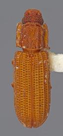
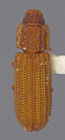
Antennaeantennae:
paired, segmental appendages, borne one on each side of head, functioning as sense organs and bearing a large number of sensilla.
11-segmented with a distinct, 2-segmented club. Antennal setation sparse. Subantennal groovessubantennal groove:
groove or concavity lying below the antennal insertion and housing the base of the antenna. Placed between the eye (if present) and the mandibular articulation, and sometimes extends below or behind the eye.
long, reaching posteriorposterior:
hinder or hindmost, opposed to anterior; hind or rear.
margin of eye. Eyes elongate, well-developed, facets fine. Pronotal discpronotal disc:
the area of the pronotum which is visible dorsally and usually delimited laterally by the two lateral carinae. Contrasted with the paired pronotal hypomera, which extend onto the ventral surface.
with network of connecting, bifurcating carinaecarina:
an elevated ridge or keel, not necessarily high or acute.
. Pronotal lateral margins subparallel, minutely serrateserrate:
sawlike, i.e., with notched edges like the teeth of a saw.
, slightly explanateexplanate:
spread out and flattened; applied to a margin.
. Procoxal cavitiesprocoxal cavities:
external closure: Externally closed when the postcoxal processes of the hypomera meet the prosternal process or meet one another.
narrowly open. Metacoxaemetacoxae:
the coxae of the metathorax.
narrowly separated, separation less than metacoxal length. Elytraelytron:
the fore wing in Coleoptera, which is more or less uniformly sclerotized and in resting position is longitudinally oriented, usually meeting the opposite elytron along the midline.
carinate, with 9 rows of regularly spaced, deep puncturespuncture:
a small impression on the cuticle, like that made by a needle.
. Tarsal formulatarsal formula:
the number of tarsomeres on the fore, mid, and hind tarsi, respectively.
4–4–4. Dorsal surface with minute setaeseta:
a sclerotized, hairlike (or scalelike) projection of cuticula arising from a single trichogen cell and surrounded at the base by a small cuticular ring.
.
The genus Microprius is extremely similar to Bitoma and seems to differ only by the length of antennal groove on the ventral side of the head (short to absent in Bitoma).
Microprius rufulus (Motschulsky, 1863Motschulsky, 1863:
Motschulsky, V. von. 1863. Essai d'un catalogue des insectes de l'ille Ceylan. Bulletin de la Societe Imperiale des Naturalistes de Moscou, 36: 421-532.)
Southwestern (CA), Northeastern (VA) USA.
This species is widespread throughout the Old World and will likely be found throughout the United States.
Microprius rufulus has been found at UV/MV light and from under the bark of a number of trees.
Abundance: Rare.
Ivie (2002a)Ivie (2002a):
Ivie, M.A. 2002a. 127. Colydiidae, pp. 445-453 In: R. H. Arnett, Jr., Jr. and M. C. Thomas (eds.), American Beetles. CRC Press, Gainesville, Florida., Ivie et al. (2001b)Ivie et al. (2001b):
Ivie, M.A., S.A. Ślipiński and P. Wegrzynowicz. 2001b. New records and synonyms in the colydiinae. Insecta Mundi, 15 (3): 185-188., Motschulsky (1863)Motschulsky (1863):
Motschulsky, V. von. 1863. Essai d'un catalogue des insectes de l'ille Ceylan. Bulletin de la Societe Imperiale des Naturalistes de Moscou, 36: 421-532.
Authors: Lord NP, Nearns EH, Miller KB. 2011–2015.
Content updated Apr 2015; taxonomy updated Feb 2026
idtools.org
