Larval Morphology Overview
Most lepidopteran larvae, including all of the pest species treated here, are characterized by a combination of the following characters (from Stehr 1987):
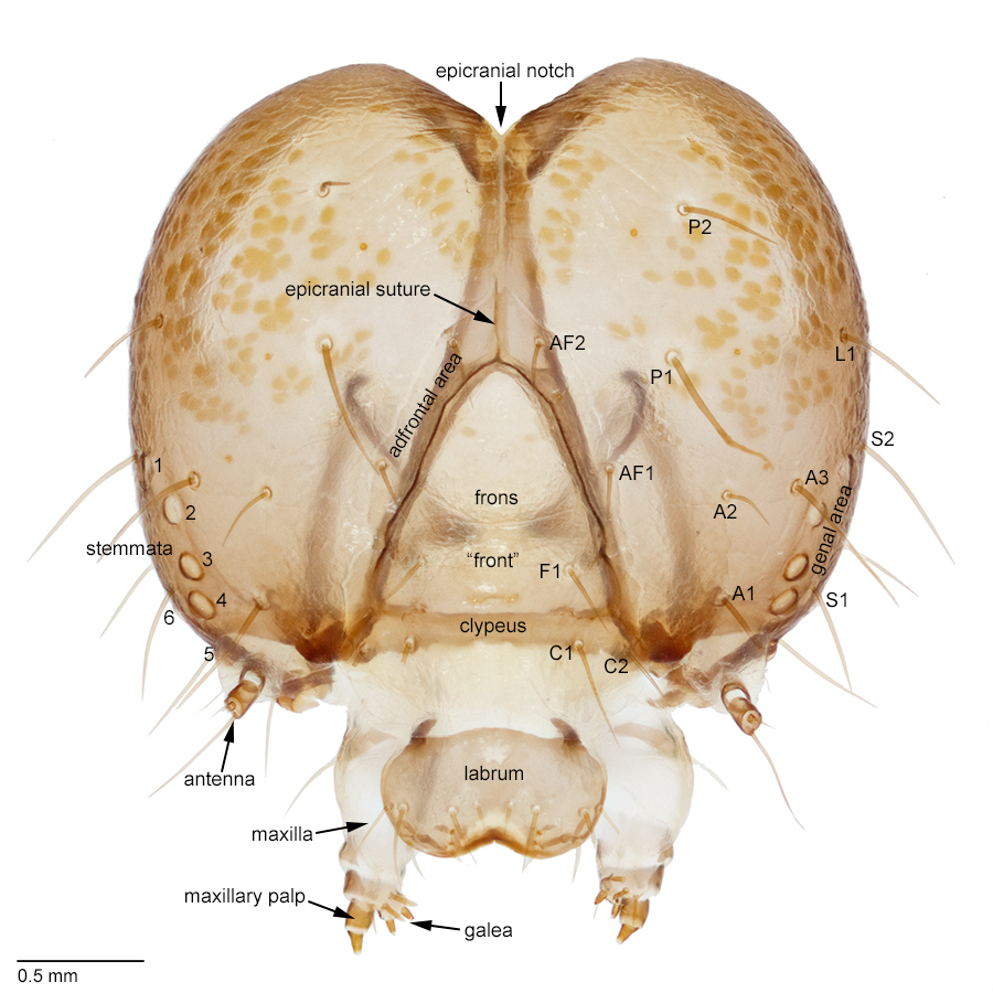
a distinct head; chewing mouthparts; one pair of antennae; six pair of stemmata; adfrontal areas; a labial spinneret; three pairs of thoracic legs;
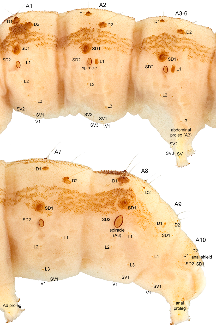
ten abdominal segments; four pairs of abdominal prolegs with crochets on segments A3-6
(reduced in some species); one pair of anal prolegs with crochets on segment A10; and spiracles on the prothorax and abdominal segments A1-8.
Setae are named according to Stehr (1987) but not all authors use the same setal homologies and the application of these names can vary slightly between
families. Two special structures used in keys are worth mentioning. The frons and clypeus are often called the "front." Because of their special
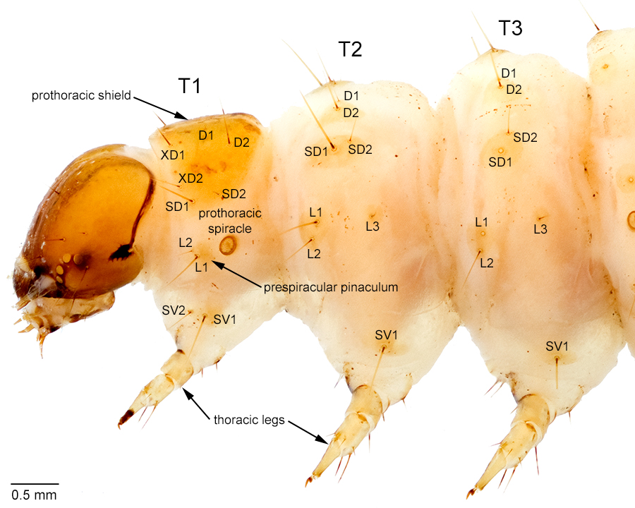
significance in identification, the L setae on the prothorax are called the prespiracular group. Their pinaculum, if present, is called the prespiracular pinaculum.
Click on the tabs at the top of this page to view detailed labeled diagrams for the head, hypopharyngeal complex, mandibles, thorax, abdomen,
and crochets. Methods for preserving and studying Lepidoptera larvae are listed on the About page under "Materials and Methods."
For more information on Lepidoptera larval morphology and study methods, consult Stehr (1987) or Peterson (1948).
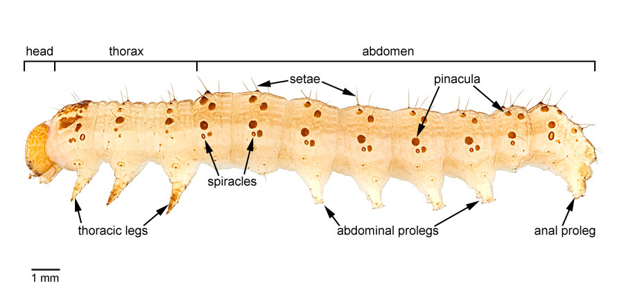
Lateral view of H. zea larva with body regions and major structures labeled.
Preserving and Studying Larvae
The following information on preserving and examining larvae is mostly repeated from the following book chapter focused on tortricids, although the techniques
can be applied to nearly any lepidopteran larva.
Passoa, S. C. 2008. Part III: Immature Stages, pp. 295-314. In: T. M. Gilligan et al. (eds.), Olethreutine moths of the midwestern United States, an identification guide.
Ohio Biological Survey, Columbus, Ohio.
Preservation techniques
Methods of preserving larvae are discussed by Peterson (1962), Winter (2000), Wagner (2005), Dugdale et al. (2005), and in detail by Stehr (1987). The objective is to fix the
tissues to prevent discoloration or rotting of samples. This can be accomplished with heat or with chemical fixing agents.
For the amateur collector, or if another preservative such as alcohol is unavilable, the simplest method for killing larvae is to drop them in
household vinegar (5% acetic acid) for 24 hours. Specimens are then transferred to alcohol for permanent preservation. Although the results are far
from ideal, this method has the advantages that vinegar is cheap, nonflammable, relatively nontoxic, and may be readily available in remote locations.
The remainder of this section discusses techniques appropriate for research purposes.
Larvae can be killed and heat-fixed by dropping them in very hot or gently boiling water, a process that causes the body to expand and become rigid.
The resulting body distention makes the crochets easy to see and prevents folds of cuticle from obscuring body setae. However, bloated larvae are difficult to
manipulate and can be punctured by forceps. Light colors tend to be poorly preserved; the process causes most individuals to bleach bright white. Dark
markings are unaffected.
Chemical killing agents and fixatives usually involve a mixture of alcohol and some organic solvent or acid. This approach allows the larva to be slightly
bent or twisted without repositioning the sample. Color preservation tends to be better than with boiling, but crochet patterns are often difficult to see
unless the larva is inflated. Perhaps the biggest disadvantage of this method is that toxic acids and organic solvents require a fume hood for use and storage.
Godfrey (1972) used a mixture of nine parts 70% ethanol and one part glacial acetic acid by volume. The specimen can be injected through the mouth or anus
using a diabetes syringe to inflate the larva as needed. In this process, use of protective eyewear is essential as specimens can burst, squirting the acid
alcohol mixture in all directions. Dugdale et al. (2005) mentions an alternative chemical fixative called Carnoy's solution, which contains chloroform as
well as the alcohol-acetic acid mixture.
After killing and fixing, the larvae should be transferred into vials containing 75-80% ethanol for storage. Proper labeling is important: place the label
inside the vial to avoid loss; use only 100% rag, acid-free paper; and write the label in pencil to avoid smearing of ink. Rubber stoppered vials are preferred
for long-term storage. Periodic inspection is needed to check for alcohol evaporation. Institutional collections usually place the vials in mason jars that are
themselves filled with alcohol. Adding a few drops of glycerine per vial provides some protection to the specimen should the alcohol completely evaporate.
Before alcohol collections became popular, most larvae were dried, mounted on pins, and stored with the associated adults. The technique involved rolling
out the body contents with a tube and then inflating and drying the cuticle under low constant heat. Details are given by Hammond (1960) and Stehr (1987).
Numerous examples of these "inflated larvae" still exist in collections, but the method is used rarely today because of the time and effort required to
prepare the specimens and the damage that results to the terminal abdominal segments. Chemical dehydration and freeze-drying alternatives are discussed by
Stehr (1987). Dried specimens have the advantage that they are easily manipulated on the pin to observe gross morphological characters, and they often show
cuticle texture better than alcohol preserved material. Dry preservation also eliminates the maintenance issues associated with alcohol collections.
Study techniques
Routine identification of larvae normally is done with a stereoscope. Larval specimens need to be completely immersed in alcohol in order to see
the setae and other morphological characters. Changing the background from white to black to gray, changing the angle or intensity
of the light source, using a ring light or dual arm fiber optic unit, using transmitted or reflected light or both at the same time and even changing the
alcohol can have a dramatic effect on the ability to see characters. Positioning the larva usually involves some kind of invention with pins, paperclips,
glass chips, glass beads, or microcontainers. No two workers use exactly the same method, but the ability to angle the larva in different views usually
needs to be invented.
Serious morphological studies of larvae require that specimens be cleared for examination under a microscope. Dugdale et al. (2005) recommends macerating
the larva in a 10% potassium hydroxide solution and then making two transverse slits to expel the body contents. One slit cuts the membrane that attaches
the head to the prothorax; the other is made between the fifth and sixth abdominal segments. The cuticle can then be stained with Chlorazol Black E and
examined under high magnification. If a slide mount is desired, longitudinal slits are made instead of two transverse ones. The cuticle is stained and
then slide mounted using typical genitalia techniques (see Robinson, 1976). Carter and Vane-Wright (1999) discussed the preservation and study of
immature Lepidoptera in general.
The following modification of the above procedure has been used by S. Passoa for many years. Maceration, staining and separation of the head follow
Dugdale et al. (2005), but the larval cuticle is prepared by making a longitudinal slit in the subdorsal region between the D and SD setae. This allows
better observation of the spacing of the V setae, an important character in some groups. The mouthparts are dissected and mounted
separately following Godfrey (1972), thus avoiding the problems mentioned by Dugdale et al. (2005) in trying to position the mandible. The spinneret
can be mounted laterally or ventrally depending on the desired view.
One option for staining larvae is to dip the larva in dilute Chlorazol Black for a
few minutes to differentially stain sclerotized parts like the pinacula. Another option
is to slide mount the larva. Small specimens can be cleared in lactophenol and then
eventually mounted in Canadian balsam after dehydrating and clearing. They also can be
directly mounted in Hoyer's medium that may or may not have a stain added. More traditional
methods include treating the larva like an adult abdomen for genital studies; clearing
the larva in potassium or sodium hydroxide and eventually mounting it in Euparal.
There are no firm rules to choose among the above methods. Access to the chemicals and
personal preference are likely the two most important considerations.
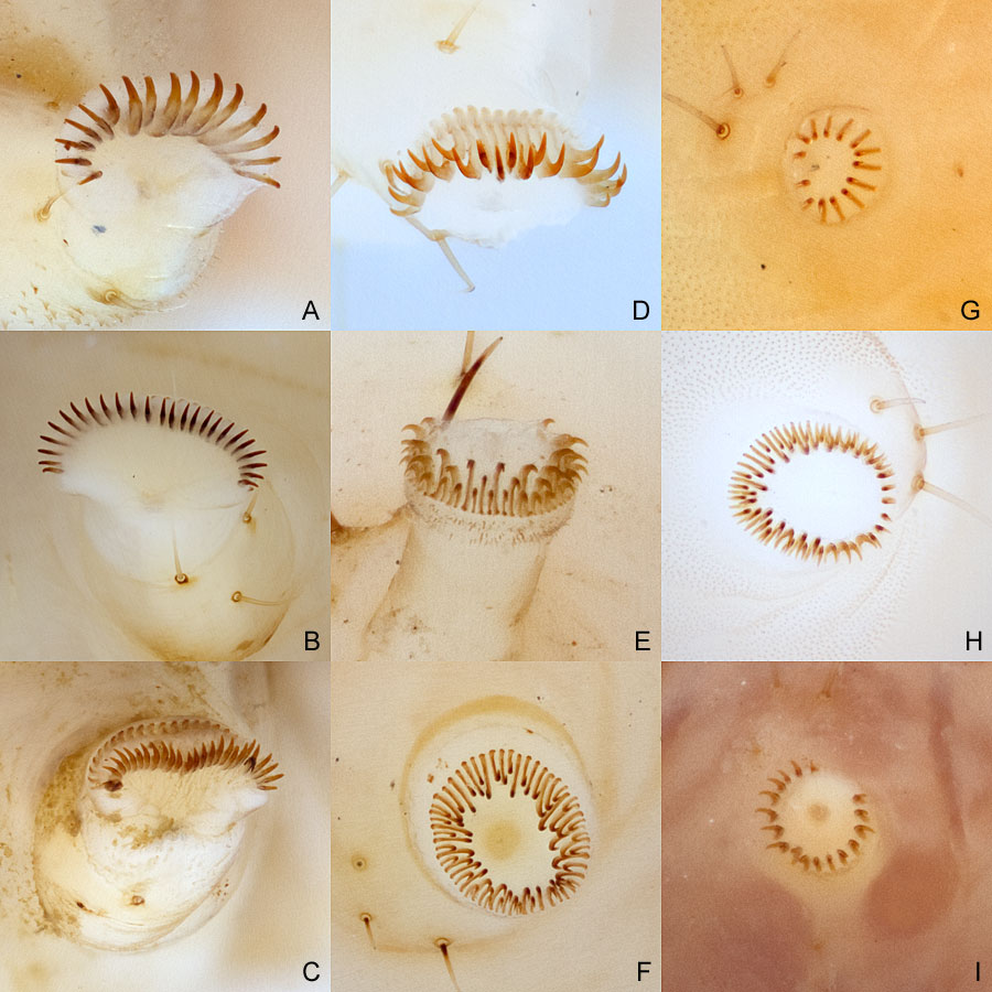
The following page illustrates miscellaneous taxonomically important structures of various species treated in LepIntercept. These images were created using
various light microscope techniques in order to demonstrate some of the tools a port identifier might use to study Lepidoptera larvae.
Specimen submission (for USDA)
Accurate identifications of intercepted Lepidoptera start with the proper submission of specimens. Passoa (2007, 2009) listed some of the issues
seen over the last two decades of receiving samples. Eggs now can be identified with molecular methods. Given that these normally come in groups of
more than one, at least some eggs should be preserved in 95% ethanol for future sequencing. Do not kill eggs and expect a species identification based on
morphology. Given the number of doubtful host/insect associations in the PestID system, one cannot help but worry about unlabeled vials by the
thousands being bounced around from officer to identifier to specialist between several different locations. At minimum, a pencil label with origin, host
(at least identified to genus), and date should be immediately dropped in the vial (not some 25 digit complicated port code). This should stay with the specimen.
No specialist should ever get unlabeled vials! Given the age of camera cell phones, there is no excuse for submitting leaf miners without, at minimum, a picture
of the mine, even if taken with a camera on a cell phone. Ideally, preserve the mine as a pressed specimen or at least cut the mine from the
leaf and send it with the larva in alcohol. Discarding a pupal cocoon and cast larval exuvium almost always makes the identification twice as
difficult to nearly impossible - these parts need to be saved if present (they may be hard to find with stem borers or in butterflies the larval exuvium is
normally not near the pupa). There is also a problem with adult label data. If an adult moth was collected in a container of produce, there is
no guarantee it was reared from that host. It is misleading to label adults with this information. The label can say "collected in a container
of ..." or if reared, then "reared from ..."; but it should not go on the form as a host or be listed on a label without qualifying the association.