Microscope techniques to view taxonomically important structures in larvae
This page illustrates miscellaneous taxonomically important structures of various species treated in LepIntercept. These images were created using
various light microscope techniques in order to demonstrate some of the tools a port identifier might use to study Lepidoptera larvae.
The value of other systems such as scanning (SEM) or transmitted electron microscopy (TEM), and more recently confocal microscopes, is well known.
An example of the use of an SEM to illustrate caterpillar morphology is found in some chapters of Stehr (1987). The use of a confocal microscope and fluorescence was
discussed by Lee et al. (2009). Microscopy with special reference to port identifiers was covered by Passoa (1997).
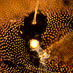
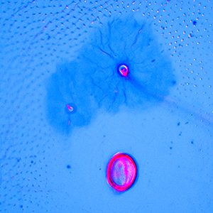
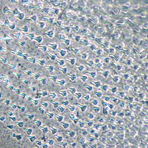
MacKay (1959) did not illustrate the SD2 seta in the codling moth or Oriental fruit moth. We illustrate the presence of this seta in
both species. The codling moth was done in Rheinberg illumination resulting in the SD2 shining red against the blue background. If no
colored stops are used, a darkfield effect is produced. This is shown in the Oriental fruit moth. In both cases the pinacula shape is
more obvious. This method of coloring with filters can save time by eliminating a need to stain. Darkfield can be produced by using a
special condenser or setting a phase condenser on Ph2 or Ph3 with a low power objective (typically 4x or 10x). An oil objective with
iris works best at higher powers.
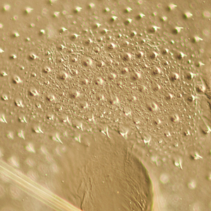
The cuticle texture of H. assulta, at least in middle instars, is uniformly composed of fine spines and small round granules. This
differs from H. armigera that has the spinules in bands from the middle to late instars. The lower power view was done in brightfield
with the condenser iris partially closed. Differential interference contrast was used to get more detail on the texture between the
granules. Another example of cuticle texture is shown with phase contrast (Ph3) on an Amorbia specimen.
The mandibles of D. considerata and D. magnifactella were both done with crossed polarizers to bring out the inner tooth character
more clearly. In the case of D. considerata, similar to a photographer who blurs the background to emphasize a subject, the depth of
field was limited to the mandibular ridge with the tooth for clarity. In fact, an advantage of differential interference contrast
over phase contrast is the shallow focus (optical sectioning) that allows dorsal and ventral surface to be easily distinguished.
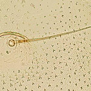
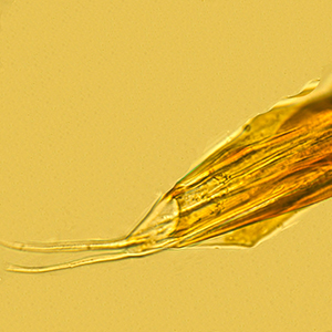
The apical setae on the spinneret of D. considerata, and the position of the SD2 seta on A8 under the middle of the SD1 pinaculum in
D. magnifactella, were both done in brightfield.
 |
 |
 |
| SD2 seta on A6 in G. molesta |
SD2 seta on A2 in C. pomonella |
Cuticle texture in H. assulta (low power) |
 |
 |
 |
| Cuticle texture in H. assulta |
Cuticle texture in Amorbia |
Inner tooth on mandible of D. considerata |
 |
 |
 |
| Inner tooth on mandible of D. magnifactella |
SD2 seta position on A8 in D. magnifactella |
Apical setae on spinneret of D. considerata |