
Family: Megachilidae
Subfamily: Megachilinae
Tribe: Osmiini
Genus: Osmia Panzer, 1806
Subgenus: O. (Osmia) Panzer, 1806
Species: Osmia rufina Cockerell, 1931
Common name: none
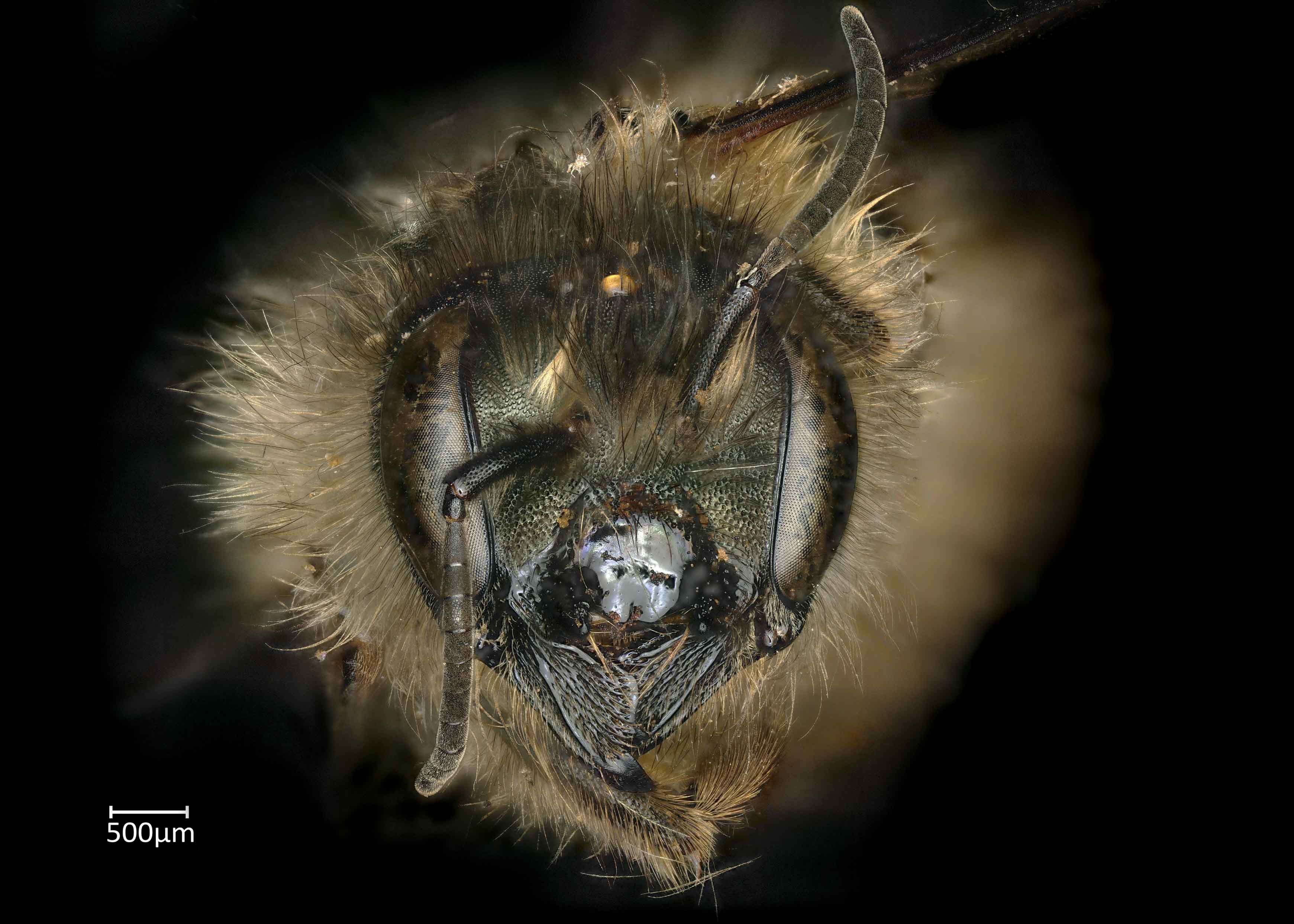
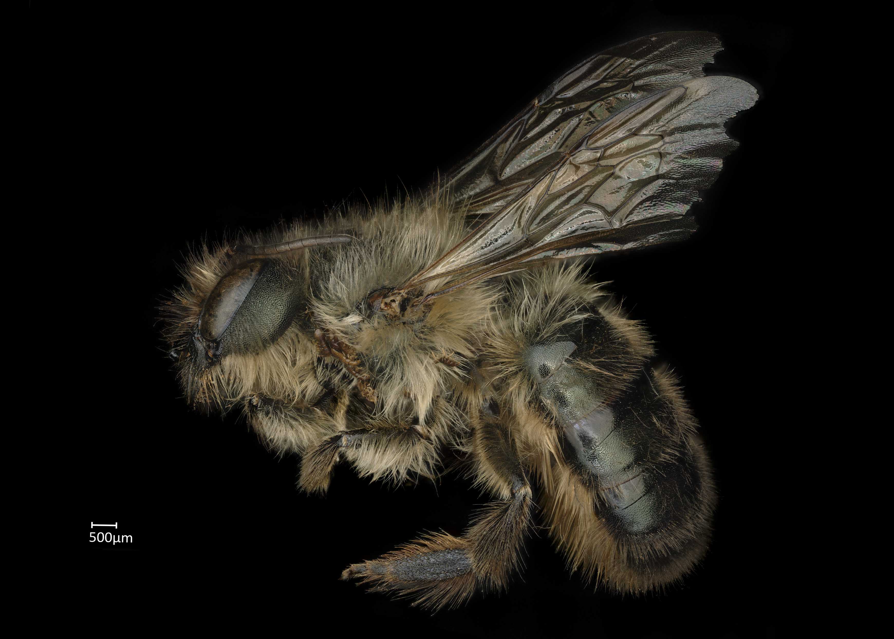
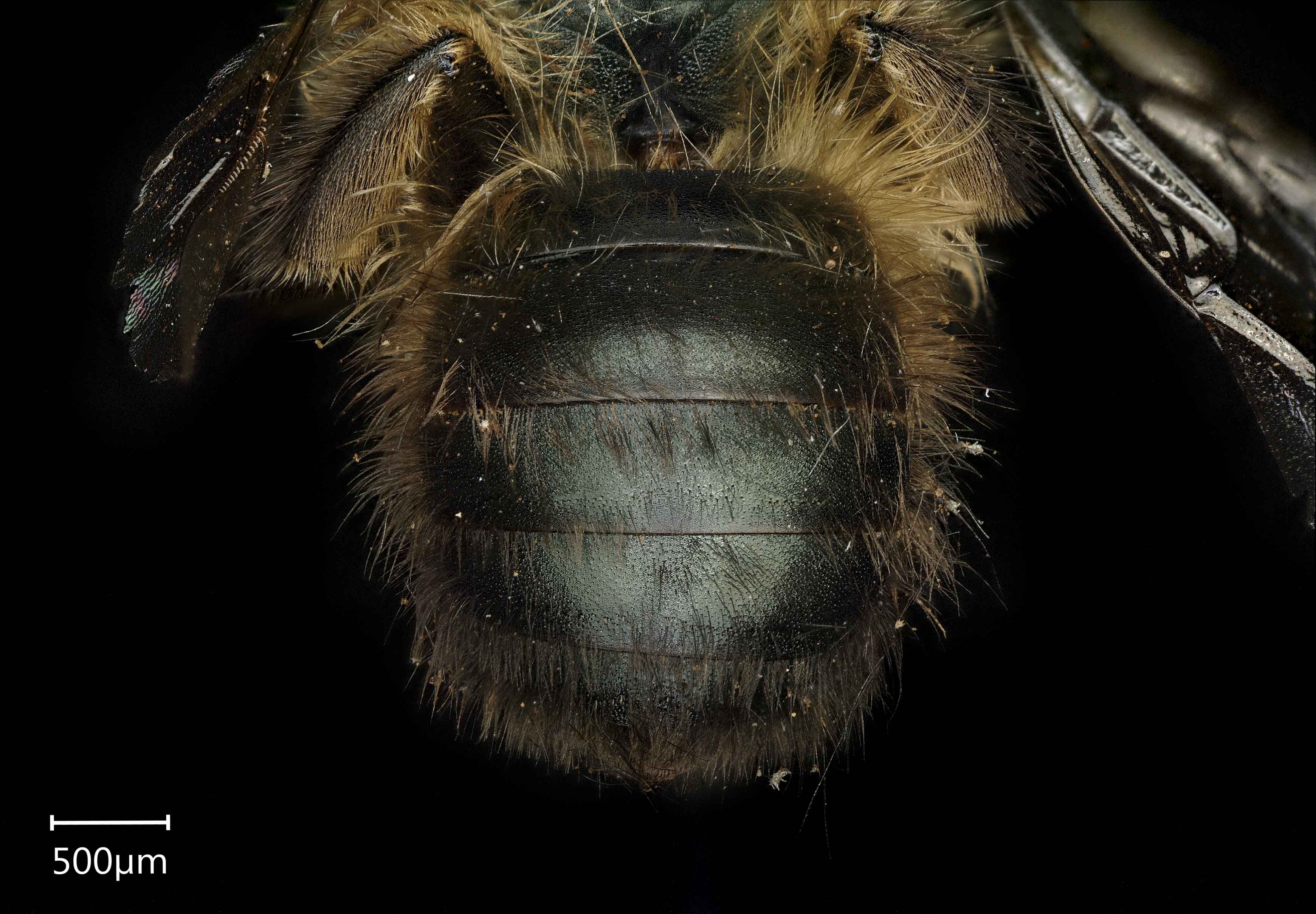
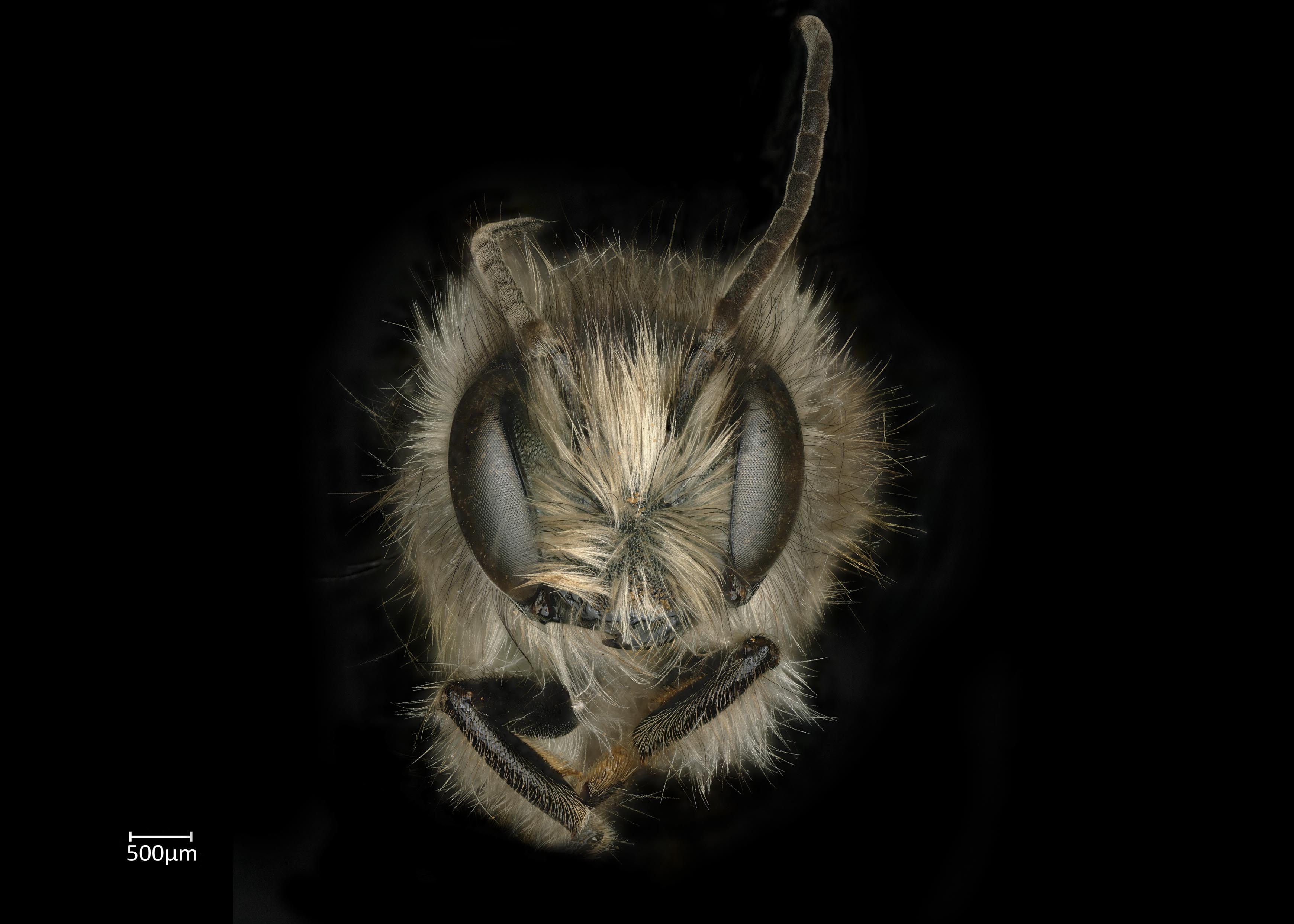
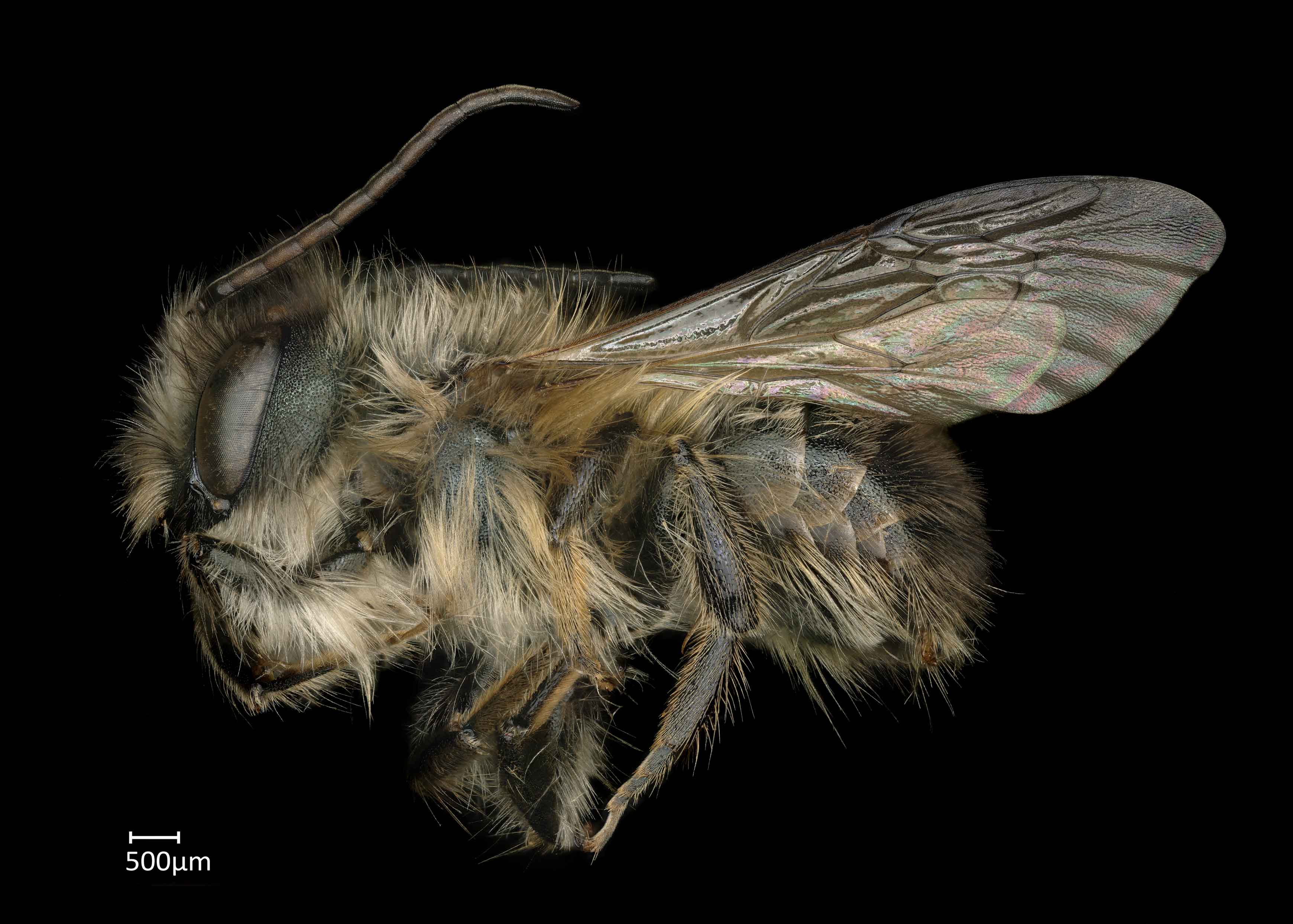
Osmia rufina are black bees with a metallic green luster (Wu 2006Wu 2006:
Wu, Y. 2006. Hymenoptera: Megachilidae. Fauna Sinica, Insecta. Vol. 4. Science Press, Beijing.). Females have dense yellow to white hair on the genagena:
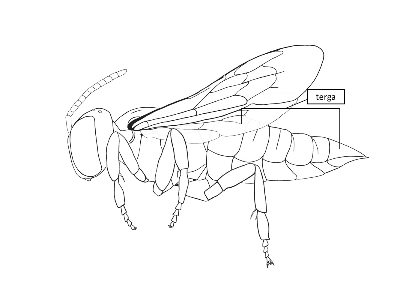
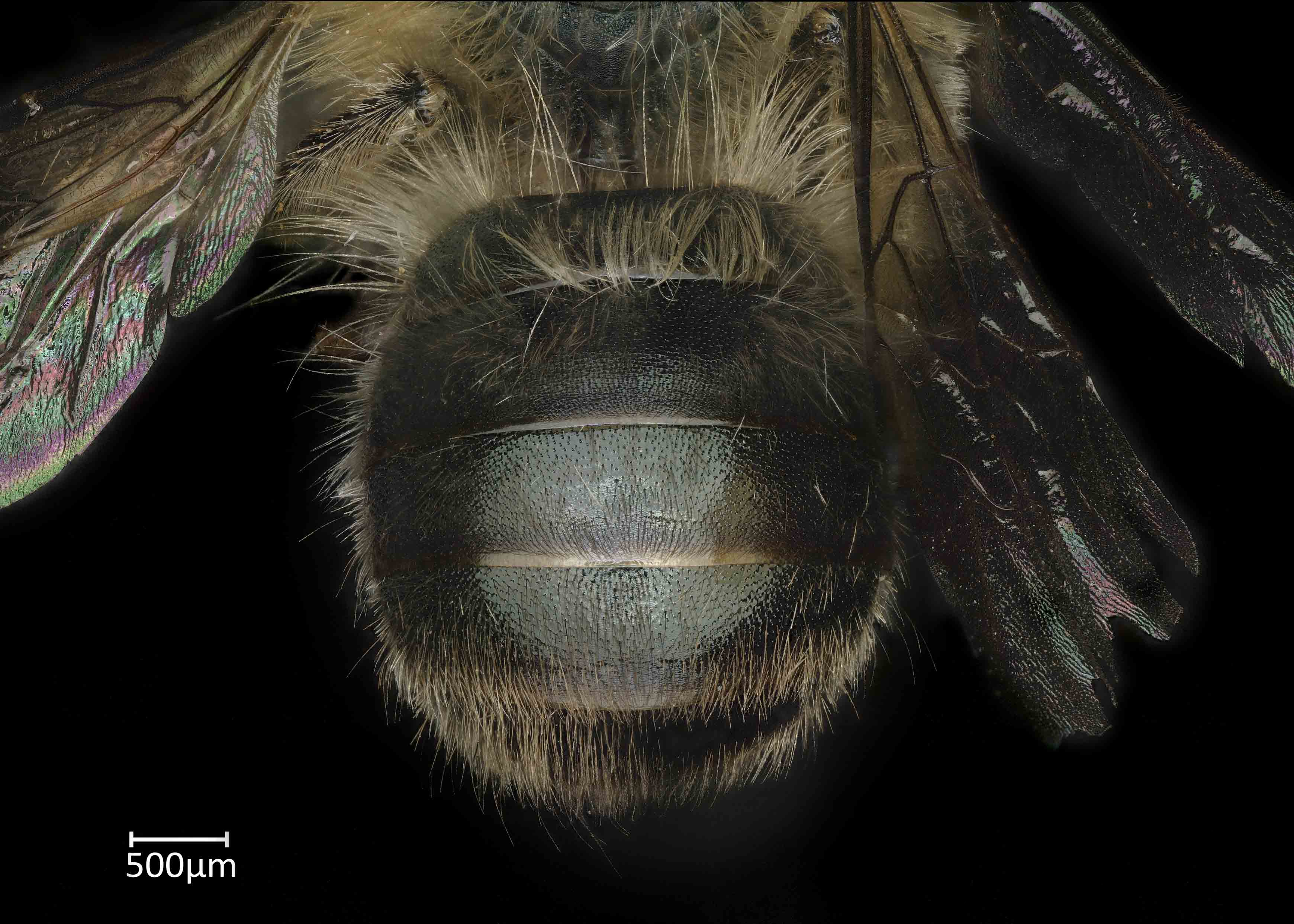
the cheek or side of the head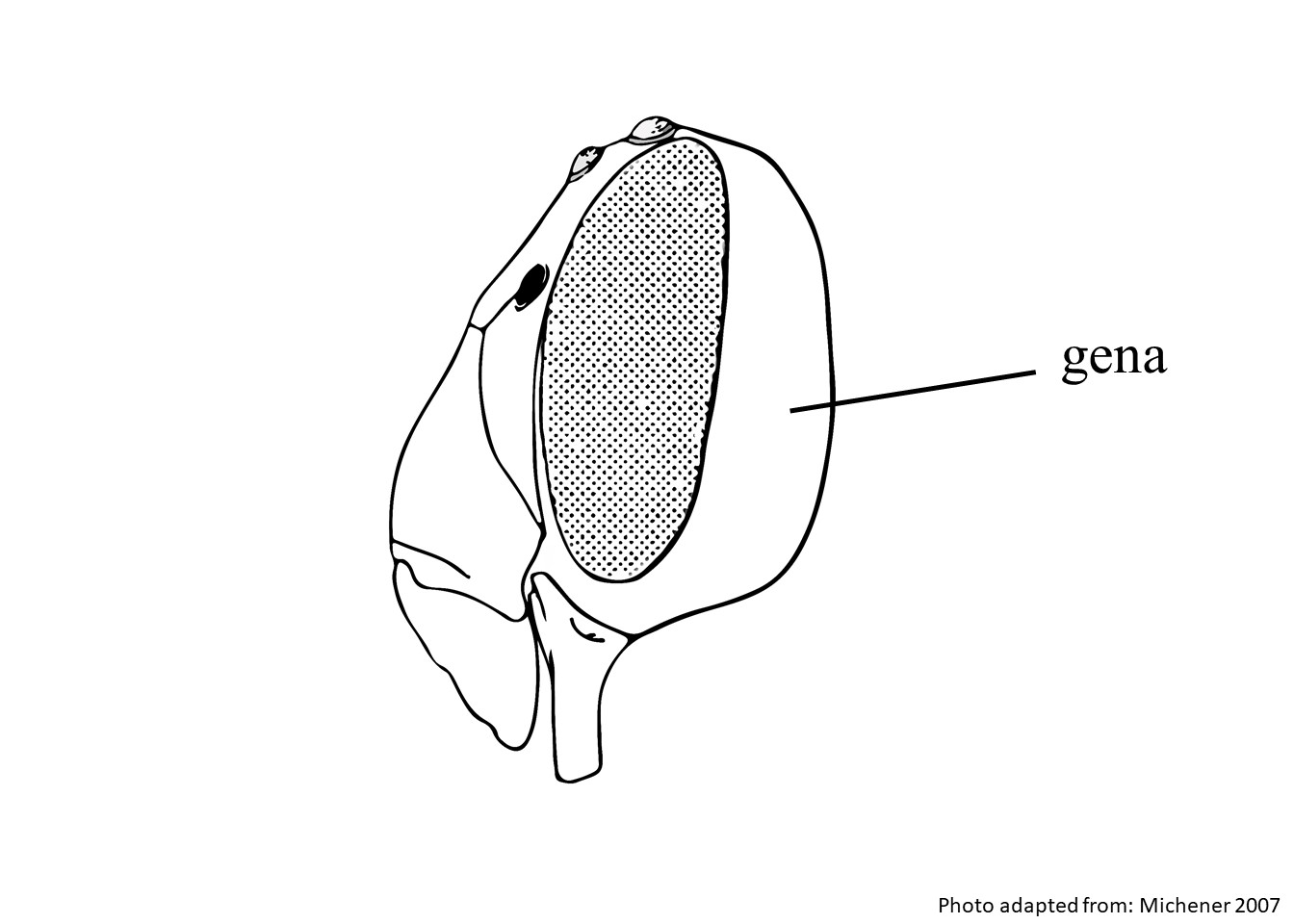 , thorax, and legs; light orange hair on T1; and T2–T5 are covered with primarily dark brown hair (Wu 2006Wu 2006:
Wu, Y. 2006. Hymenoptera: Megachilidae. Fauna Sinica, Insecta. Vol. 4. Science Press, Beijing.; Fig 3). Males have white to pale yellow hair on the face, thorax, and abdomen (Wu 2006Wu 2006:
Wu, Y. 2006. Hymenoptera: Megachilidae. Fauna Sinica, Insecta. Vol. 4. Science Press, Beijing.; Fig 4–6). Female body length is 11–13 mm ,and male body length is 8–9 mm (Wu 2006Wu 2006:
Wu, Y. 2006. Hymenoptera: Megachilidae. Fauna Sinica, Insecta. Vol. 4. Science Press, Beijing.).
(modified from Wu 2006Wu 2006:
Wu, Y. 2006. Hymenoptera: Megachilidae. Fauna Sinica, Insecta. Vol. 4. Science Press, Beijing.)
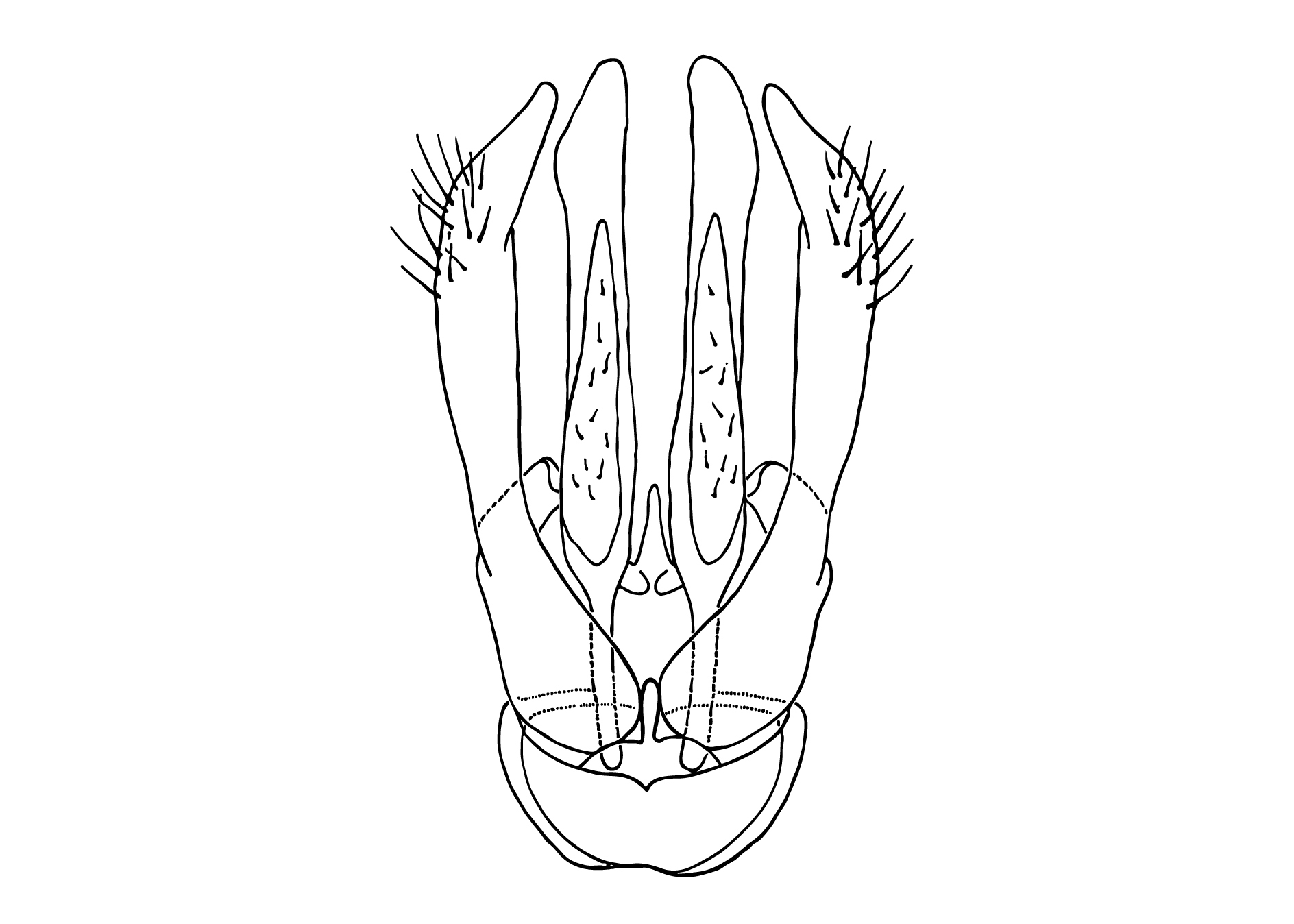
 without apicalapical:
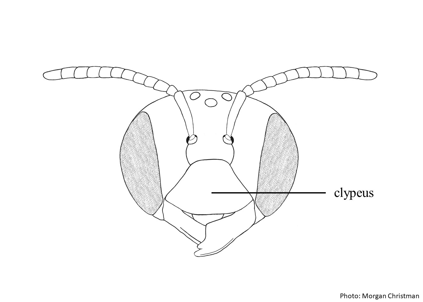 with short, triangular-shaped laterallateral: apicalapical:
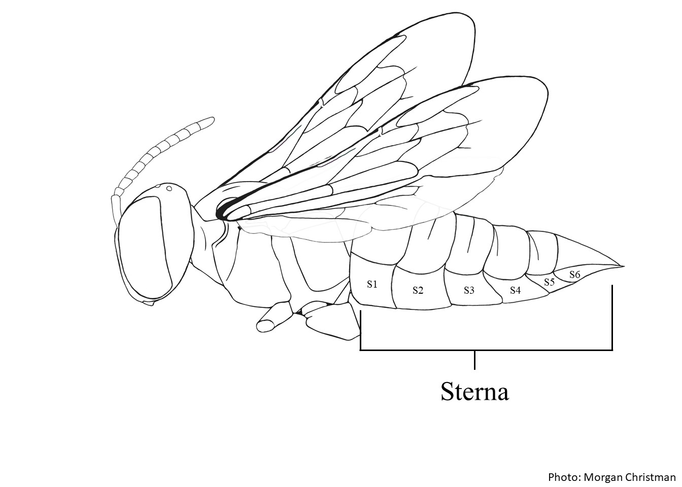 large, mostly covering the medially emarginateemarginate: without median apicalapical: apicallyapically:
Osmia rufina look similar to O. bicornis and O. pedicornis. Females of these species all have laterallateral:
relating, pertaining, or attached to the side
horns and bidentatebidentate:
having two teeth
median apicalapical:
near or at the apex or end of any structure
projection on the clypeusclypeus:
a section of the face below the antennae, demarcated by the epistomal sutures. O. rufina females can be differentiated by the much shorter, rounded median apicalapical:
near or at the apex or end of any structure
projections on the clypeusclypeus:
a section of the face below the antennae, demarcated by the epistomal sutures and the short, triangular-shaped laterallateral:
relating, pertaining, or attached to the side
horns on the clypeusclypeus:
a section of the face below the antennae, demarcated by the epistomal sutures. Males are similar in size and hair color and can be difficult to distinguish without dissecting them to see the genitaliagenitalia:
all the genital structures collectively
and hidden sternasterna:
the plates on the underside of the abdomen, often abbreviated when referring to a specific segment to S1, S2, S3, S4, S5, S6, S7, or S8
. They can, however, be distinguished by the shape of S8S8:
the plates on the underside of the abdomen, often abbreviated when referring to a specific segment to S1, S2, S3, S4, S5, S6, S7, or S8
and the apexapex:
end of any structure
of the gonocoxites.
unknown
Osmia rufina are associated with Fabaceae and Platycladus orientalis (Wu 2006Wu 2006:
Wu, Y. 2006. Hymenoptera: Megachilidae. Fauna Sinica, Insecta. Vol. 4. Science Press, Beijing.).
unknown
Osmia rufina specimens have been recorded in Beijing, Fujian, and Sichuan in China (Wu 2006Wu 2006:
Wu, Y. 2006. Hymenoptera: Megachilidae. Fauna Sinica, Insecta. Vol. 4. Science Press, Beijing.).
Distribution map generated by Discover Life -- click on map for details, credits, and terms of use.
Wu. Y. 2006. Hymenoptera: Megachilidae. Fauna Sinica, Insecta: 44: 1-474.
Authors: S. Burrows, C. Ritner, M. Christman, L. Spears, A. Smith-Pardo, S. Price, R. Ramirez, T. Griswold, A. Redford
Edition 3 - last updated November 2021
tool images at ITP Node
idtools.org

.jpg)