
Family: Megachilidae
Subfamily: Megachilinae
Tribe: Anthidiini
Genus: Anthidium Fabricius, 1804
Subgenus: A. (Anthidium) Fabricius, 1804
Species: Anthidium parkeri Gonzalez and Griswold, 2013
Common name: none
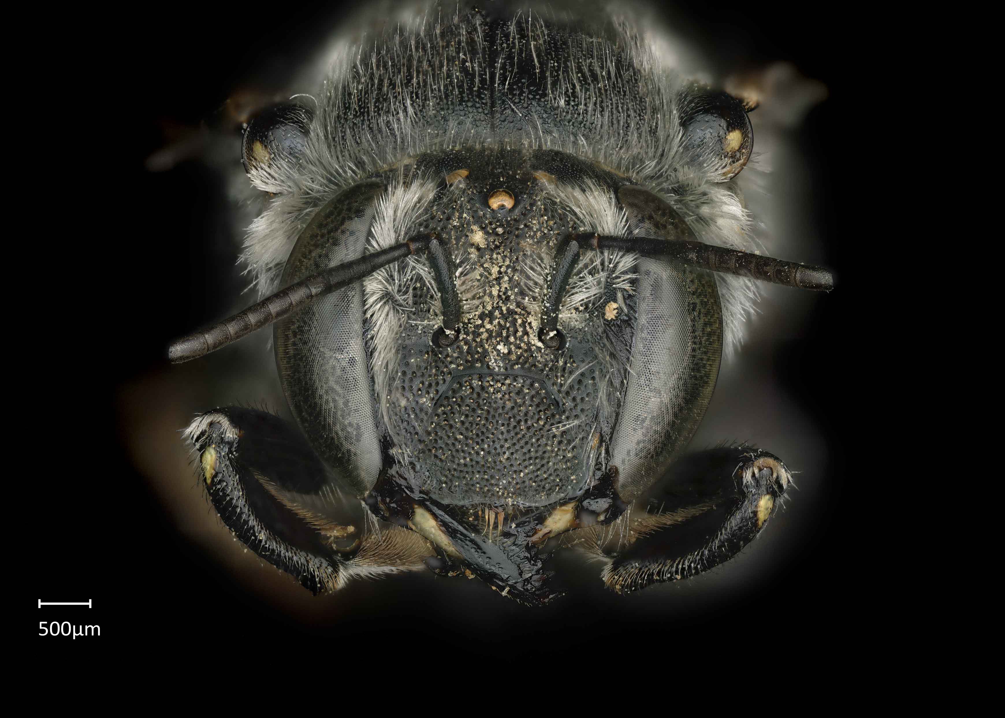
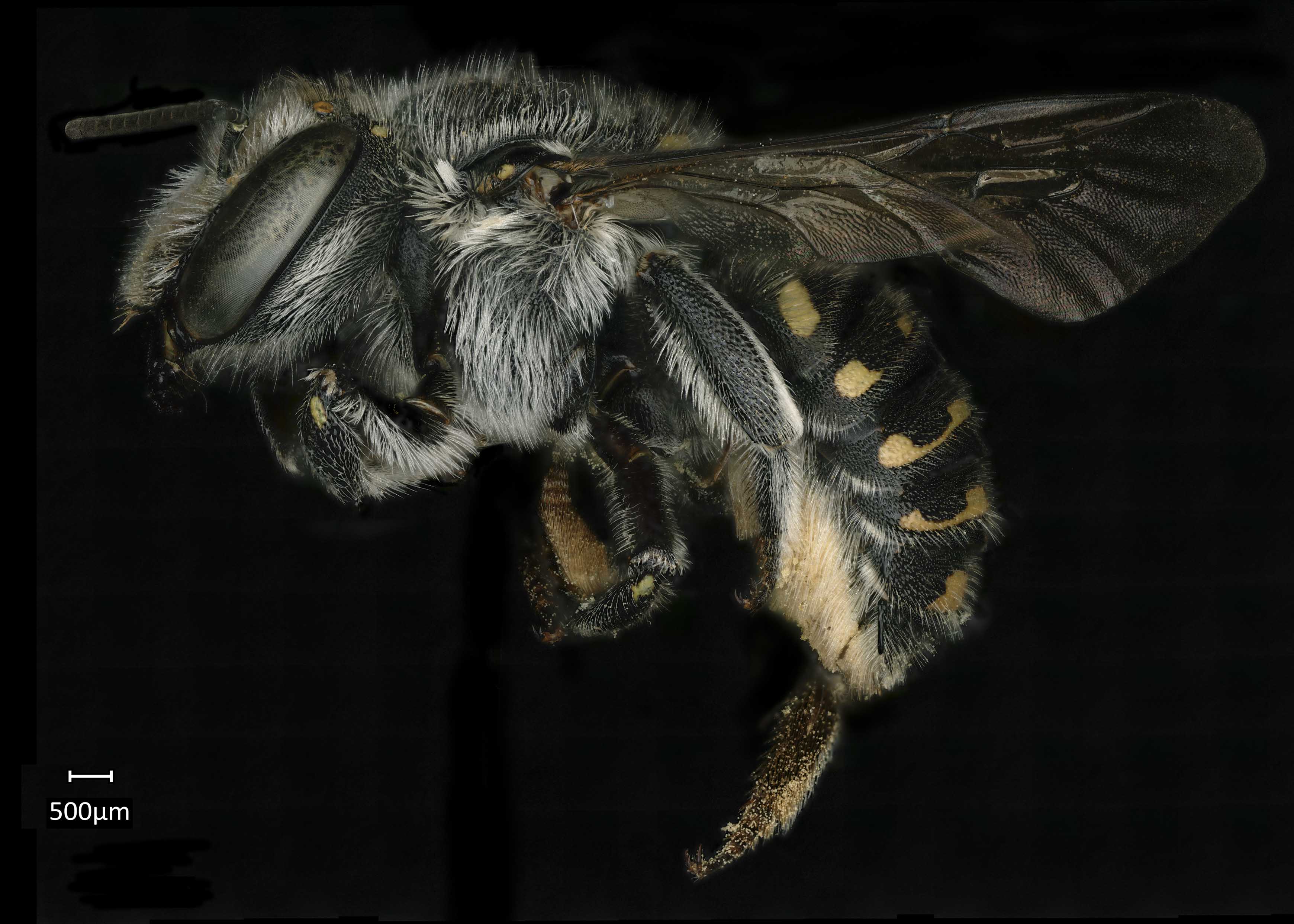
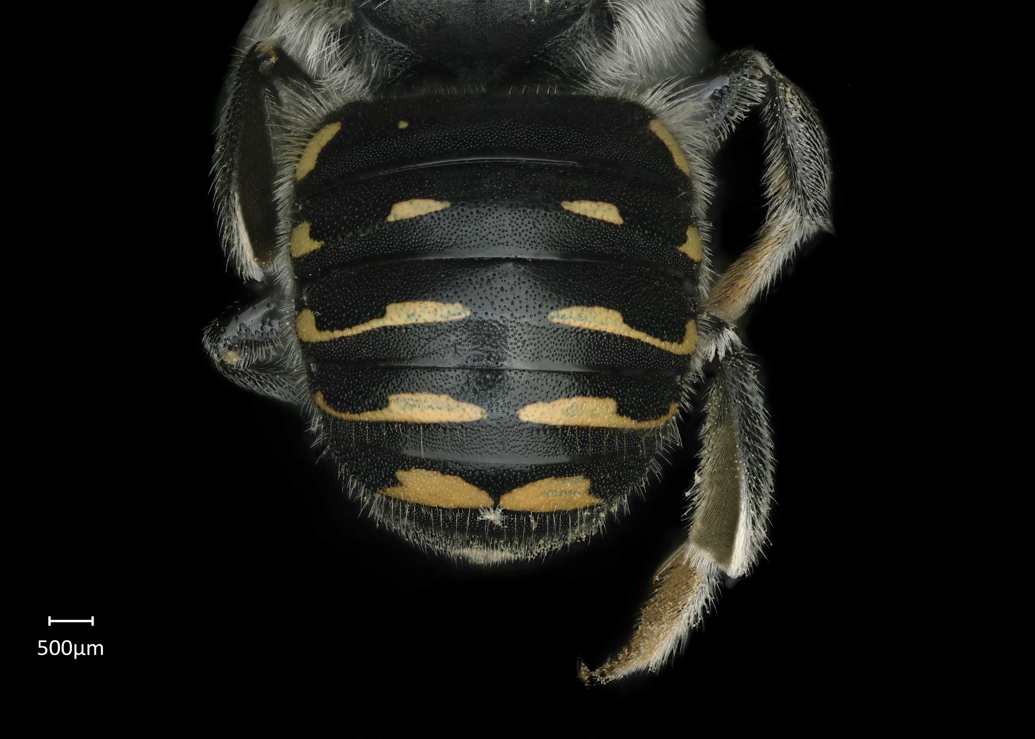
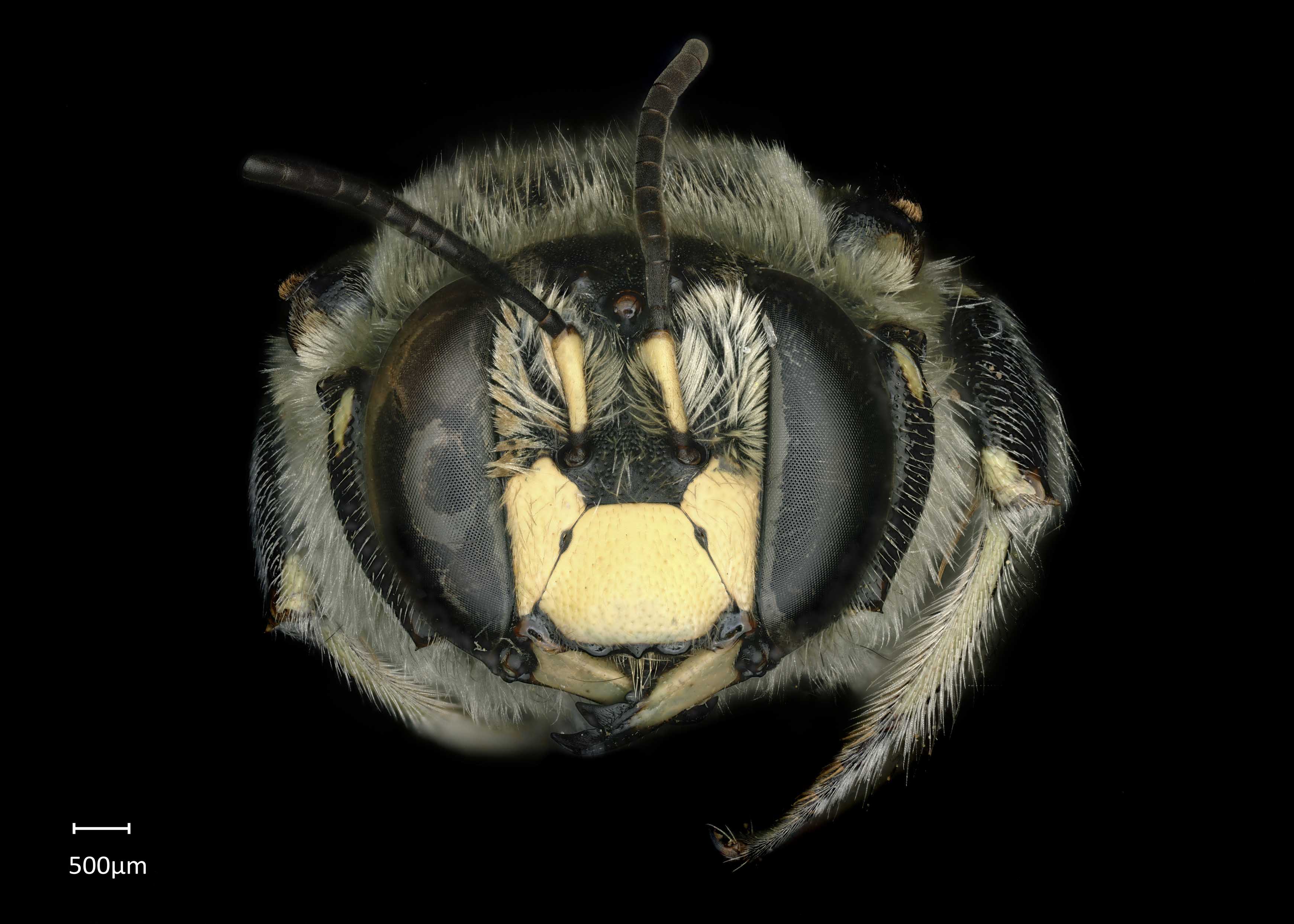
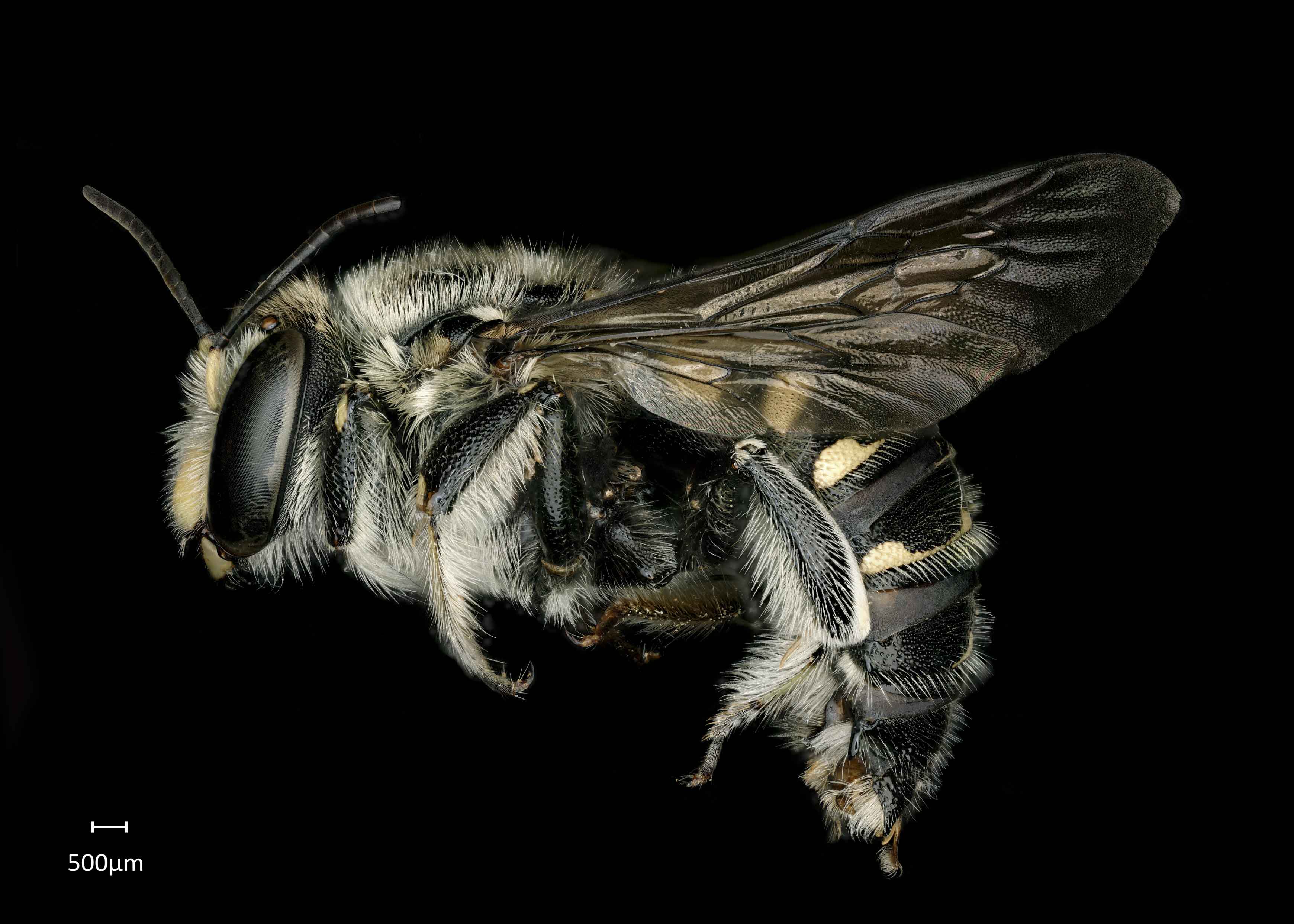
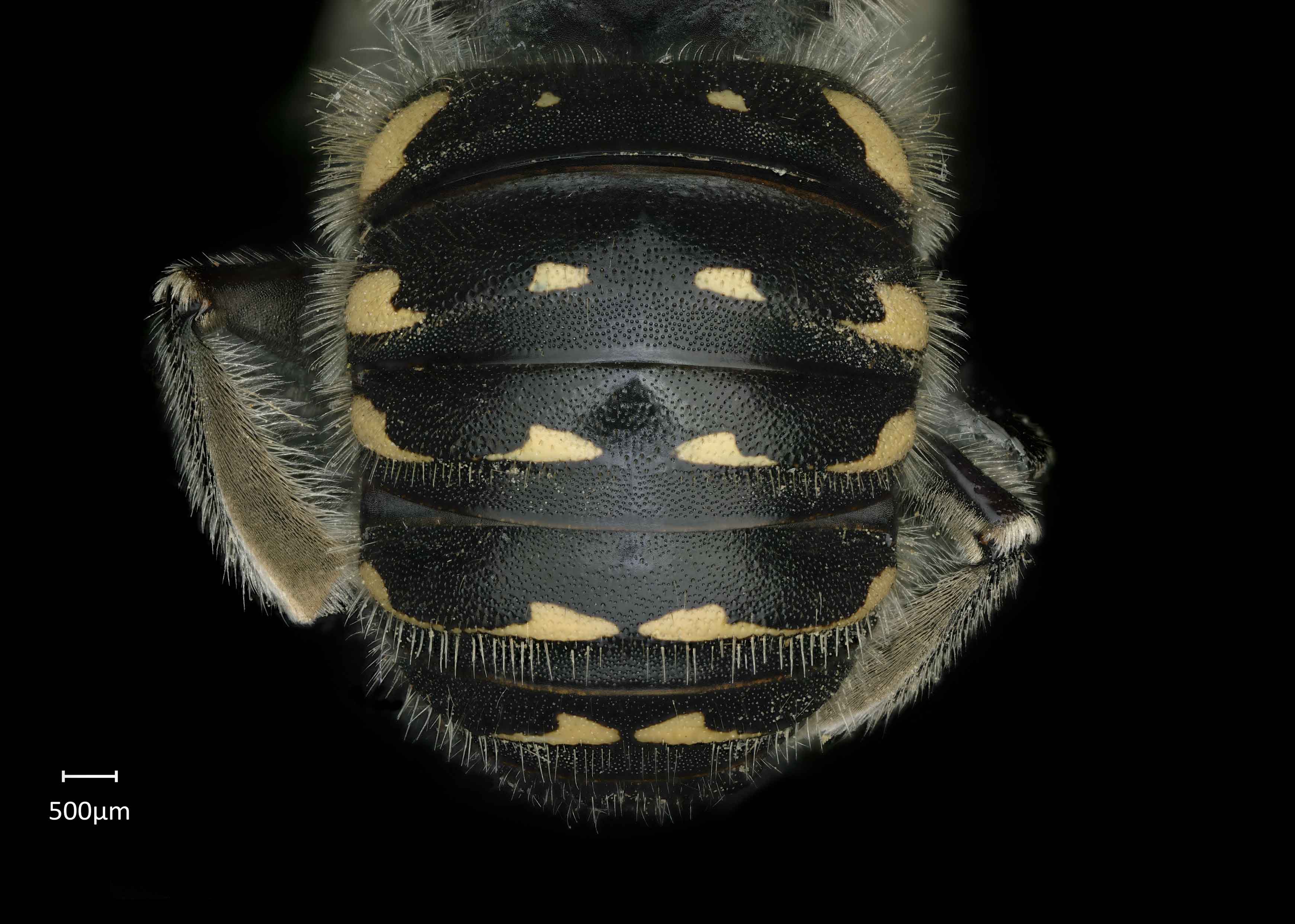
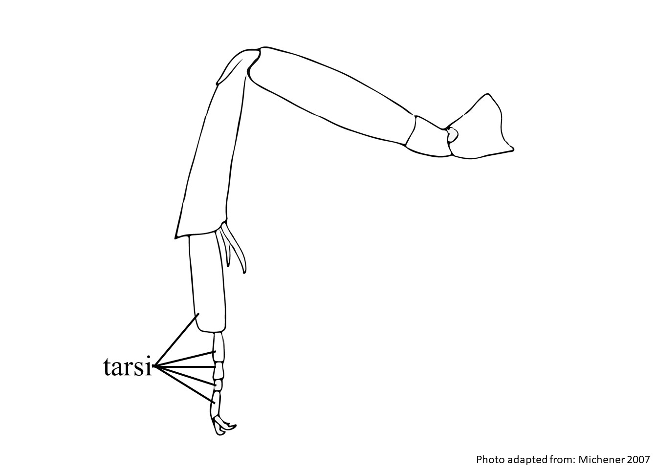
Anthidium (Anthidium) parkeri are black with brown tarsitarsi:
the group of segments at the end of the leg following the tibia
 and yellow maculations (Gonzalez and Griswold 2013Gonzalez and Griswold 2013:
Gonzalez, V.H. and T.L. Griswold. 2013. Wool carder bees of the genus Anthidium in the Western Hemisphere (Hymenoptera: Megachilidae): diversity, host plant associations, phylogeny, and biogeography. Zoological Journal 168: 221ndash;425.). Females have white pubescencepubescence:
short, fine hair
except for some brown hairs on the clypeusclypeus:
a section of the face below the antennae, demarcated by the epistomal sutures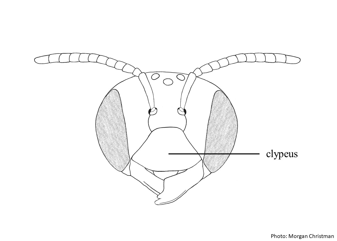 , supraclypeal areasupraclypeal area:
the region of the head between the antennal sockets and clypeus, demarcated on the sides by the subantennal sutures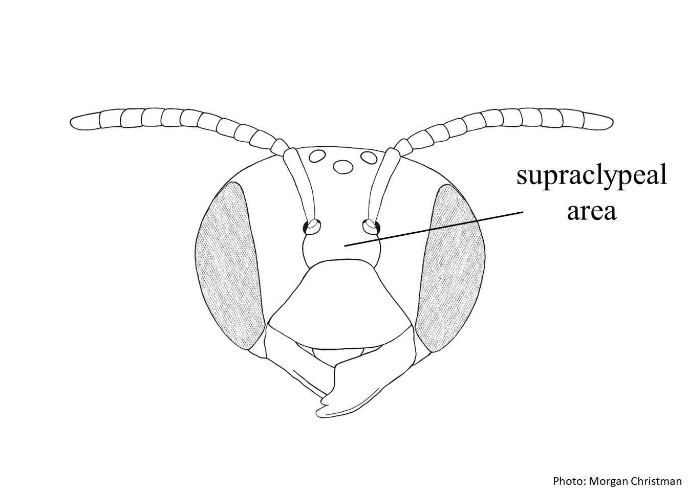 , fronsfrons:
the area between the antennae and ocelli on the bee's head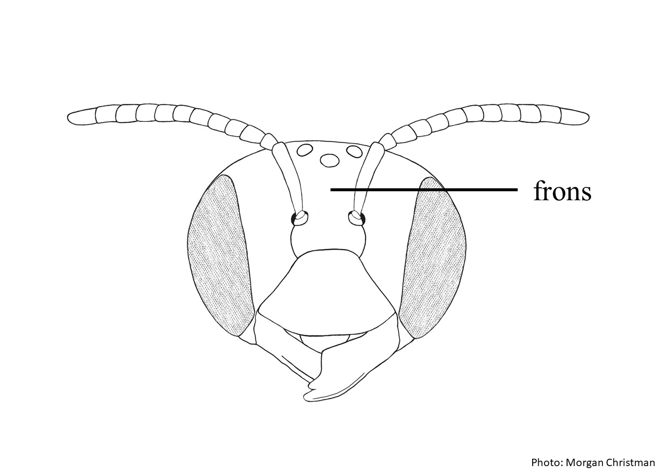 , vertexvertex:
the area between the ocelli and the back of the head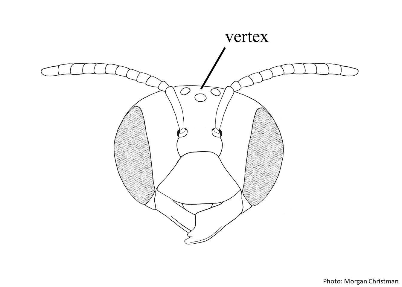 , inner basitarsibasitarsi:
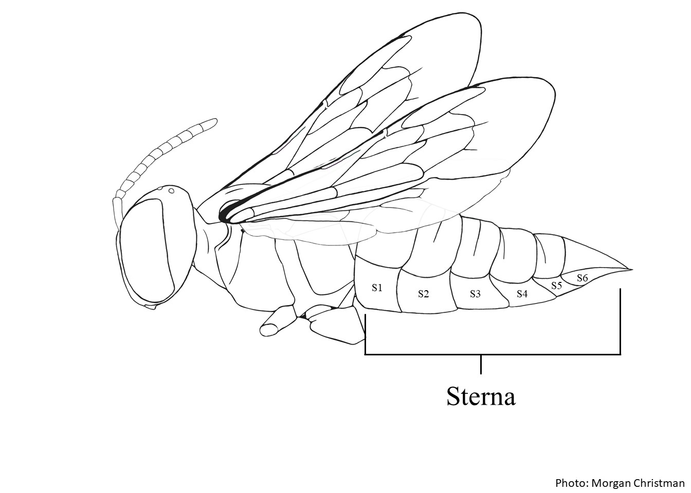
the segment of the tarsus that is the nearest to the body of the bee, usually the largest of all the tarsal segments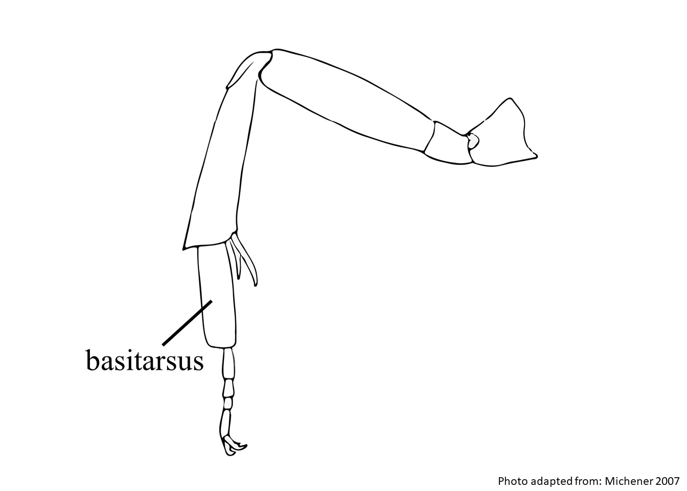 , T1–T4 discs, and center of S6S6:
the plates on the underside of the abdomen, often abbreviated when referring to a specific segment to S1, S2, S3, S4, S5, S6, S7, or S8
 . Females have a body length of 10.0–12.3 mm, and males range in length from 11.5–16.9 mm (Gonzalez and Griswold 2013Gonzalez and Griswold 2013:
Gonzalez, V.H. and T.L. Griswold. 2013. Wool carder bees of the genus Anthidium in the Western Hemisphere (Hymenoptera: Megachilidae): diversity, host plant associations, phylogeny, and biogeography. Zoological Journal 168: 221ndash;425.).
(modified from Gonzalez and Griswold 2013Gonzalez and Griswold 2013:
Gonzalez, V.H. and T.L. Griswold. 2013. Wool carder bees of the genus Anthidium in the Western Hemisphere (Hymenoptera: Megachilidae): diversity, host plant associations, phylogeny, and biogeography. Zoological Journal 168: 221ndash;425.)
, supraclypeal areasupraclypeal area:, paraocular areaparaocular area: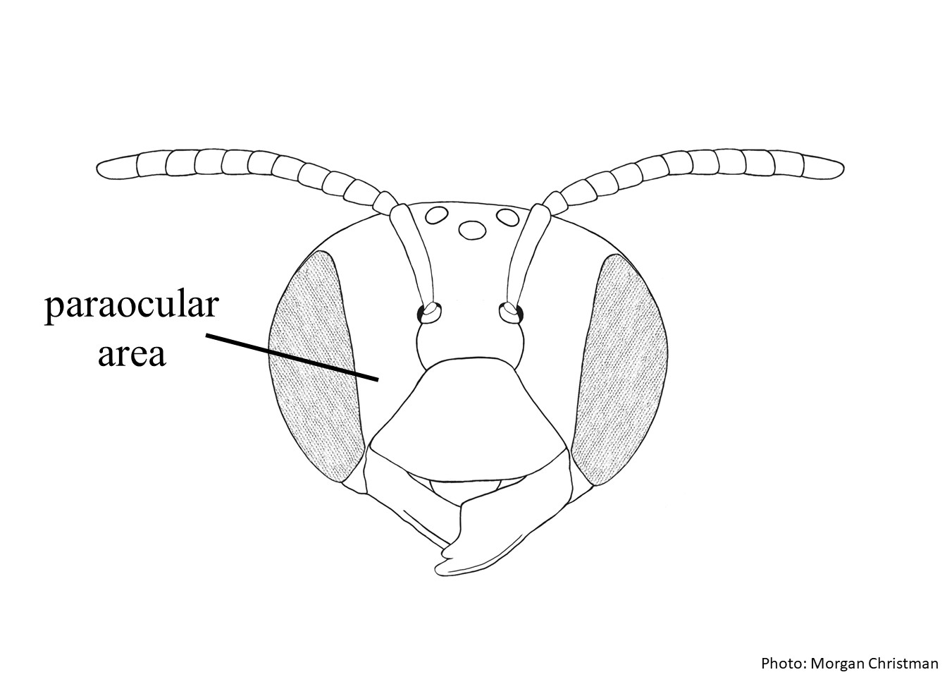 , and fronsfrons: are dull between coarse, sparse punctures.
 has basalbasal:
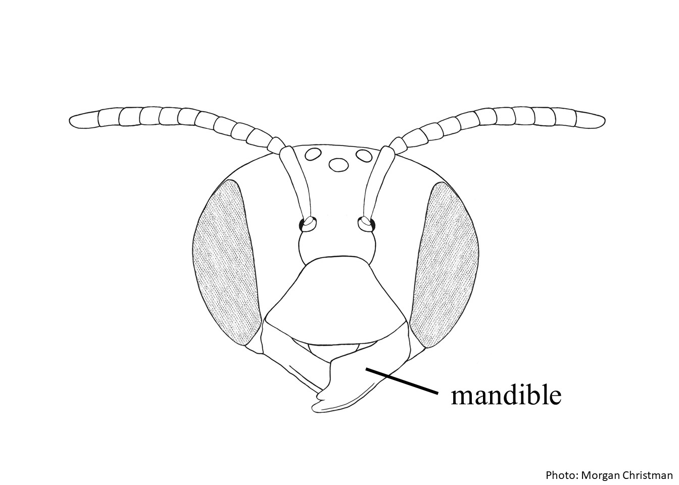 is elongate with 8–9 teeth.
 triangle is dull and finely lineolatelineolate:
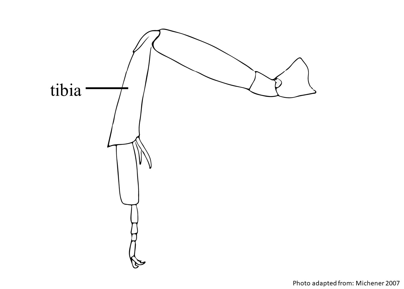 with anterioranterior:
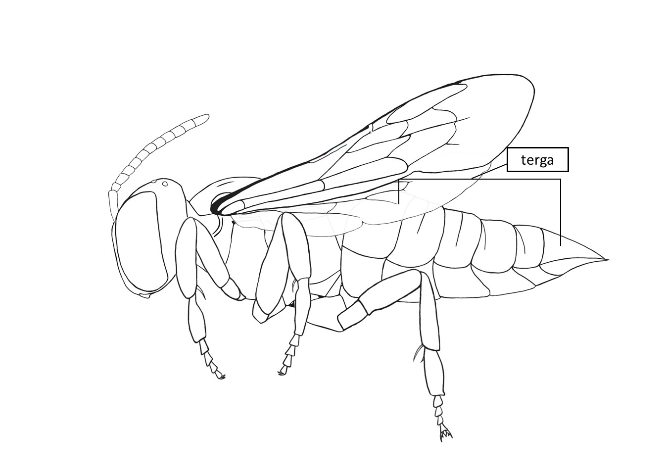 has a small laterallateral: apicalapical: have small, acute, laterally directed laterallateral: apicalapical: laterallateral:. laterallateral:
Anthidium parkeri may be confused with A. maculosumdue to the presence of anterioranterior:
toward the head or on the head side of a segment being described
hind tibial carinae and dull fronsfrons:
the area between the antennae and ocelli on the bee's head with coarse and sparse punctures. Female A. parkeri can be differentiated from A. maculosumby the absence of dense tomentumtomentum:
a form of pubescence composed of short matted, woolly hair
on the outer surface of the basitarsibasitarsi:
the segment of the tarsus that is the nearest to the body of the bee, usually the largest of all the tarsal segments, dull clypeusclypeus:
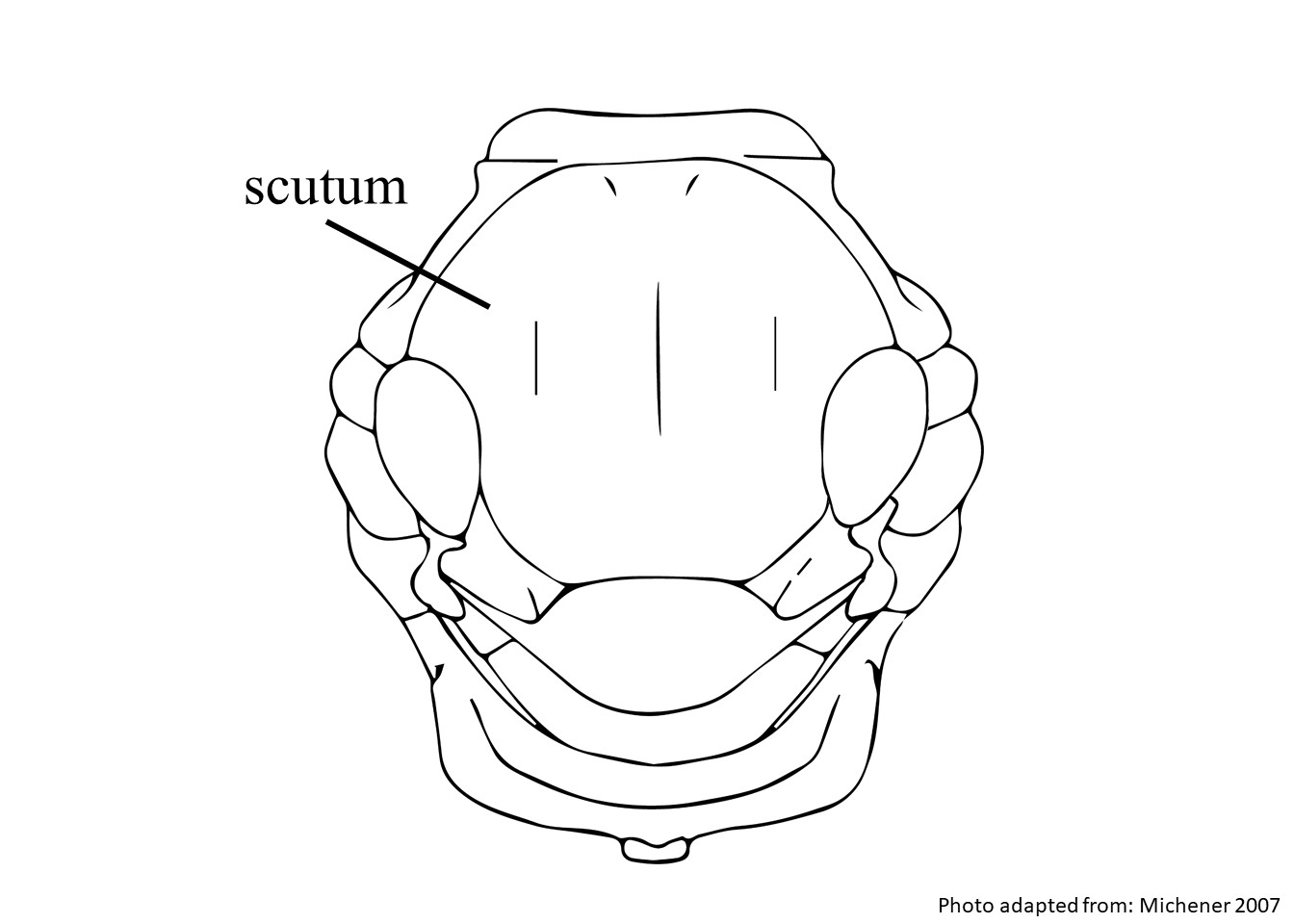
a section of the face below the antennae, demarcated by the epistomal sutures, and shiny scutumscutum:
the large segment on top of the thorax located between the wings and behind the head
 and scutellumscutellum:
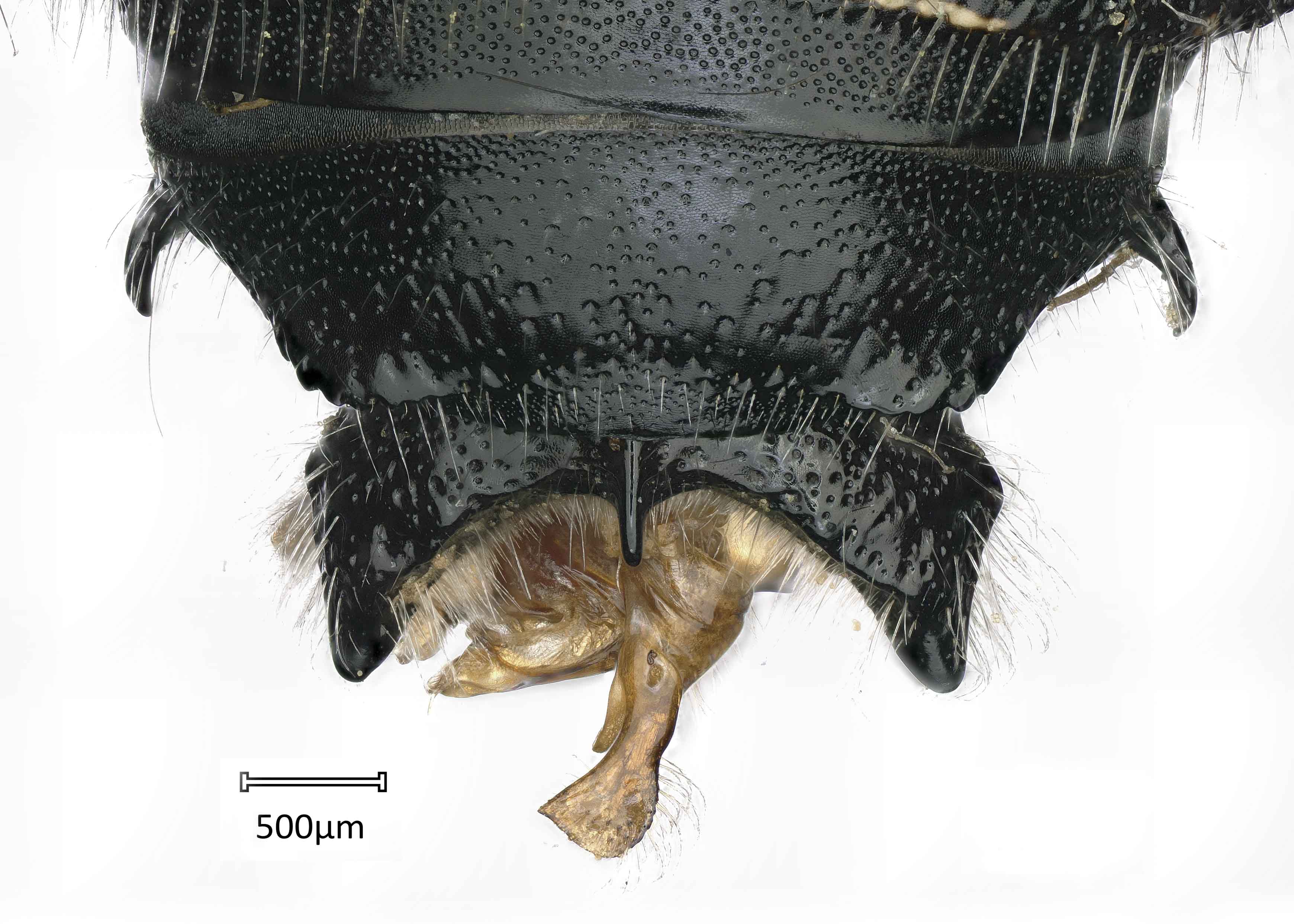
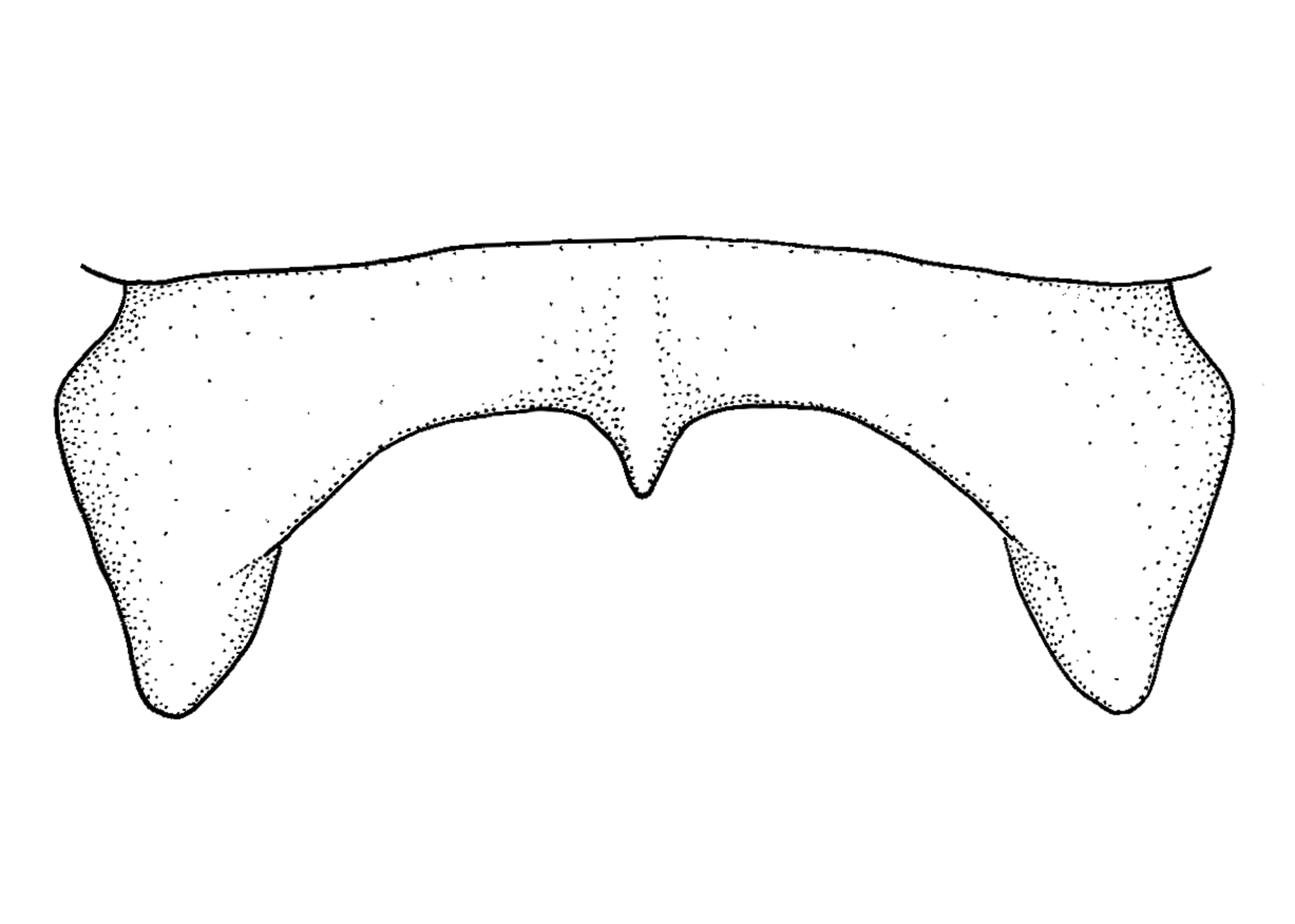
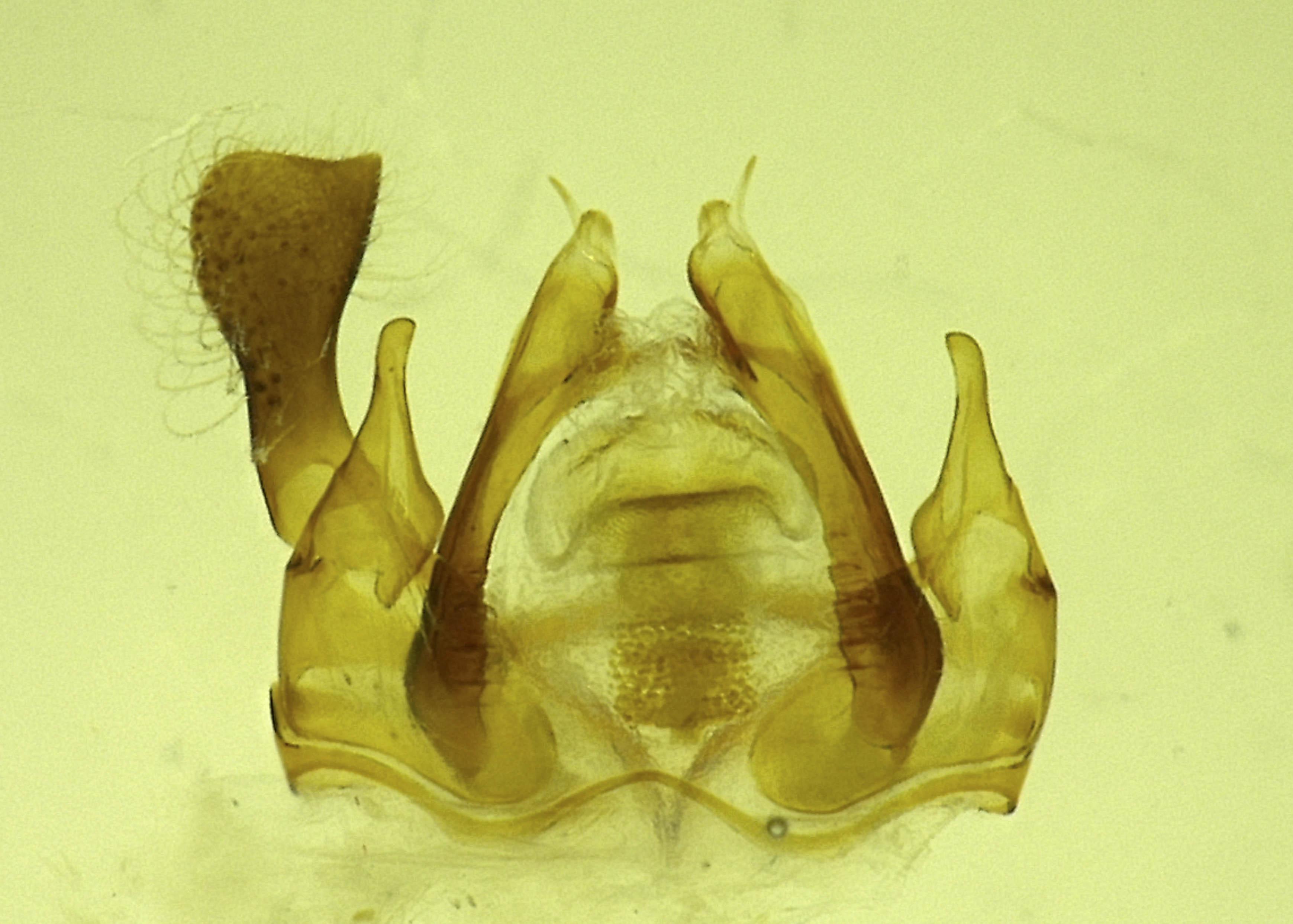
shield shaped plate behind scutum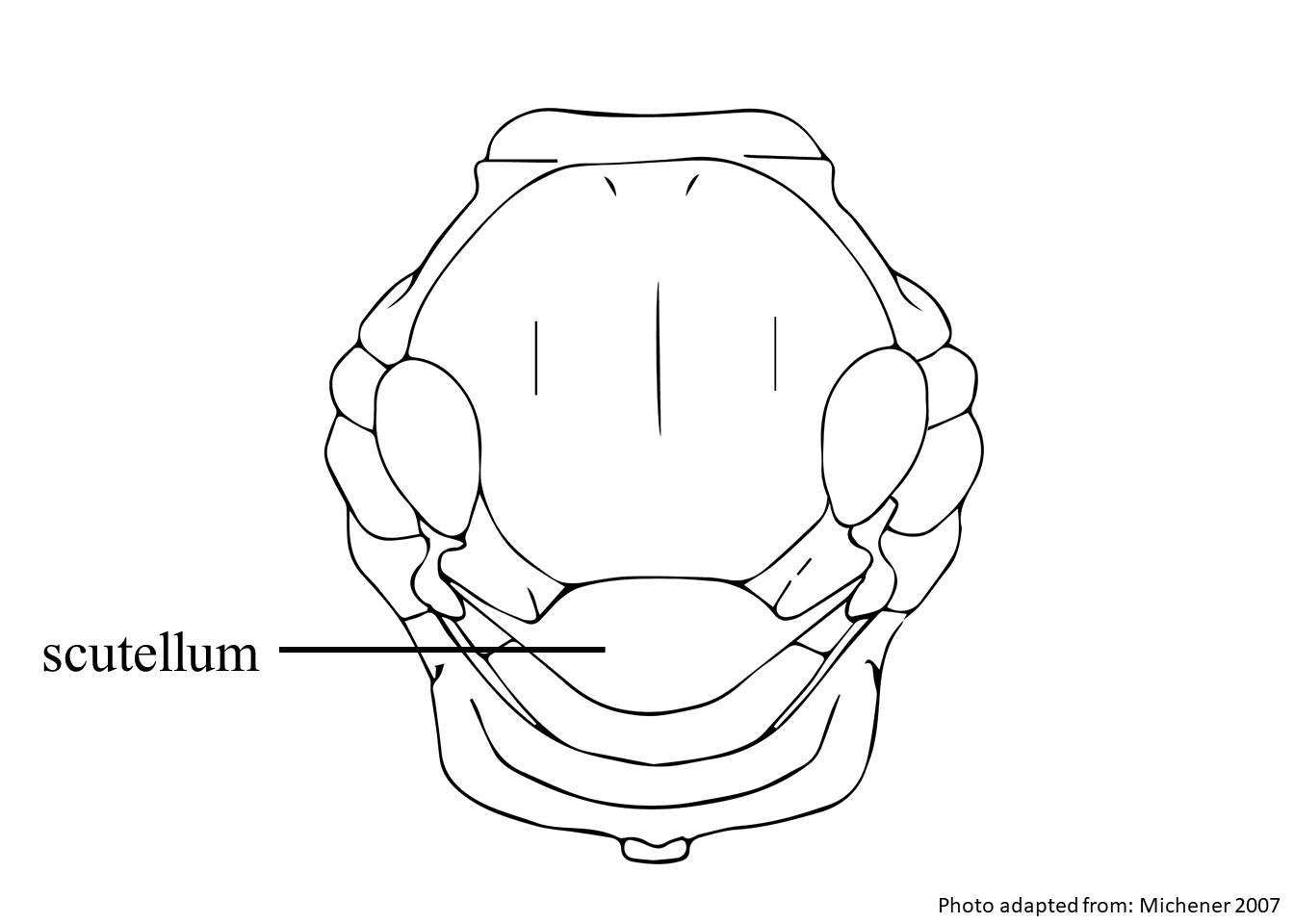 with coarse, sparse punctures. Male A. parkeri can be differentiated from A. maculosumby a small medial denticledenticle:
a small tooth-like projection
on the apicalapical:
near or at the apex or end of any structure
margin of the clypeusclypeus:
a section of the face below the antennae, demarcated by the epistomal sutures, apicallyapically:
near or at the apex or end of any structure
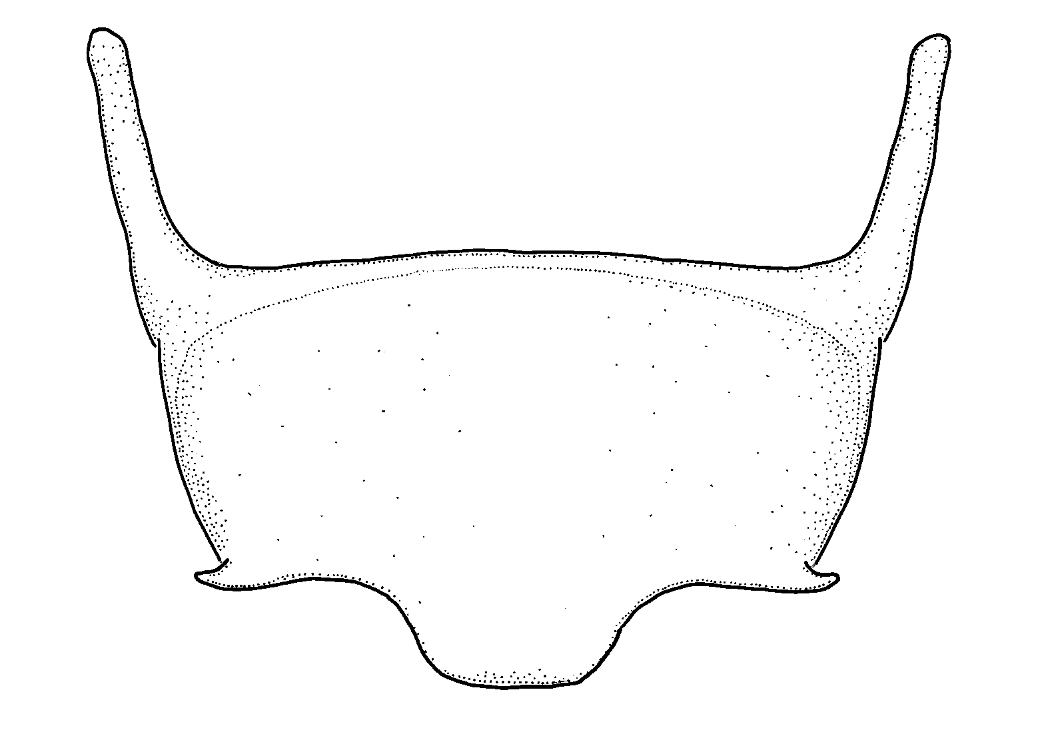
subtruncate median lobe of S6S6:
the plates on the underside of the abdomen, often abbreviated when referring to a specific segment to S1, S2, S3, S4, S5, S6, S7, or S8
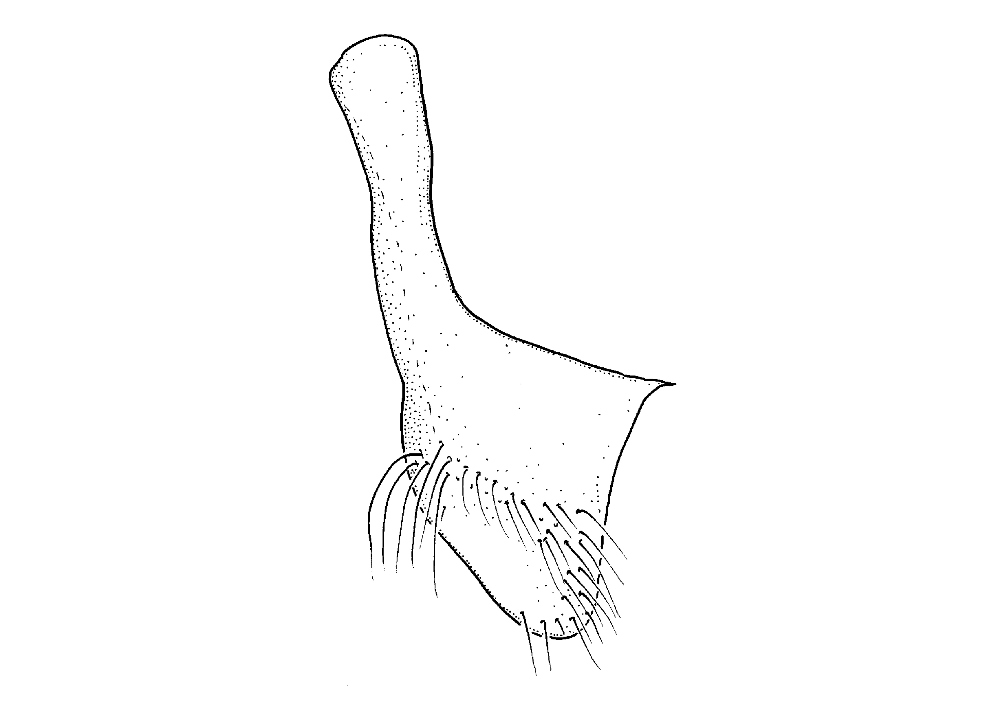
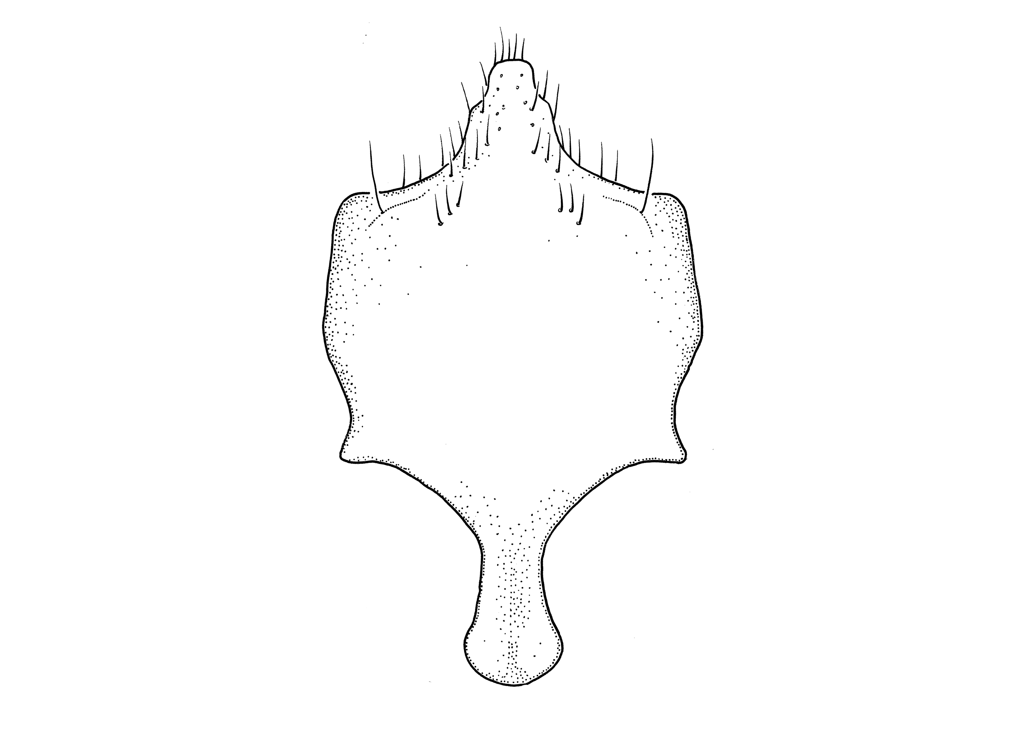
, and shape of S7S7:
the plates on the underside of the abdomen, often abbreviated when referring to a specific segment to S1, S2, S3, S4, S5, S6, S7, or S8
and S8S8:
the plates on the underside of the abdomen, often abbreviated when referring to a specific segment to S1, S2, S3, S4, S5, S6, S7, or S8
(Gonzalez and Griswold 2013Gonzalez and Griswold 2013:
Gonzalez, V.H. and T.L. Griswold. 2013. Wool carder bees of the genus Anthidium in the Western Hemisphere (Hymenoptera: Megachilidae): diversity, host plant associations, phylogeny, and biogeography. Zoological Journal 168: 221ndash;425.).
Anthidium parkeri adults have been recorded in flight in March, and from late June to December (Gonzalez and Griswold 2013Gonzalez and Griswold 2013:
Gonzalez, V.H. and T.L. Griswold. 2013. Wool carder bees of the genus Anthidium in the Western Hemisphere (Hymenoptera: Megachilidae): diversity, host plant associations, phylogeny, and biogeography. Zoological Journal 168: 221ndash;425.).
Anthidium parkeri has been observed visiting Vitex pyramidata (Lamiaceae) and Cuphea paucipetala (Lythraceae) (Gonzalez and Griswold 2013Gonzalez and Griswold 2013:
Gonzalez, V.H. and T.L. Griswold. 2013. Wool carder bees of the genus Anthidium in the Western Hemisphere (Hymenoptera: Megachilidae): diversity, host plant associations, phylogeny, and biogeography. Zoological Journal 168: 221ndash;425.).
Nesting behavior is unknown.
Anthidium parkeri are native to Mexico and northern Guatemala. In Mexico, they are found in Sinaloa and from Durango to Chiapas. They primarily occur in dry and pine-oak forests (Gonzalez and Griswold 2013Gonzalez and Griswold 2013:
Gonzalez, V.H. and T.L. Griswold. 2013. Wool carder bees of the genus Anthidium in the Western Hemisphere (Hymenoptera: Megachilidae): diversity, host plant associations, phylogeny, and biogeography. Zoological Journal 168: 221ndash;425.). None are known to occur in the U.S. or Canada.
Distribution map generated by Discover Life -- click on map for details, credits, and terms of use.
Gonzalez, V.H. and T.L. Griswold. 2013. Wool carder bees of the genus Anthidium in the Western Hemisphere (Hymenoptera: Megachilidae): diversity, host plant associations, phylogeny, and biogeography. Zoological Journal of the Linnean Society 168: 221-425.
Authors: S. Burrows, C. Ritner, M. Christman, L. Spears, A. Smith-Pardo, S. Price, R. Ramirez, T. Griswold, A. Redford
Edition 3 - last updated November 2021
tool images at ITP Node
idtools.org