
Family: Megachilidae
Subfamily: Megachilinae
Tribe: Anthidiini
Genus: Anthidium Fabricius, 1804
Subgenus: A. (Anthidium) Fabricius, 1804
Species: Anthidium michenerorum Gonzalez and Griswold, 2013
Common name: none
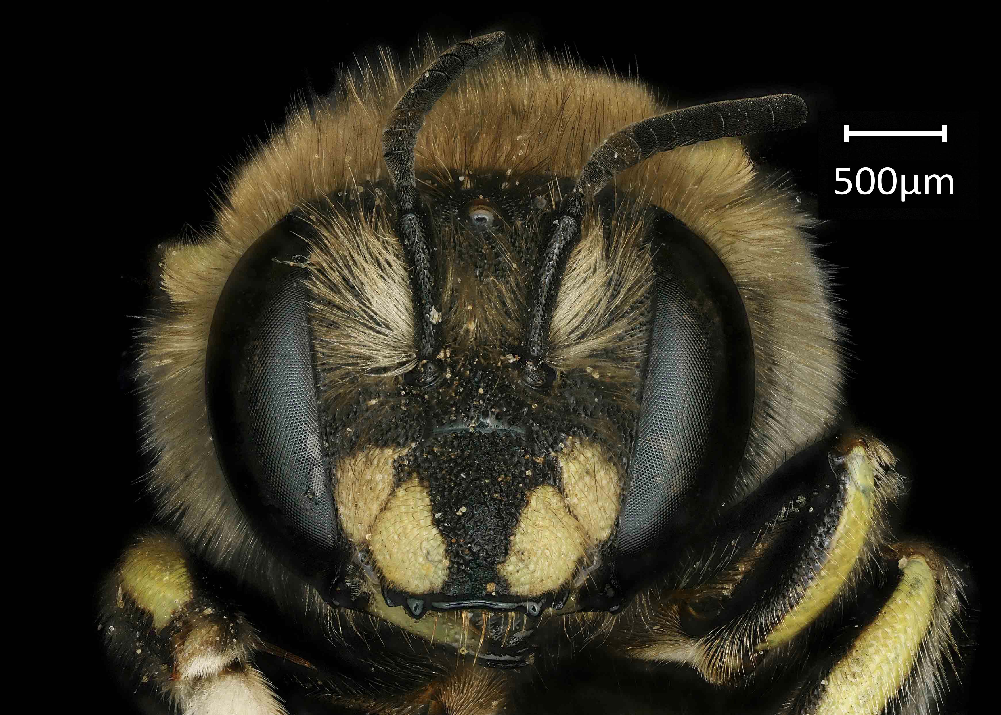
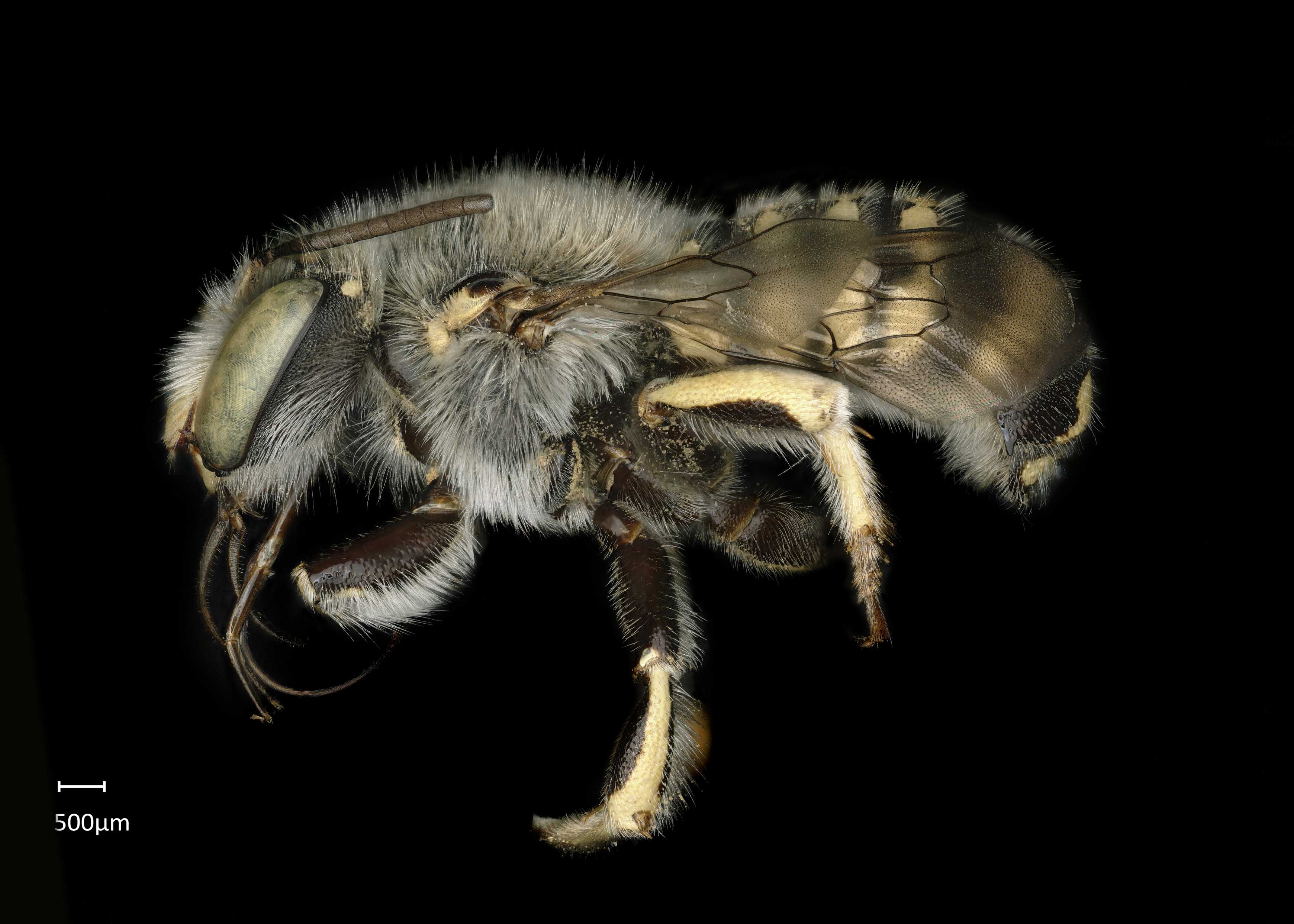
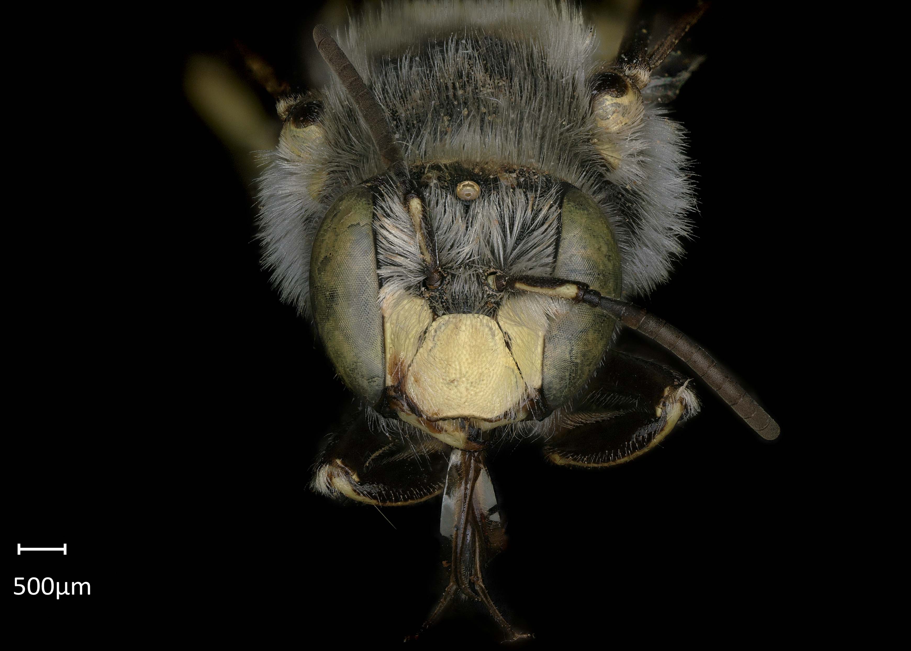
Anthidium (Anthidium) michenerorum are black with dark reddish-brown coloration from the coxaecoxae:
the basal segment of the leg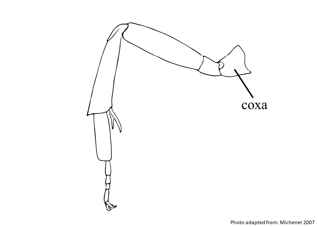 to the tibiaetibiae:
the segment of the leg, between the femur and the tarsus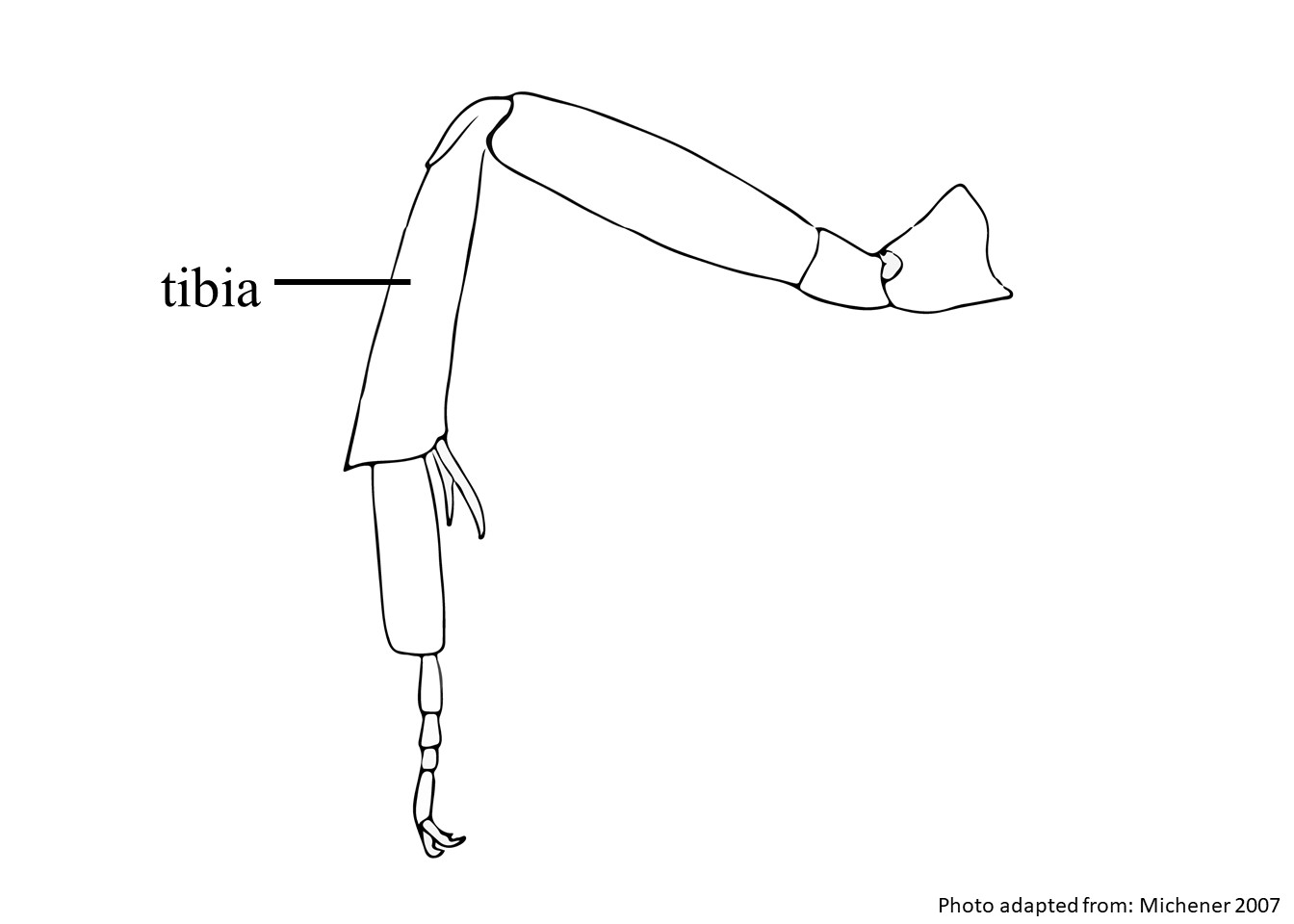 , light ferruginousferruginous:
rust-colored
coloration on the distitarsidistitarsi:
the fifth tarsal segment, furthest from the body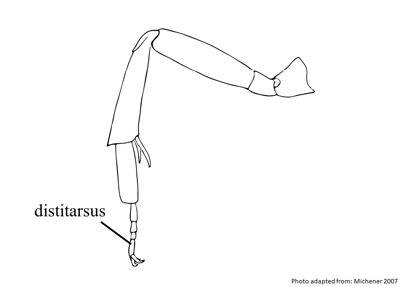 , and yellow or cream maculations (Gonzalez and Griswold 2013Gonzalez and Griswold 2013:
Gonzalez, V.H. and T.L. Griswold. 2013. Wool carder bees of the genus Anthidium in the Western Hemisphere (Hymenoptera: Megachilidae): diversity, host plant associations, phylogeny, and biogeography. Zoological Journal 168: 221ndash;425.). Females have white pubescencepubescence:
short, fine hair
except for yellow to ferruginousferruginous:
rust-colored
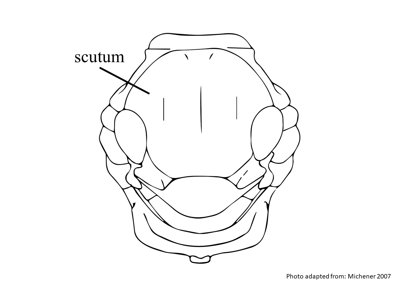
hairs on the vertexvertex:
the area between the ocelli and the back of the head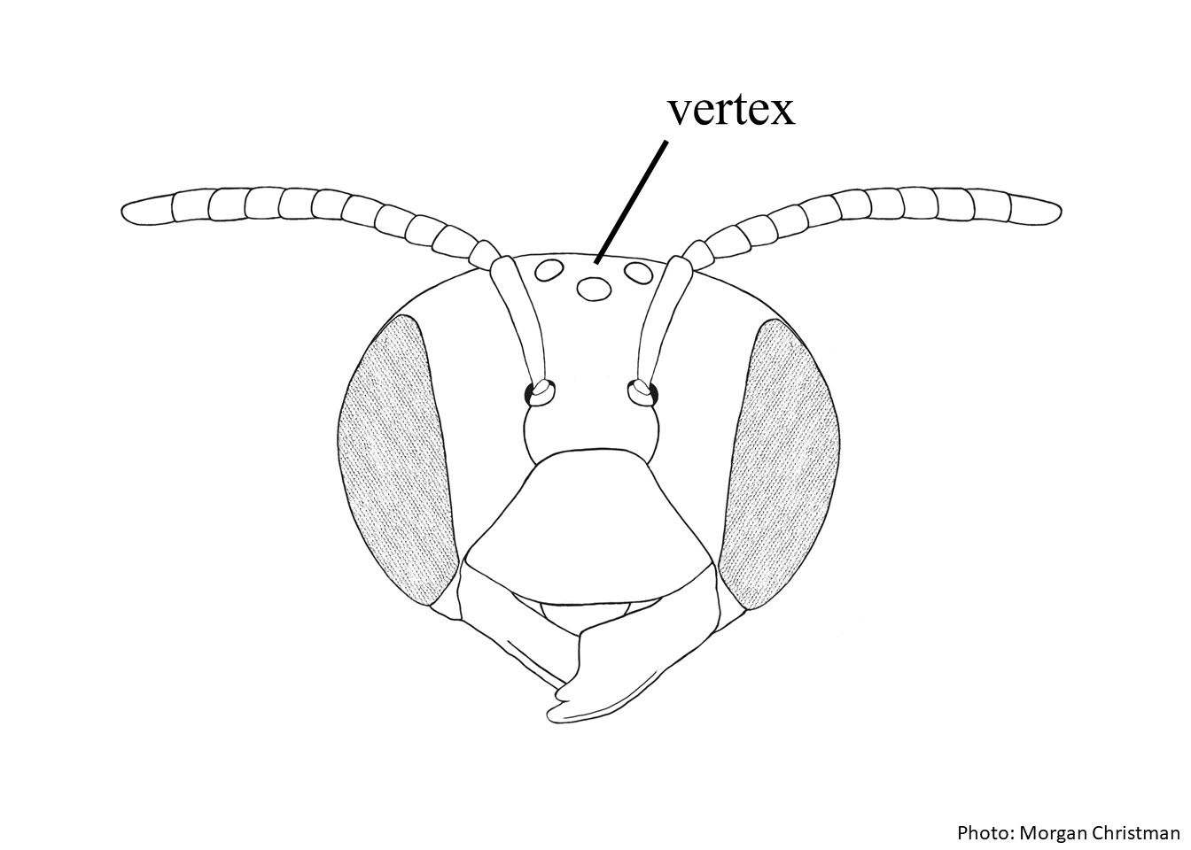 , scutumscutum:
the large segment on top of the thorax located between the wings and behind the head
 , axillaaxilla:
the triangular or rounded point on the thorax where thoracic muscles meet the forewing of an insect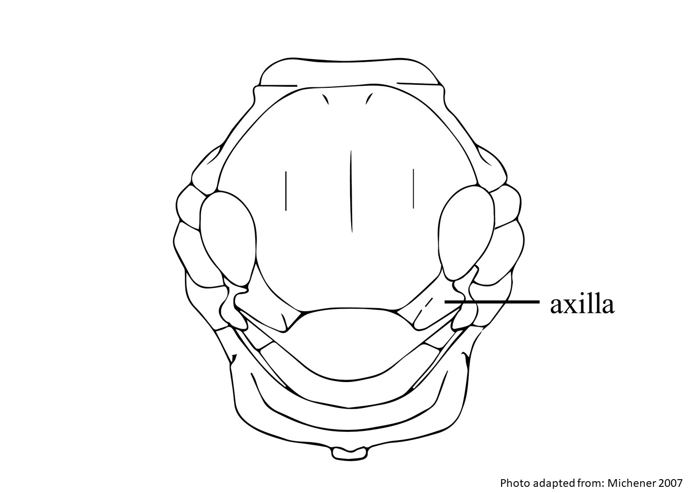 , scutellumscutellum:
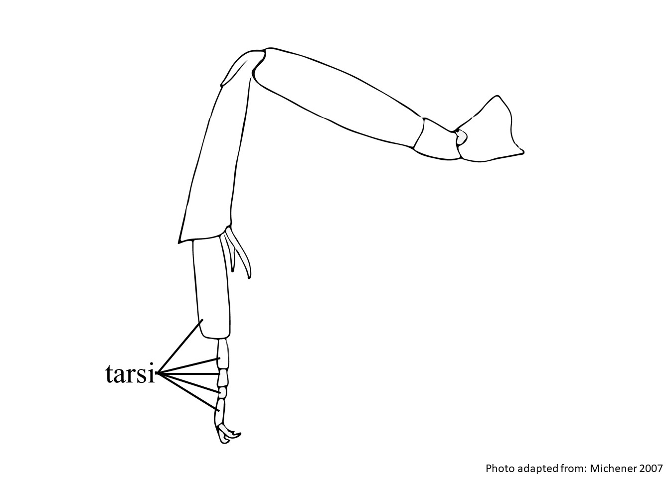
shield shaped plate behind scutum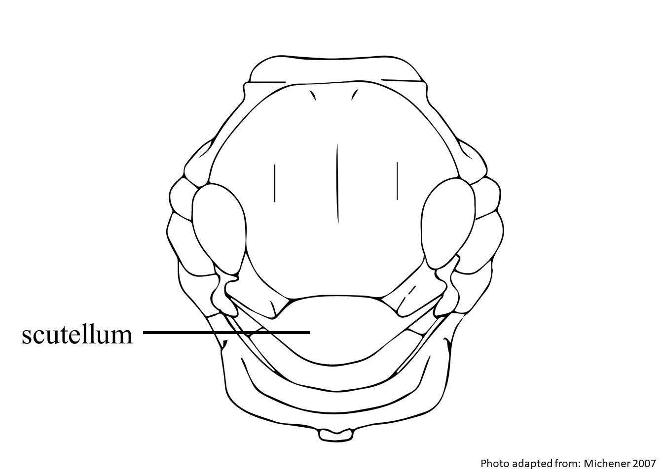 , inner tarsitarsi:
the group of segments at the end of the leg following the tibia
 , and apexapex:
end of any structure
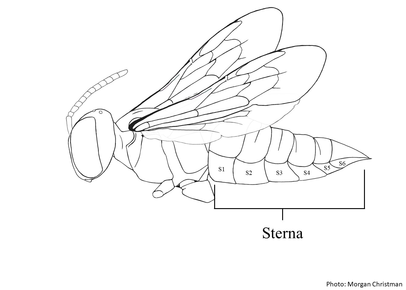
of S6S6:
the plates on the underside of the abdomen, often abbreviated when referring to a specific segment to S1, S2, S3, S4, S5, S6, S7, or S8
 . Females have a body length of 9.5–9.7 mm (Gonzalez and Griswold 2013Gonzalez and Griswold 2013:
Gonzalez, V.H. and T.L. Griswold. 2013. Wool carder bees of the genus Anthidium in the Western Hemisphere (Hymenoptera: Megachilidae): diversity, host plant associations, phylogeny, and biogeography. Zoological Journal 168: 221ndash;425.). Males have white pubescencepubescence:
short, fine hair
, and range in body length from 9.4–14.3 mm (Gonzalez and Griswold 2013Gonzalez and Griswold 2013:
Gonzalez, V.H. and T.L. Griswold. 2013. Wool carder bees of the genus Anthidium in the Western Hemisphere (Hymenoptera: Megachilidae): diversity, host plant associations, phylogeny, and biogeography. Zoological Journal 168: 221ndash;425.).
(modified from Gonzalez and Griswold 2013Gonzalez and Griswold 2013:
Gonzalez, V.H. and T.L. Griswold. 2013. Wool carder bees of the genus Anthidium in the Western Hemisphere (Hymenoptera: Megachilidae): diversity, host plant associations, phylogeny, and biogeography. Zoological Journal 168: 221ndash;425.)
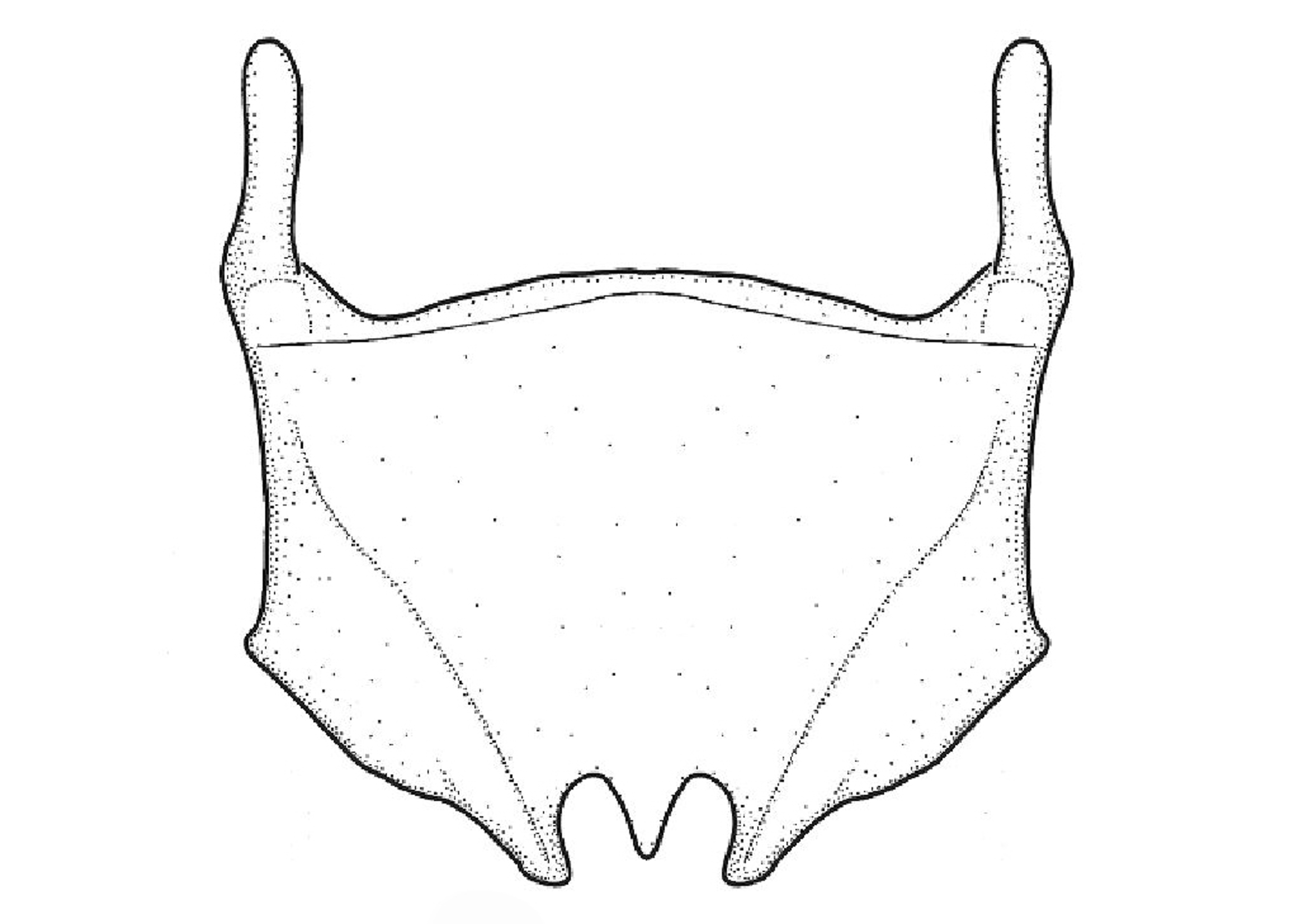
 lacks basalbasal:
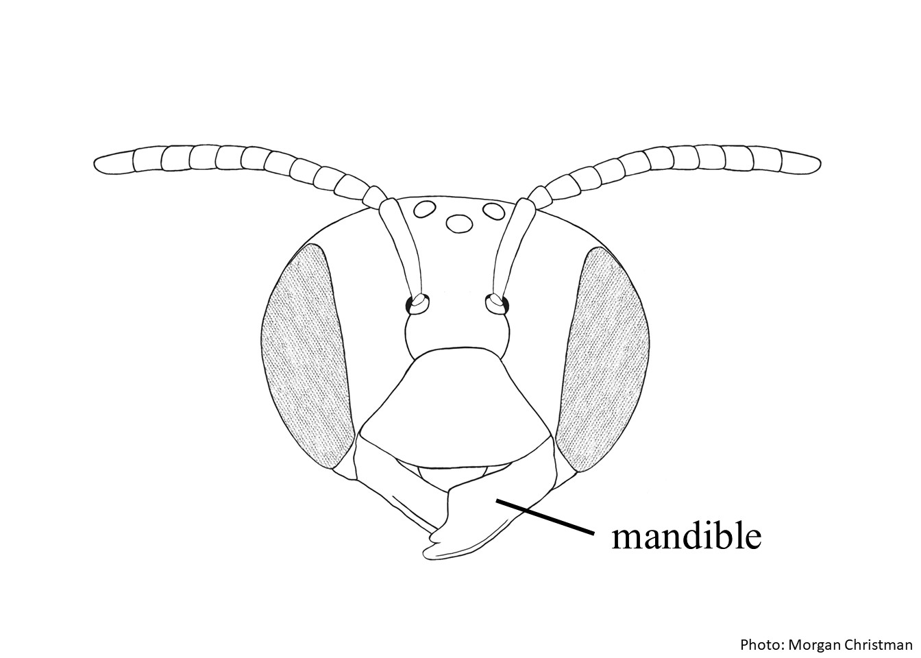 has six teeth.
 triangle is shiny and smooth. without anterioranterior:
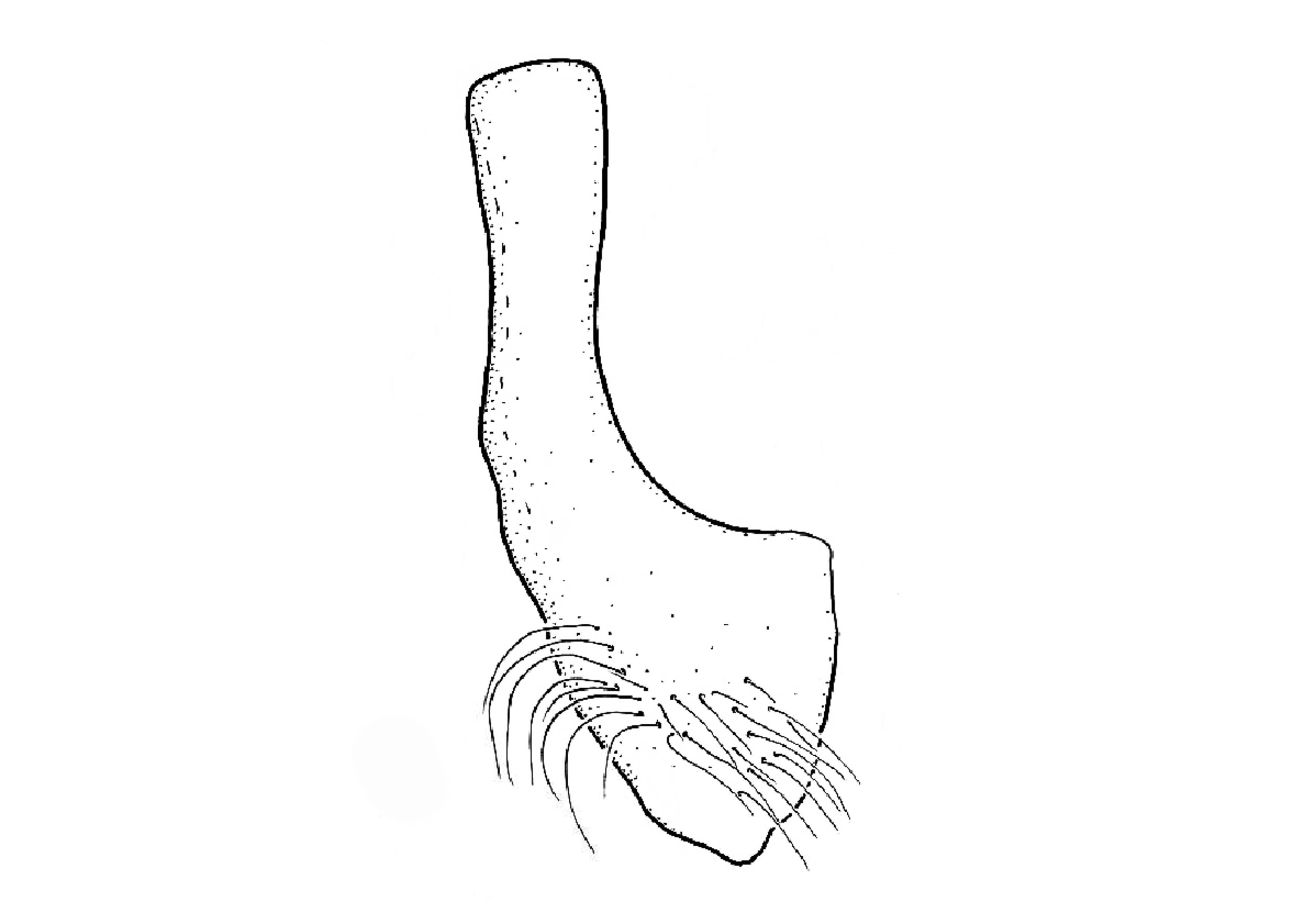
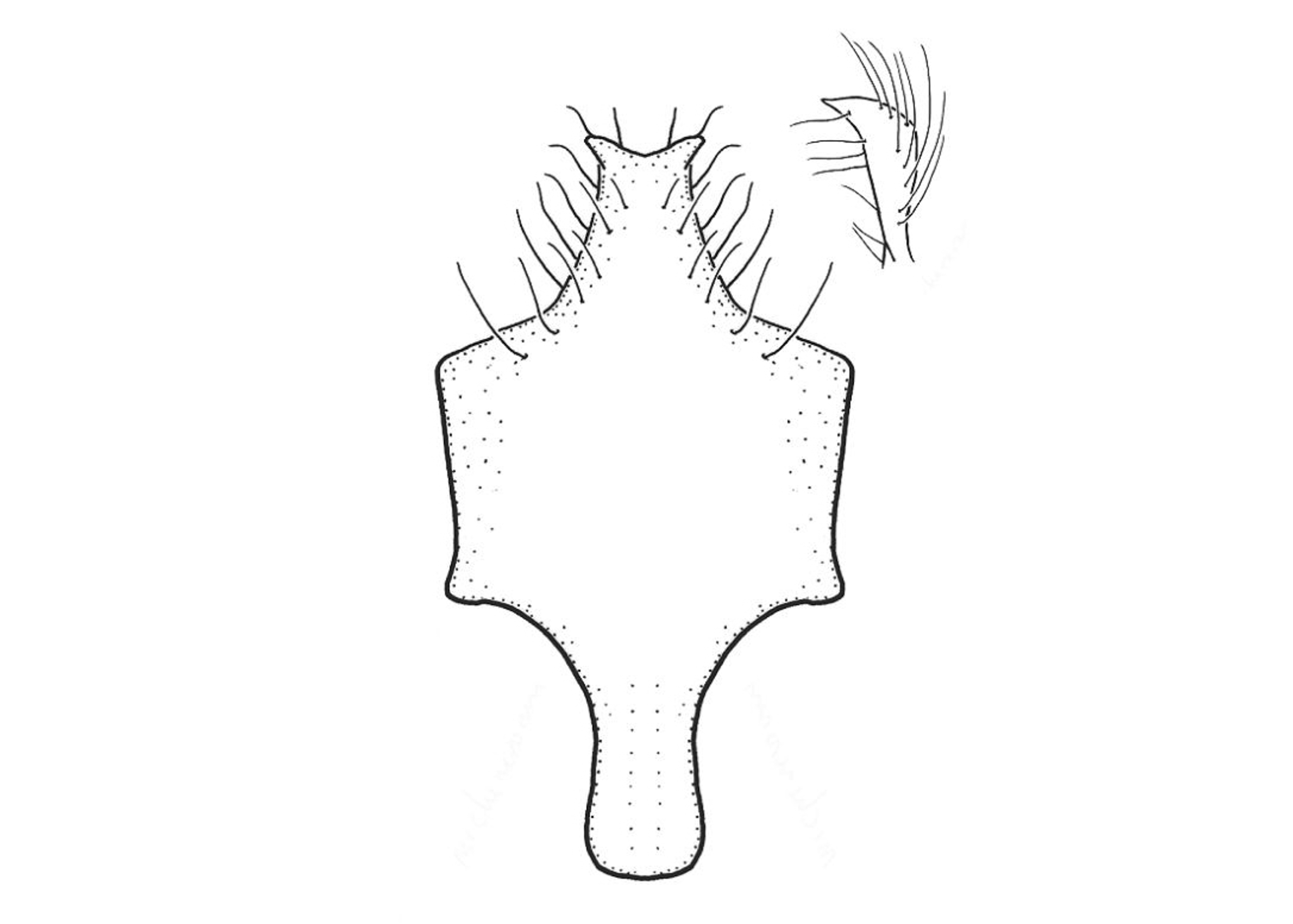
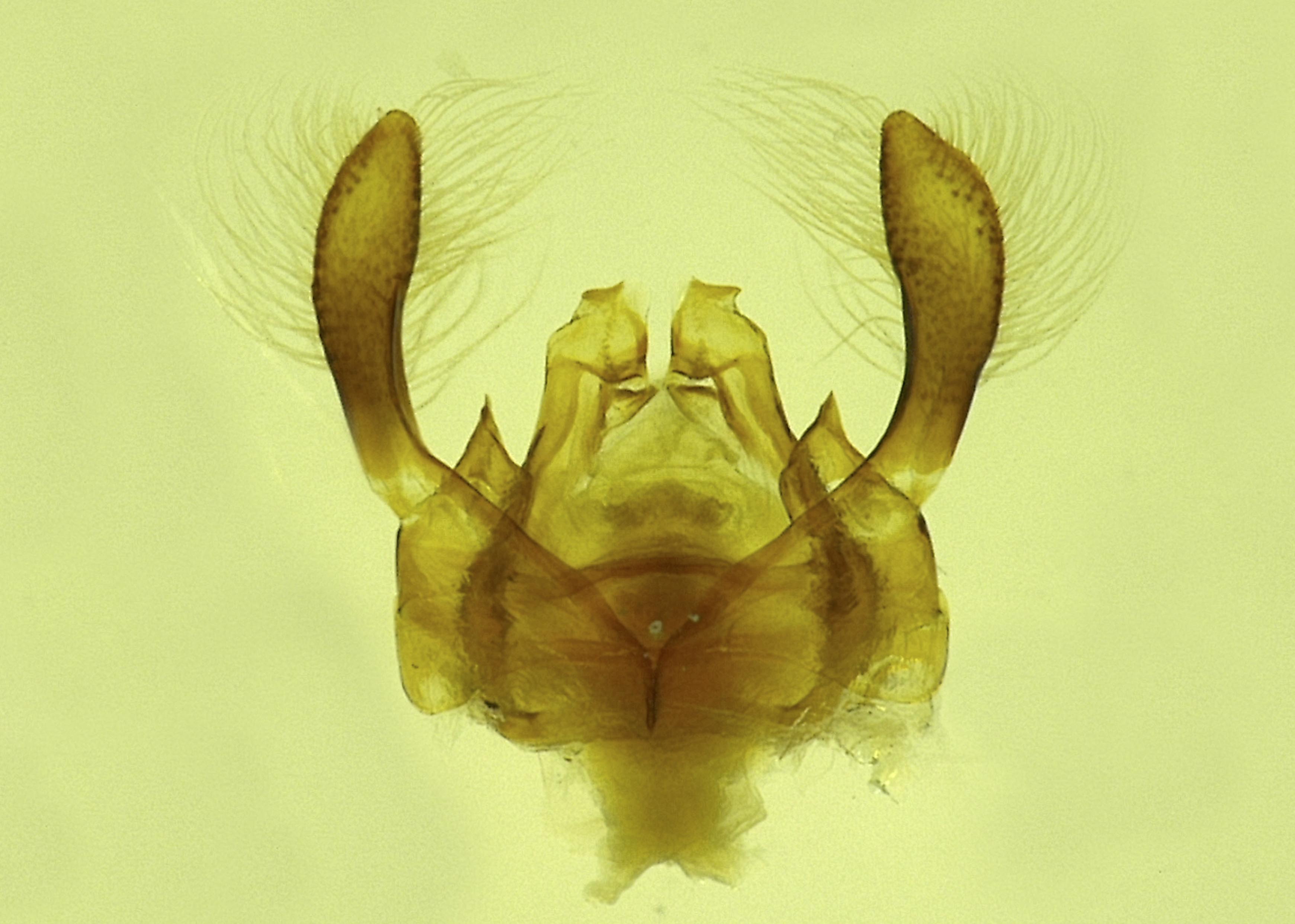
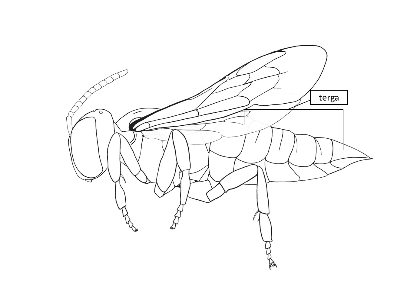 with a median apicalapical: preapical projections are longer than those in females. median apicalapical: apicalapical: submedian and median lobes are small and digitiform, giving S6S6: an almost trilobed appearance. is pointed apicallyapically: apicalapical: laterallateral: median spine. laterallateral:
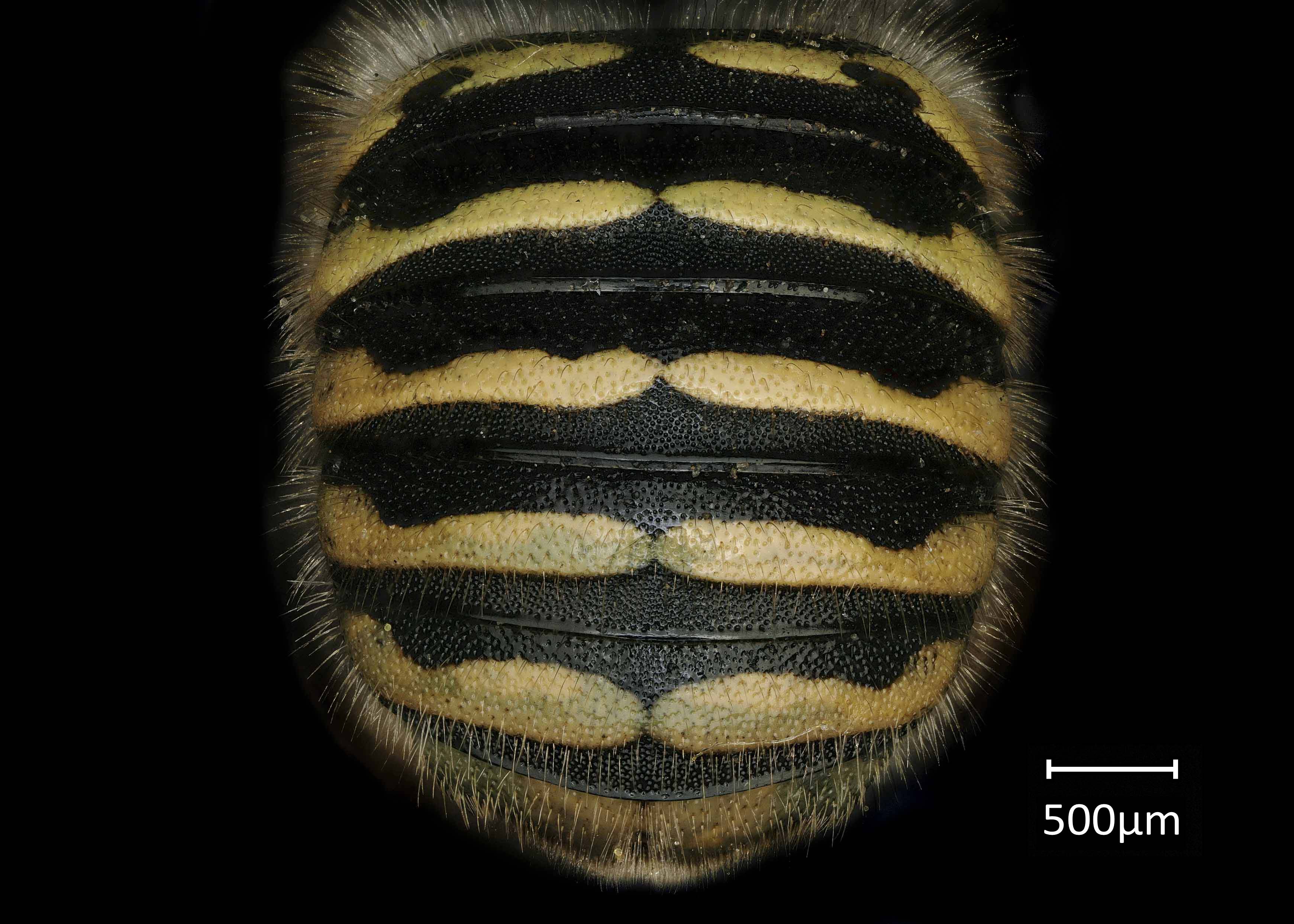
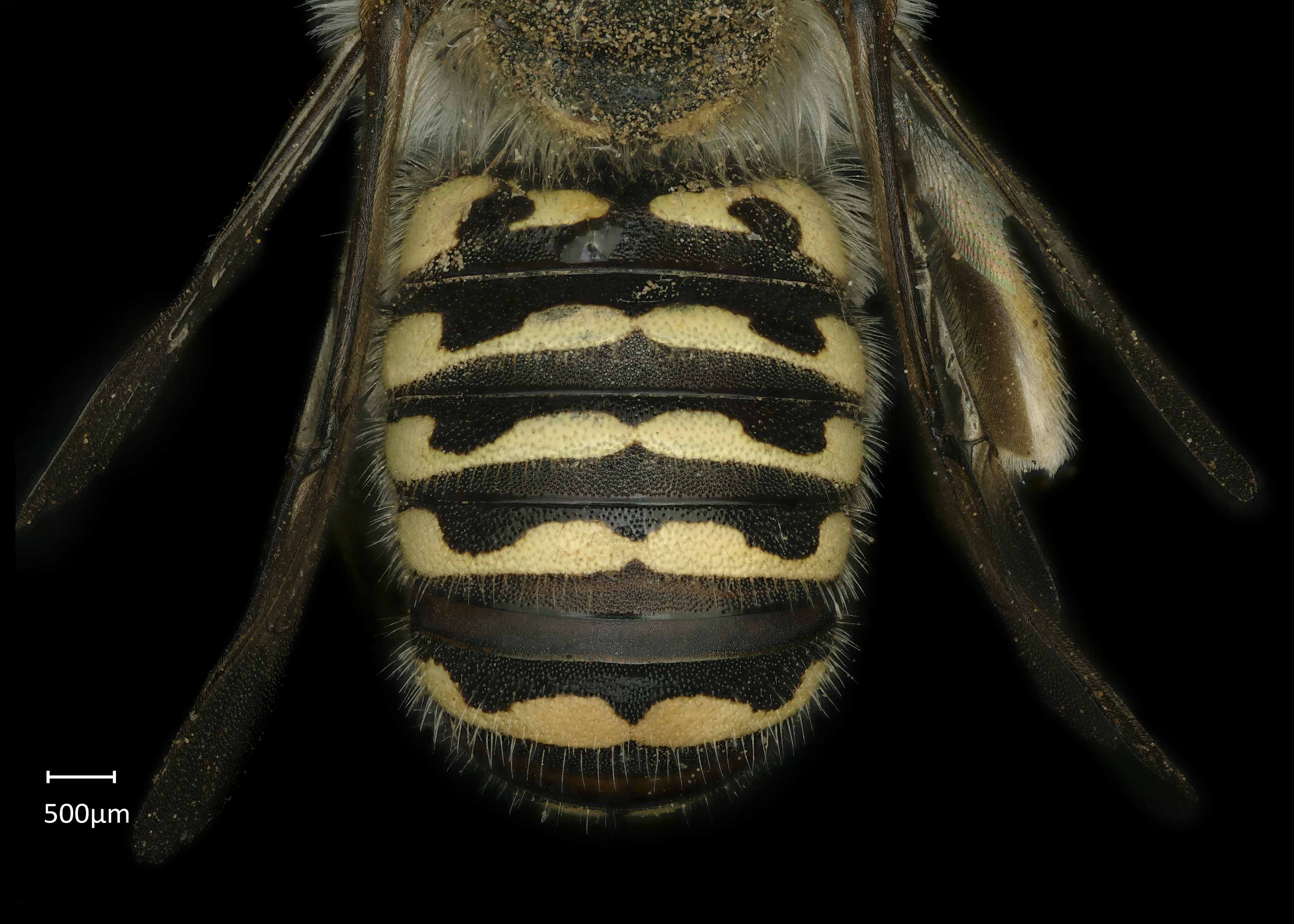
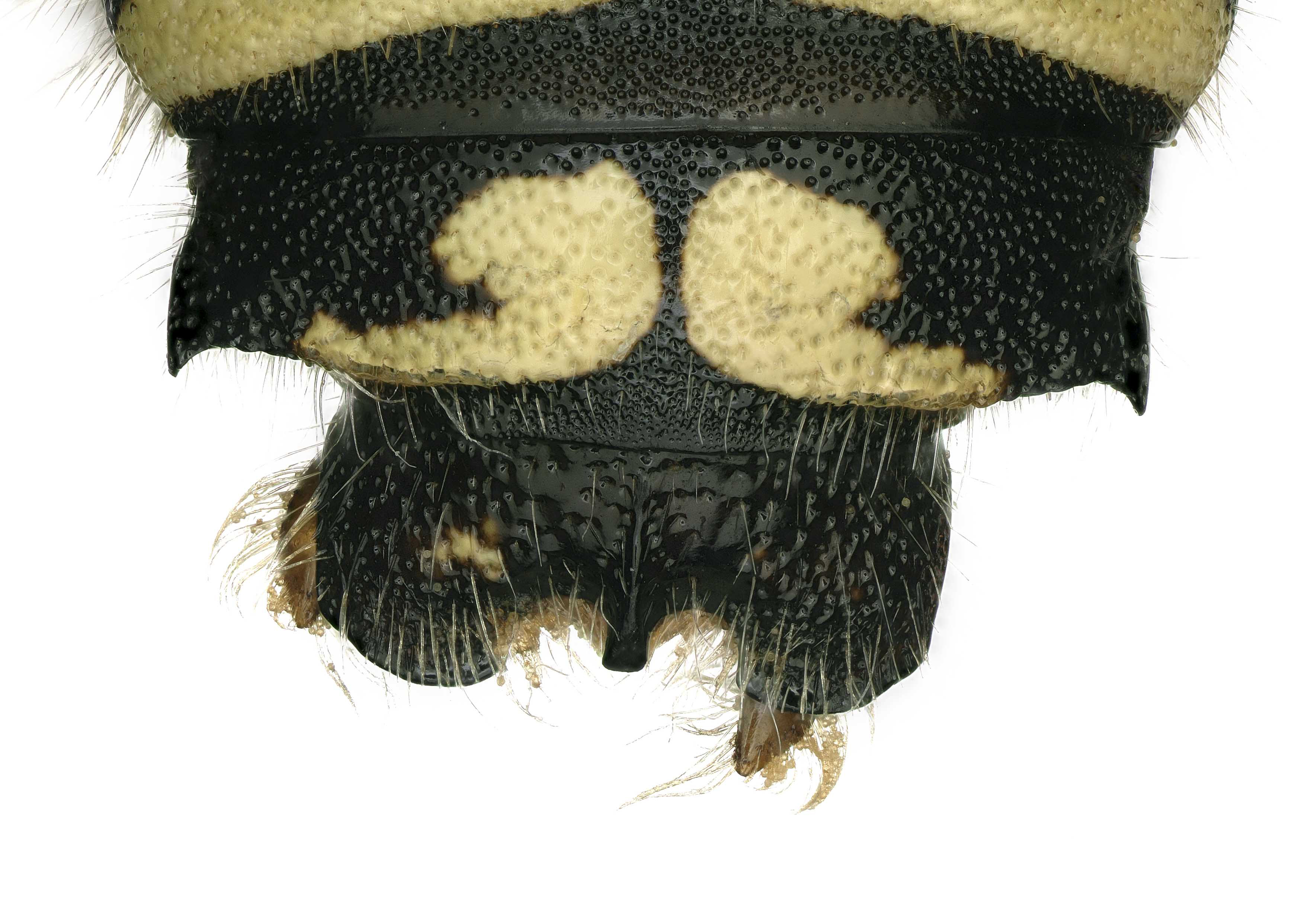
Anthidium michenerorum can be easily distinguished from all other Anthidium species in the northwestern U.S. by the coarse and densely punctatepunctate:
studded with tiny holes
terga with doubly carinatecarinate:
having keels or carinae
apicalapical:
near or at the apex or end of any structure
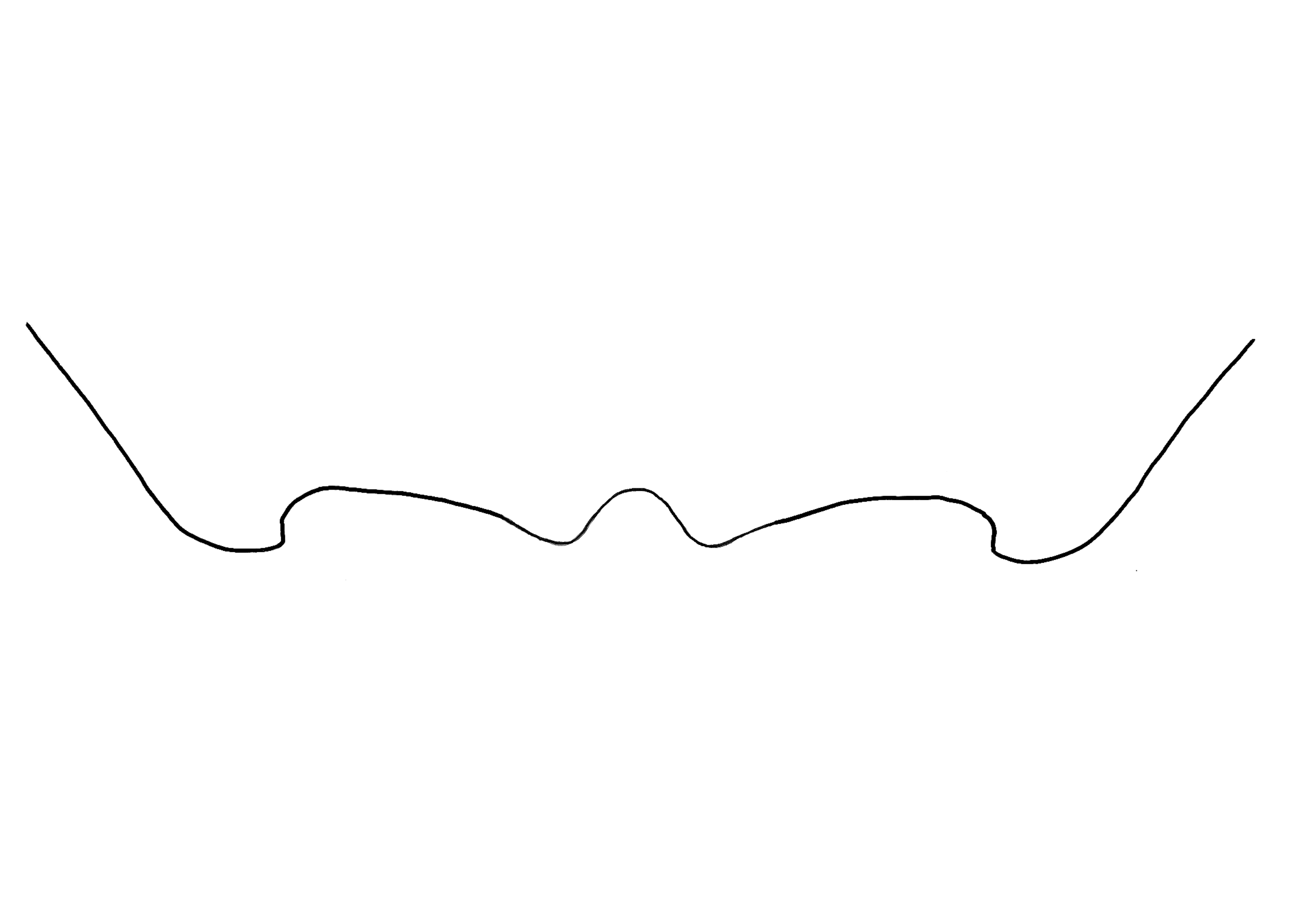
margins. Female A. michenerorum can be distinguished from other Anthidium by the shape of T6T6:
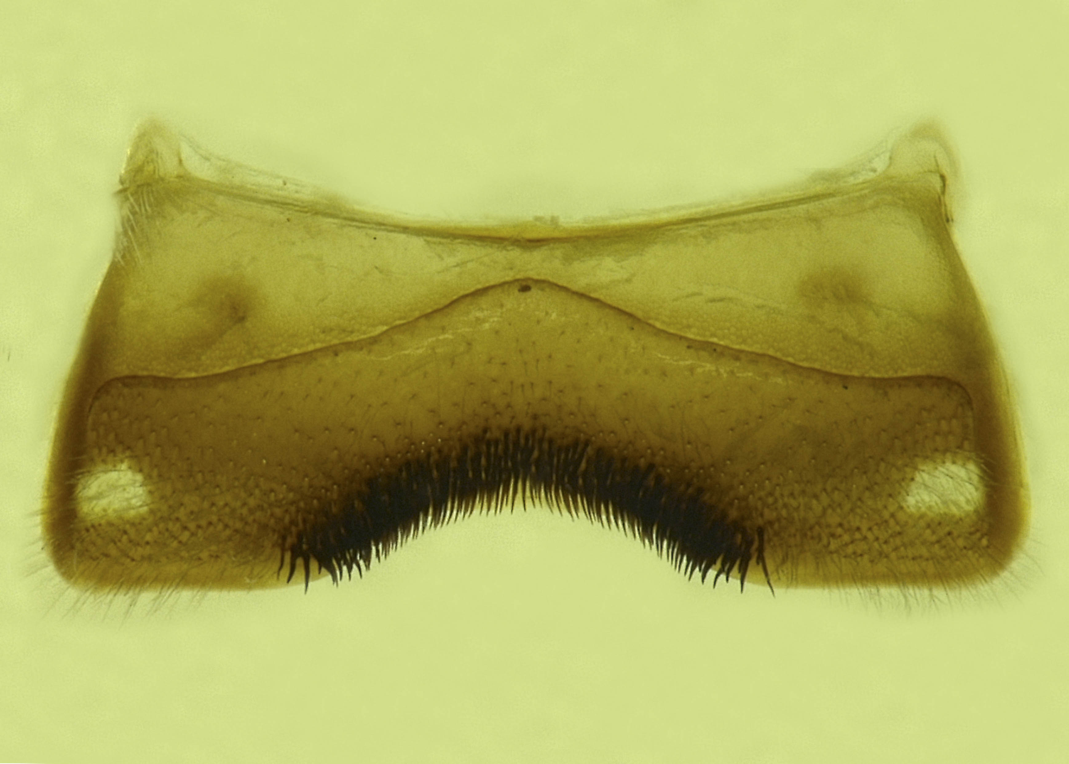
the segments on the top side of the abdomen, often abbreviated when referring to a specific segment to T1, T2, T3, T4, T5, T6, or T7. Male A. michenerorum can be distinguished from other Anthidium by the deeply concave S4S4:
the plates on the underside of the abdomen, often abbreviated when referring to a specific segment to S1, S2, S3, S4, S5, S6, S7, or S8
with a broad median apicalapical:
near or at the apex or end of any structure
brush and the shape of S6S6:
the plates on the underside of the abdomen, often abbreviated when referring to a specific segment to S1, S2, S3, S4, S5, S6, S7, or S8
(Gonzalez and Griswold 2013Gonzalez and Griswold 2013:
Gonzalez, V.H. and T.L. Griswold. 2013. Wool carder bees of the genus Anthidium in the Western Hemisphere (Hymenoptera: Megachilidae): diversity, host plant associations, phylogeny, and biogeography. Zoological Journal 168: 221ndash;425.).
Anthidium michenerorum adults have been recorded in flight from April to early June (Gonzalez and Griswold 2013Gonzalez and Griswold 2013:
Gonzalez, V.H. and T.L. Griswold. 2013. Wool carder bees of the genus Anthidium in the Western Hemisphere (Hymenoptera: Megachilidae): diversity, host plant associations, phylogeny, and biogeography. Zoological Journal 168: 221ndash;425.).
Anthidium michenerorum has been observed visiting Astragalus gracilis, Astragalus racemosus, and Psoralea cuspidata (Fabaceae) (Gonzalez and Griswold 2013Gonzalez and Griswold 2013:
Gonzalez, V.H. and T.L. Griswold. 2013. Wool carder bees of the genus Anthidium in the Western Hemisphere (Hymenoptera: Megachilidae): diversity, host plant associations, phylogeny, and biogeography. Zoological Journal 168: 221ndash;425.).
Nesting behavior is unknown.
Anthidium michenerorum occur in the southern Great Plains in Kansas, Oklahoma, and Texas. They are restricted to grassland ecosystems (Gonzalez and Griswold 2013Gonzalez and Griswold 2013:
Gonzalez, V.H. and T.L. Griswold. 2013. Wool carder bees of the genus Anthidium in the Western Hemisphere (Hymenoptera: Megachilidae): diversity, host plant associations, phylogeny, and biogeography. Zoological Journal 168: 221ndash;425.).
Distribution map generated by Discover Life -- click on map for details, credits, and terms of use.
Gonzalez, V.H. and T.L. Griswold. 2013. Wool carder bees of the genus Anthidium in the Western Hemisphere (Hymenoptera: Megachilidae): diversity, host plant associations, phylogeny, and biogeography. Zoological Journal of the Linnean Society 168: 221-425.
Authors: S. Burrows, C. Ritner, M. Christman, L. Spears, A. Smith-Pardo, S. Price, R. Ramirez, T. Griswold, A. Redford
Edition 3 - last updated November 2021
tool images at ITP Node
idtools.org