
Family: Megachilidae
Subfamily: Megachilinae
Tribe: Anthidiini
Genus: Anthidium Fabricius, 1804
Subgenus: A. (Anthidium) Fabricius, 1804
Species: Anthidium chamelense Gonzalez and Griswold, 2013
Common name: none
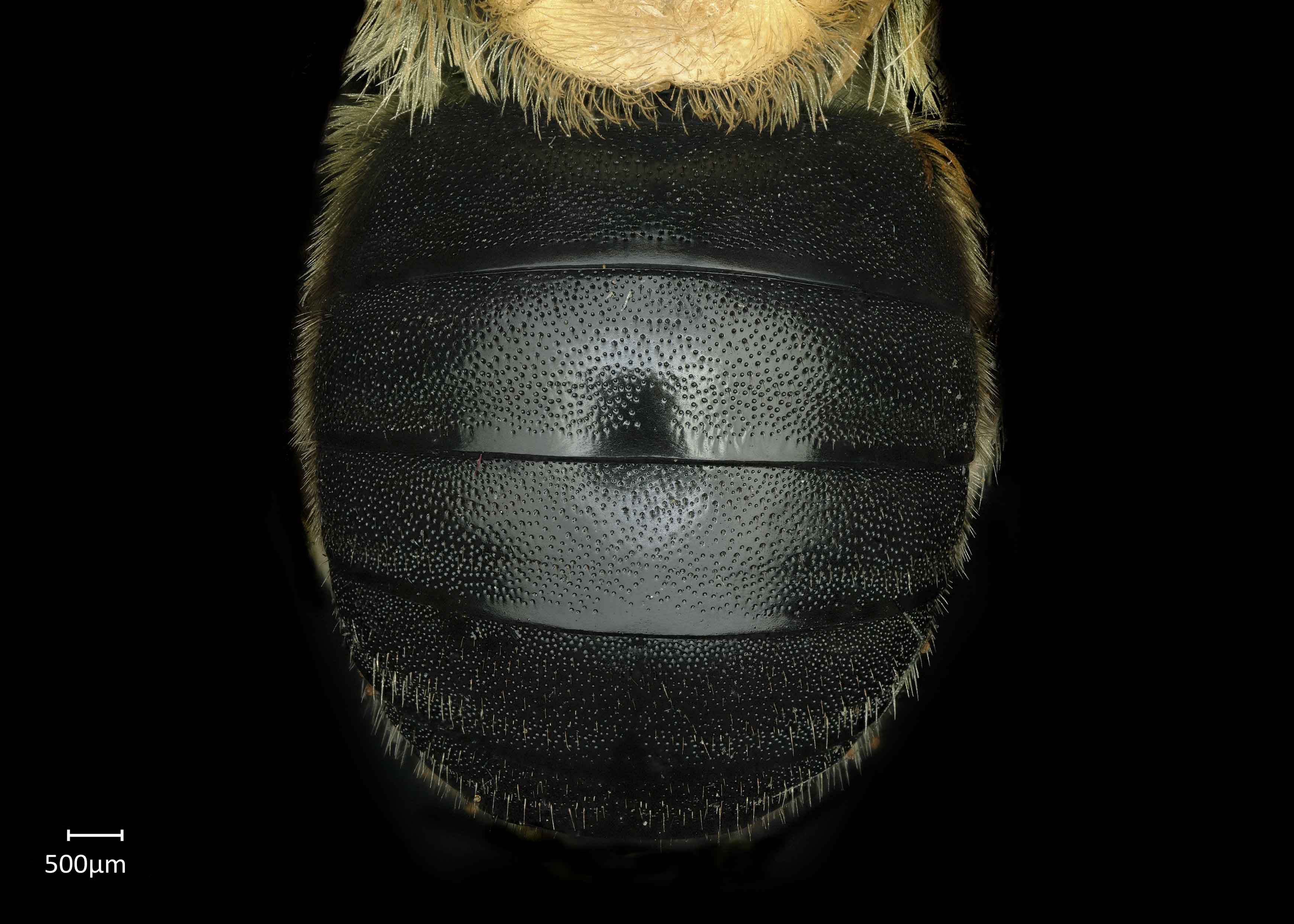
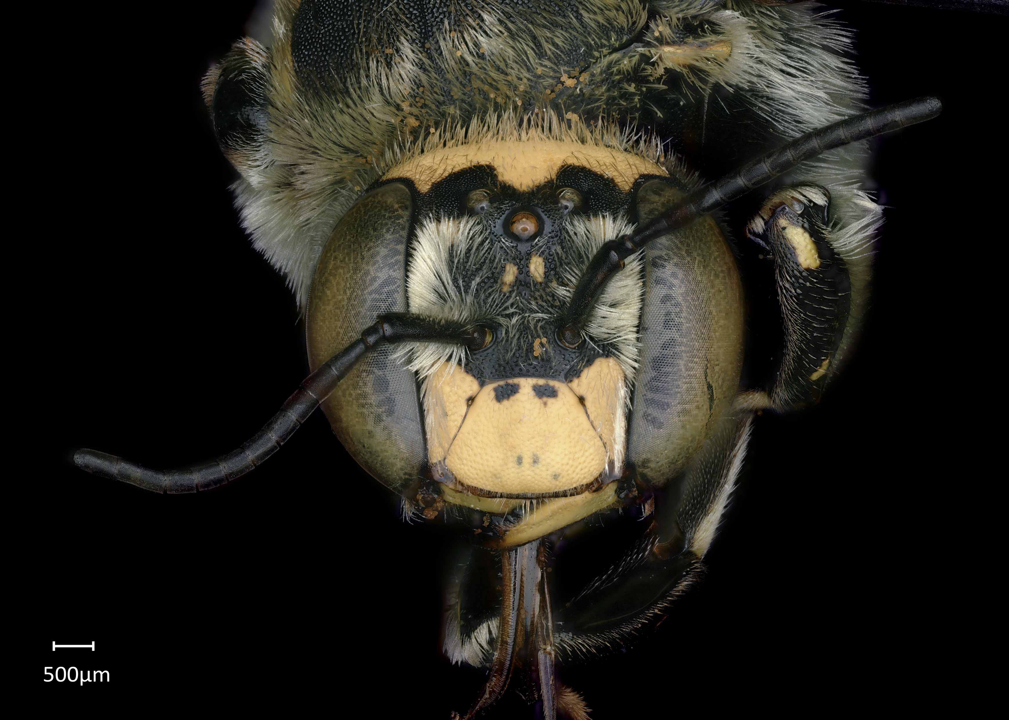
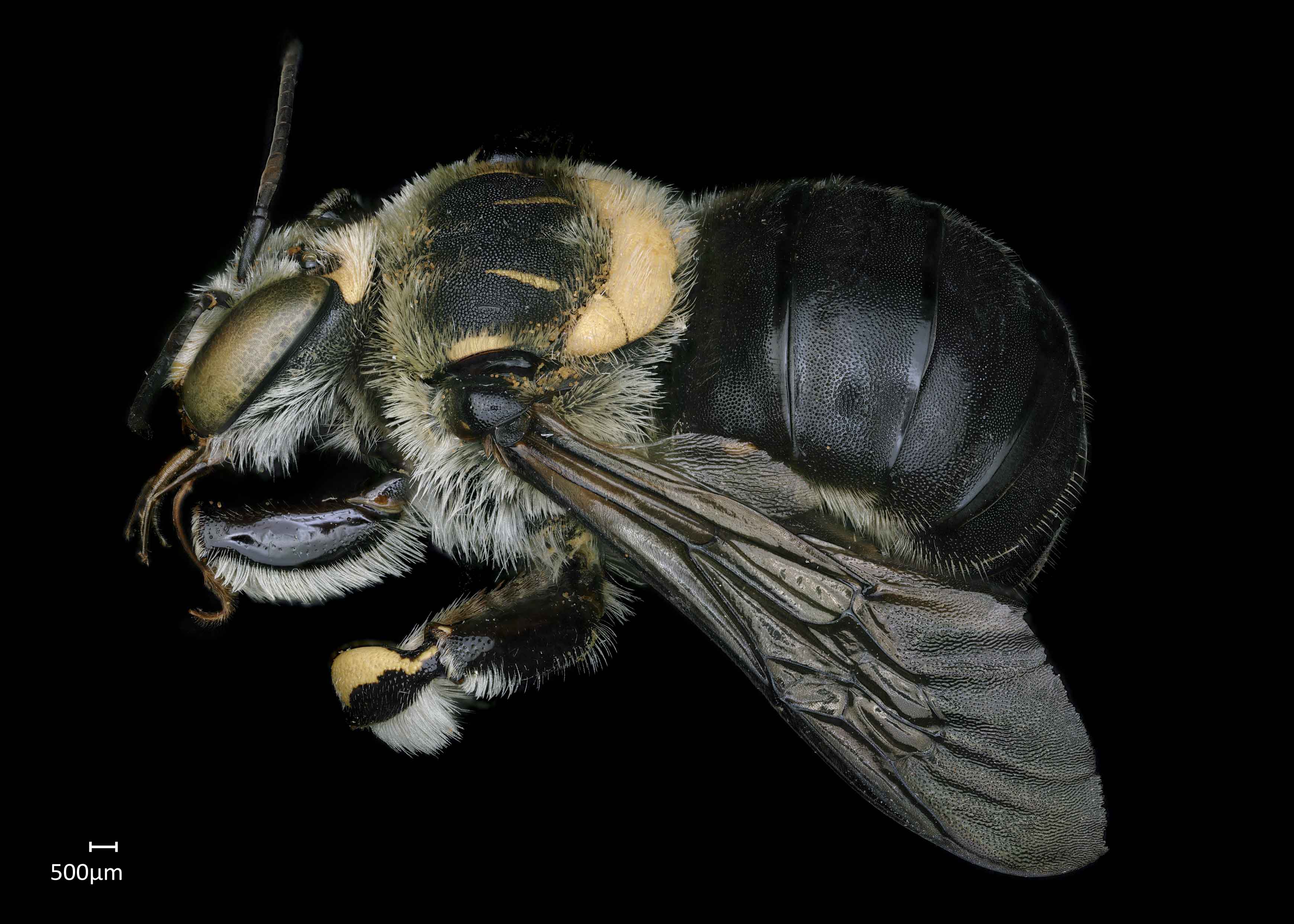
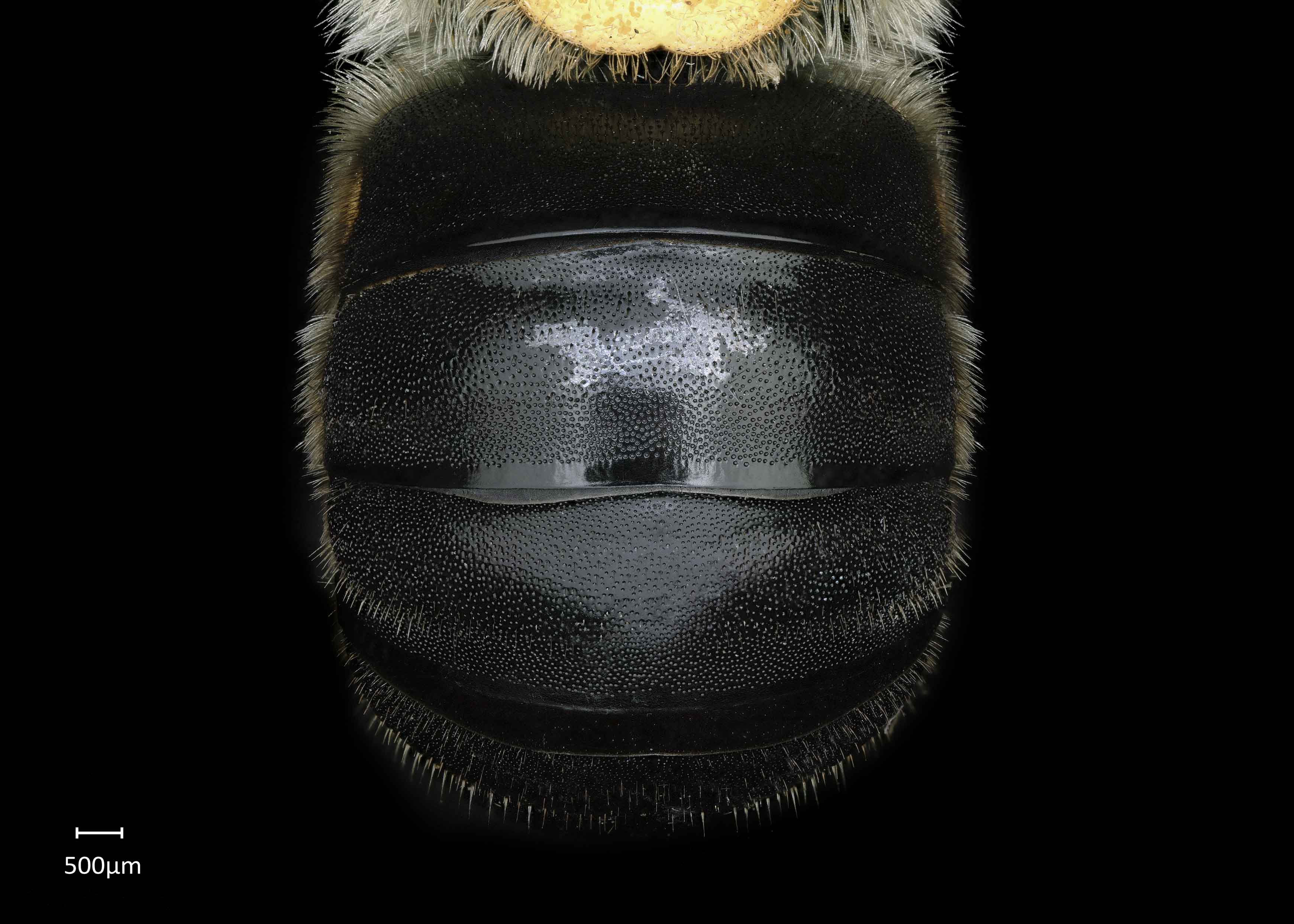
Anthidium (Anthidium) chamelense have an entirely, or almost entirely, black abdomen with an entirely yellow scutellumscutellum:
shield shaped plate behind scutum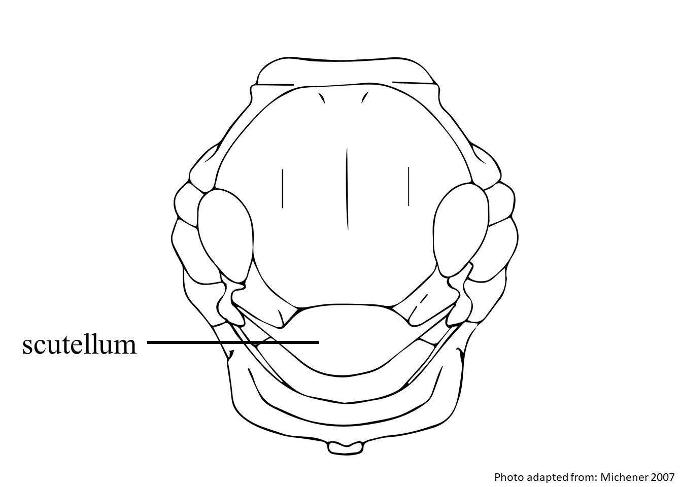 and axillae (Gonzalez and Griswold 2013Gonzalez and Griswold 2013:
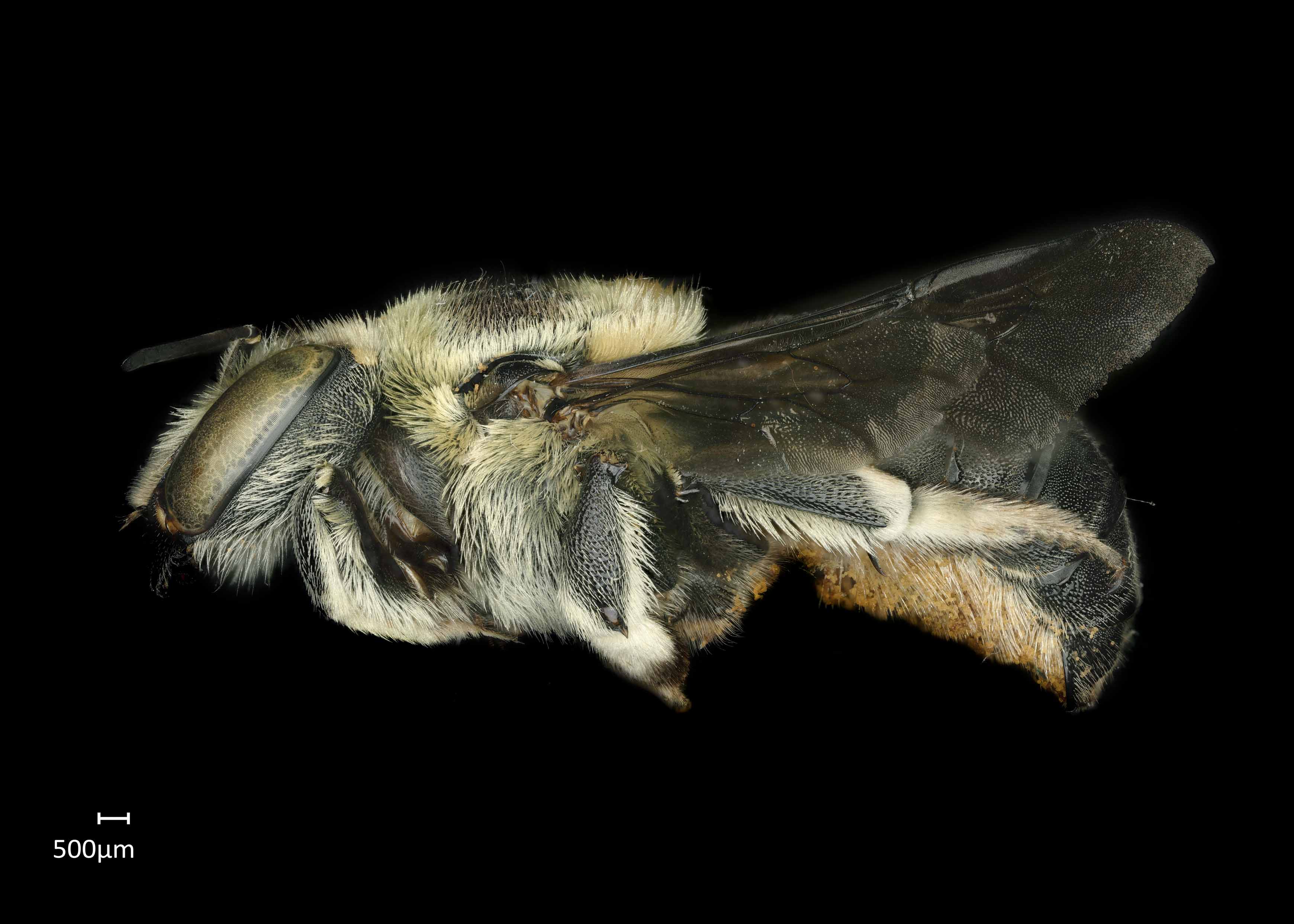
Gonzalez, V.H. and T.L. Griswold. 2013. Wool carder bees of the genus Anthidium in the Western Hemisphere (Hymenoptera: Megachilidae): diversity, host plant associations, phylogeny, and biogeography. Zoological Journal 168: 221ndash;425.). Females have white pubescencepubescence:
short, fine hair
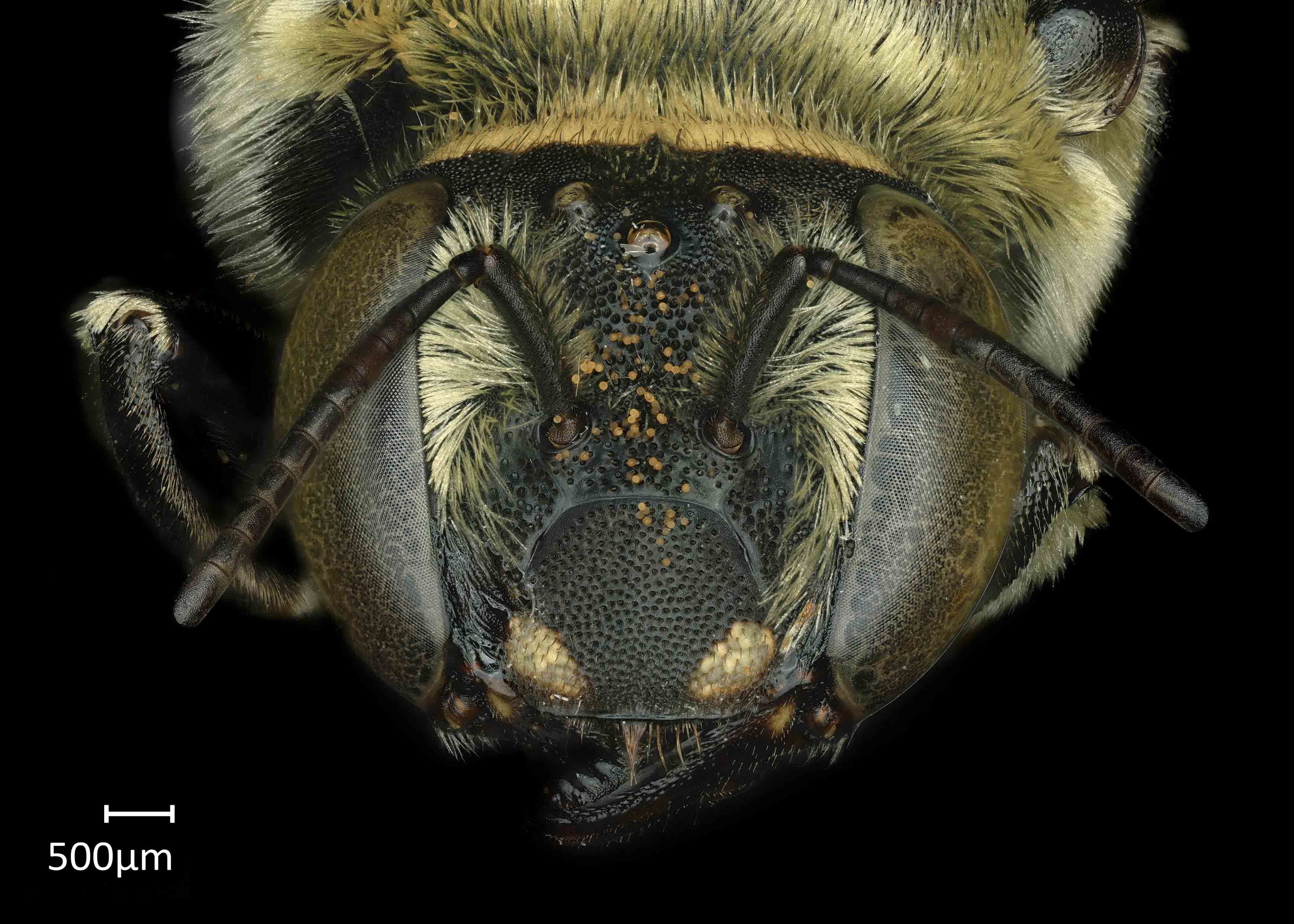
with limited dark brown to black hairs on their clypeusclypeus:
a section of the face below the antennae, demarcated by the epistomal sutures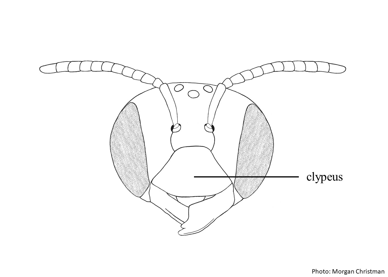 , supraclypeal areasupraclypeal area:
the region of the head between the antennal sockets and clypeus, demarcated on the sides by the subantennal sutures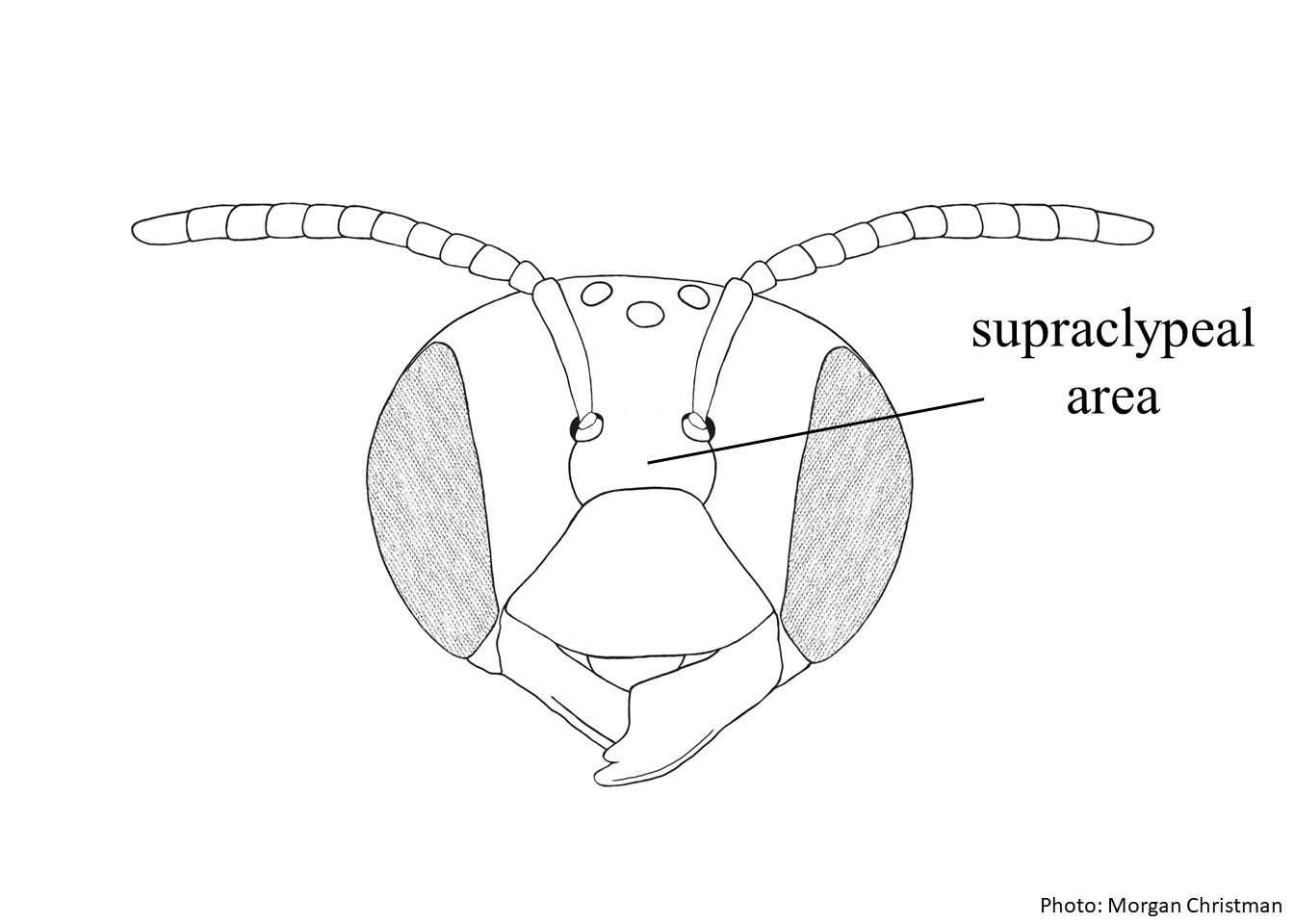 , vertexvertex:
the area between the ocelli and the back of the head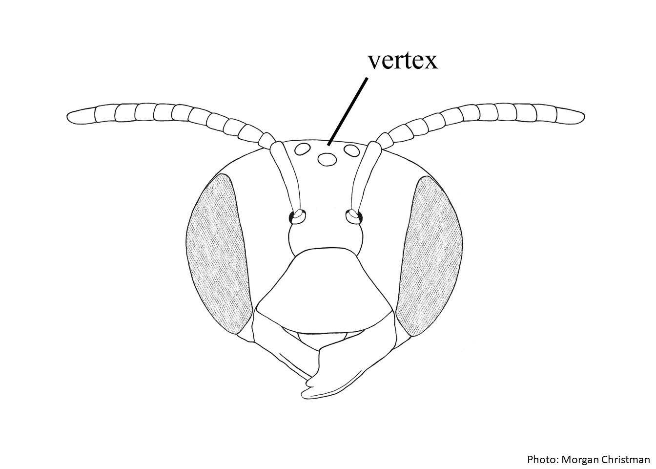 , discdisc:
a generic term for the middle surface of a plate (usually in reference to an abdominal segment)
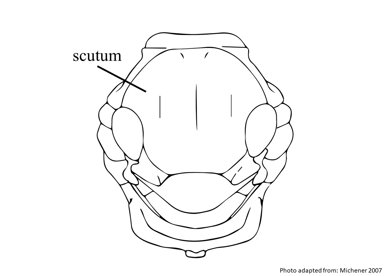
of the scutumscutum:
the large segment on top of the thorax located between the wings and behind the head
 , coxaecoxae:
the basal segment of the leg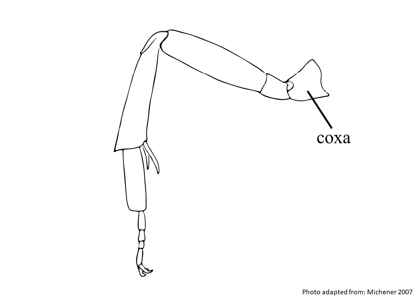 , trochanters, outer mid basitarsusbasitarsus:
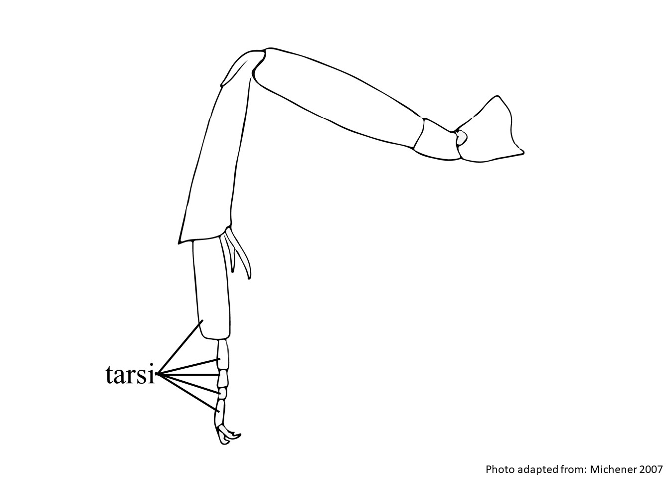
the segment of the tarsus that is the nearest to the body of the bee, usually the largest of all the tarsal segments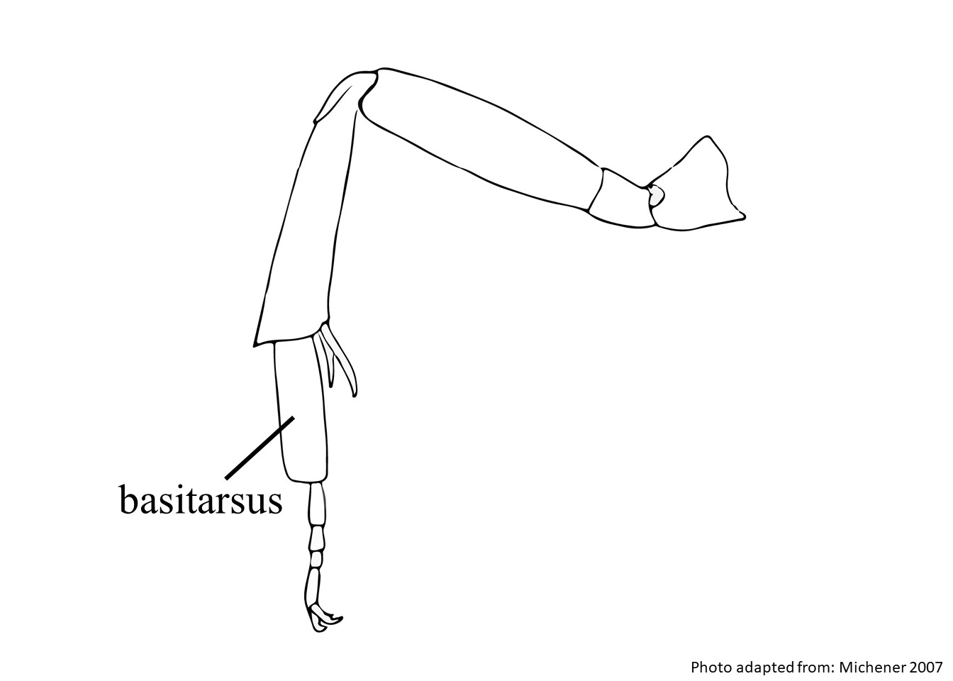 , inner tarsitarsi:
the group of segments at the end of the leg following the tibia
 , and tergaterga:
the segments on the top side of the abdomen, often abbreviated when referring to a specific segment to T1, T2, T3, T4, T5, T6, or T7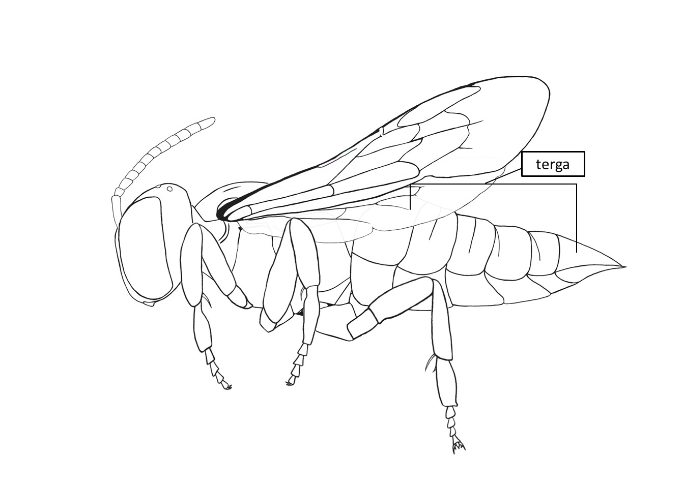 (except T6). Females range in body length from 16.2–20 mm. Males have the same color pubescencepubescence:
short, fine hair
as females, and range in body length from 18.5–21.5 mm (Gonzalez and Griswold 2013Gonzalez and Griswold 2013:
Gonzalez, V.H. and T.L. Griswold. 2013. Wool carder bees of the genus Anthidium in the Western Hemisphere (Hymenoptera: Megachilidae): diversity, host plant associations, phylogeny, and biogeography. Zoological Journal 168: 221ndash;425.).
(modified from Gonzalez and Griswold 2013Gonzalez and Griswold 2013:
Gonzalez, V.H. and T.L. Griswold. 2013. Wool carder bees of the genus Anthidium in the Western Hemisphere (Hymenoptera: Megachilidae): diversity, host plant associations, phylogeny, and biogeography. Zoological Journal 168: 221ndash;425.)
are covered with dense tomentumtomentum:, supraclypeal areasupraclypeal area:, and fronsfrons: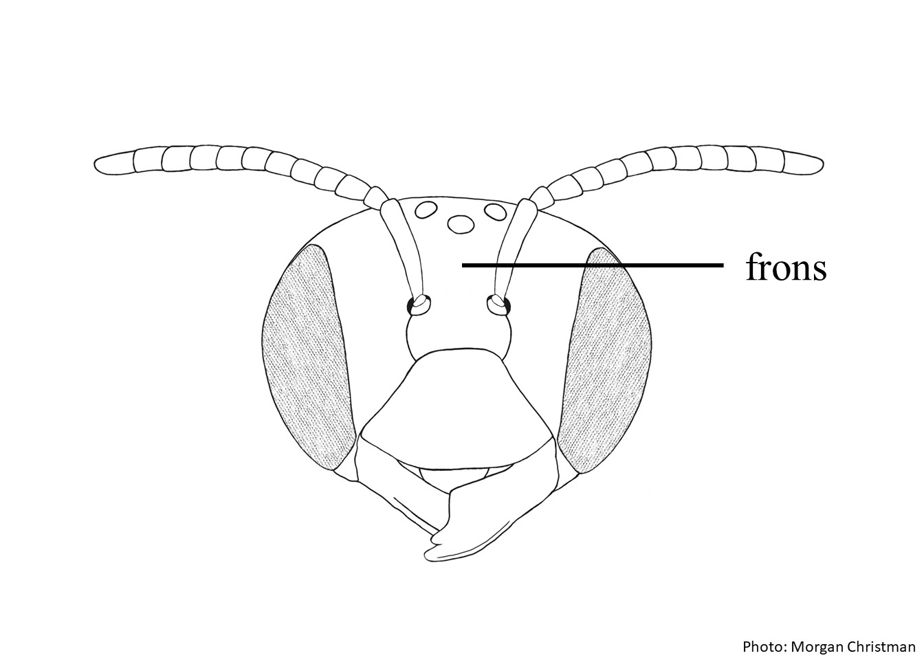 are covered with simple, stiff, and apicallyapically:
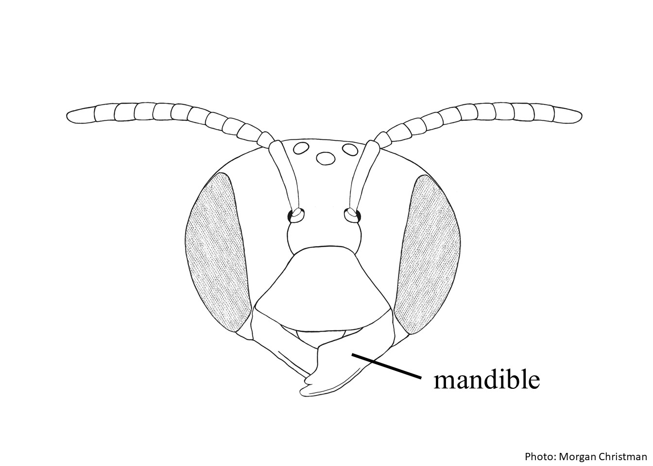 has 7–8 teeth.
 triangle is dull and finely punctatepunctate:
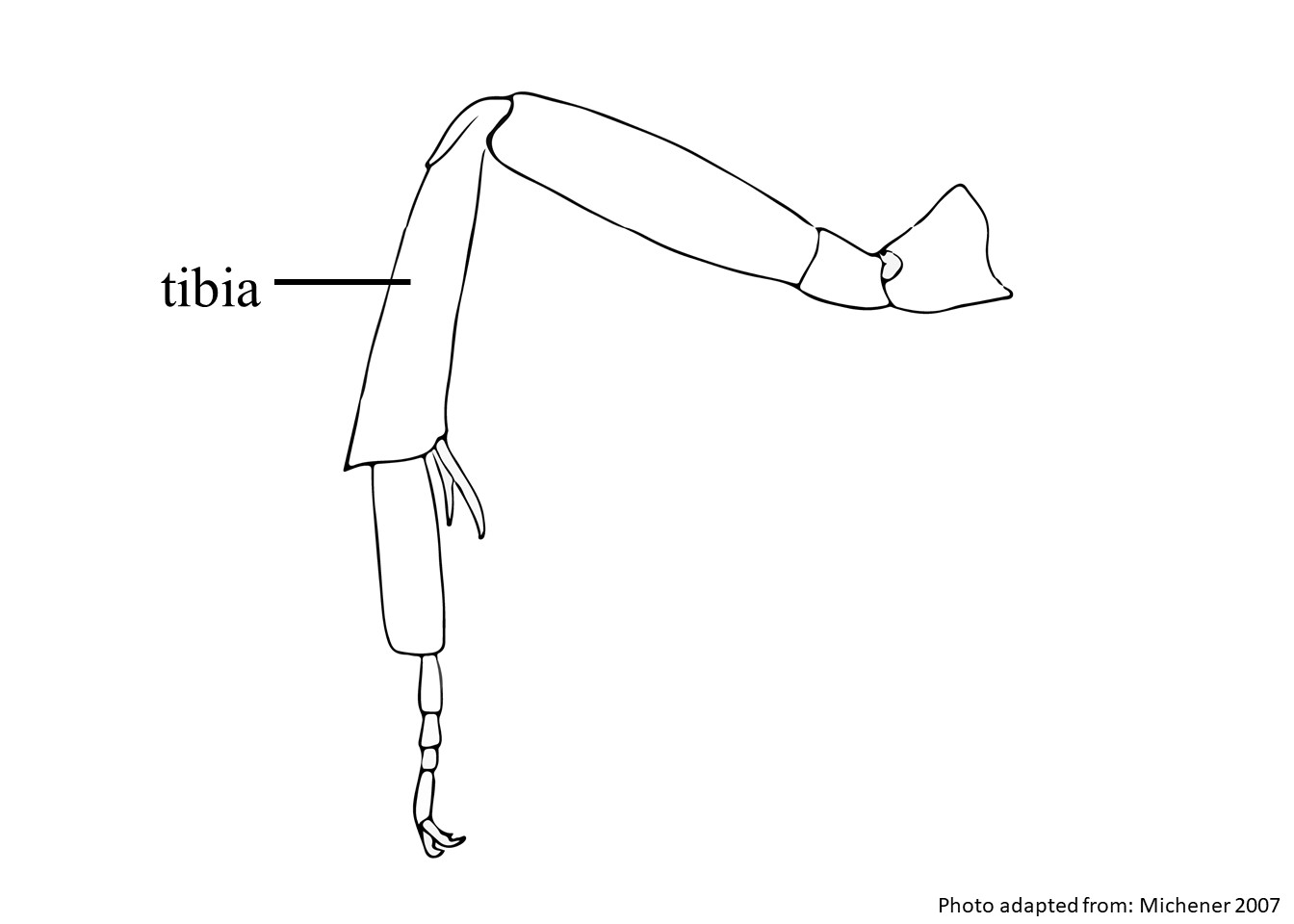 with anterioranterior: have long, dense hairs. slightly convexconvex: are ventrally depressed.
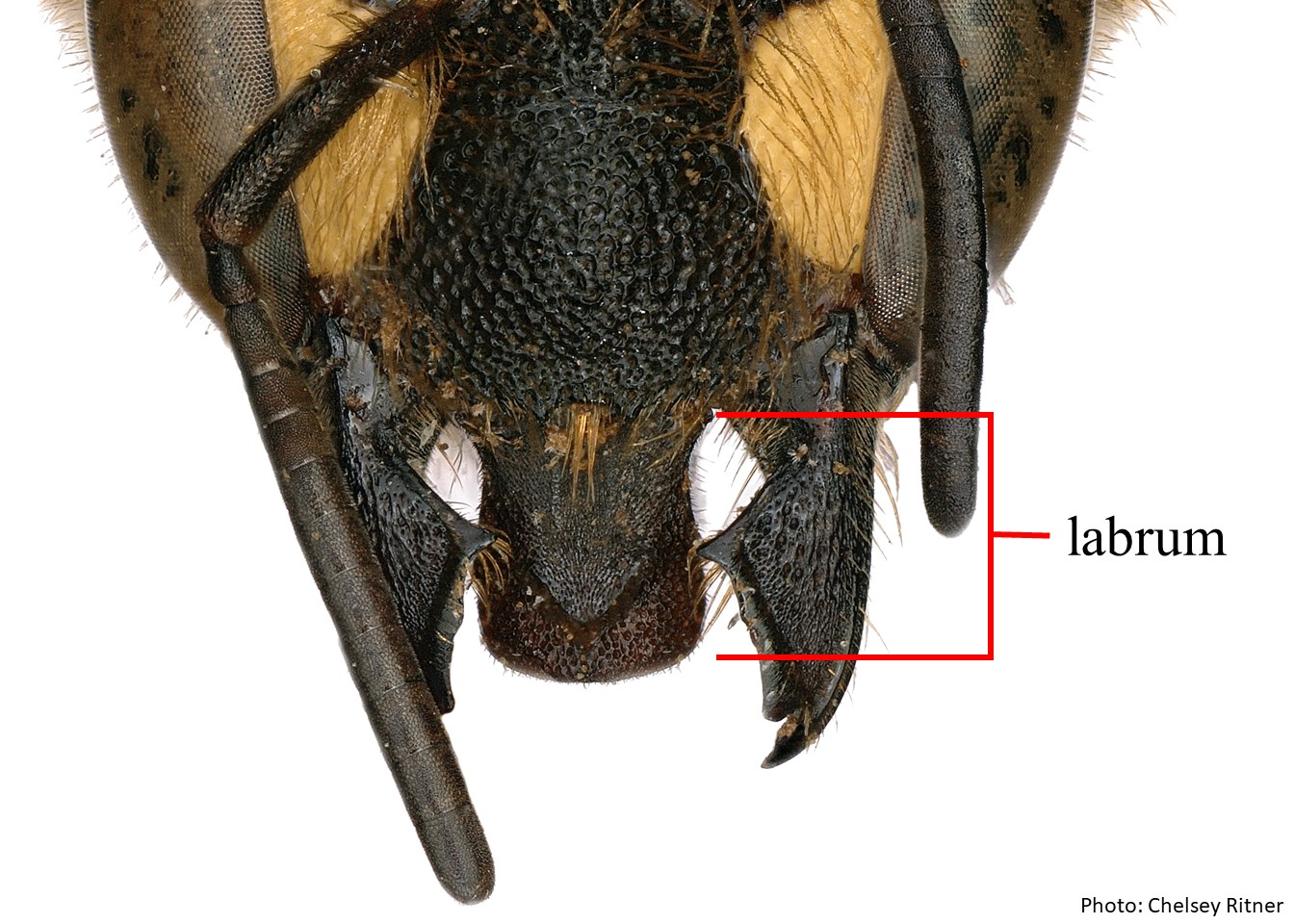 has low or absent basalbasal: is elongate with three large teeth, sometimes with the upper mandibularmandibular:.
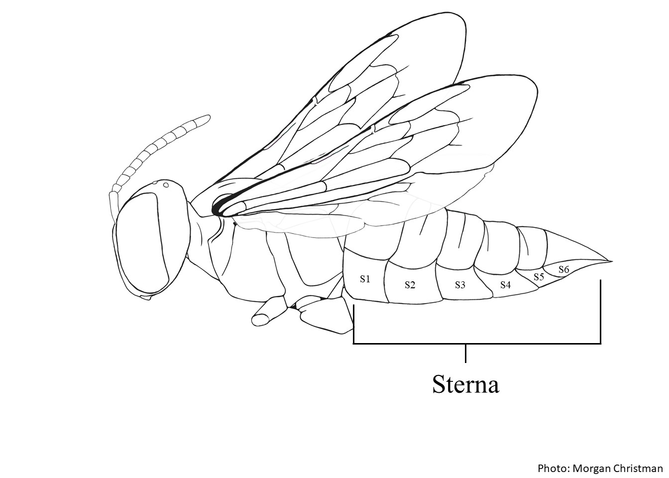 distaldistal: is straight with long, acute laterallateral: laterallateral: median spine is blunt.
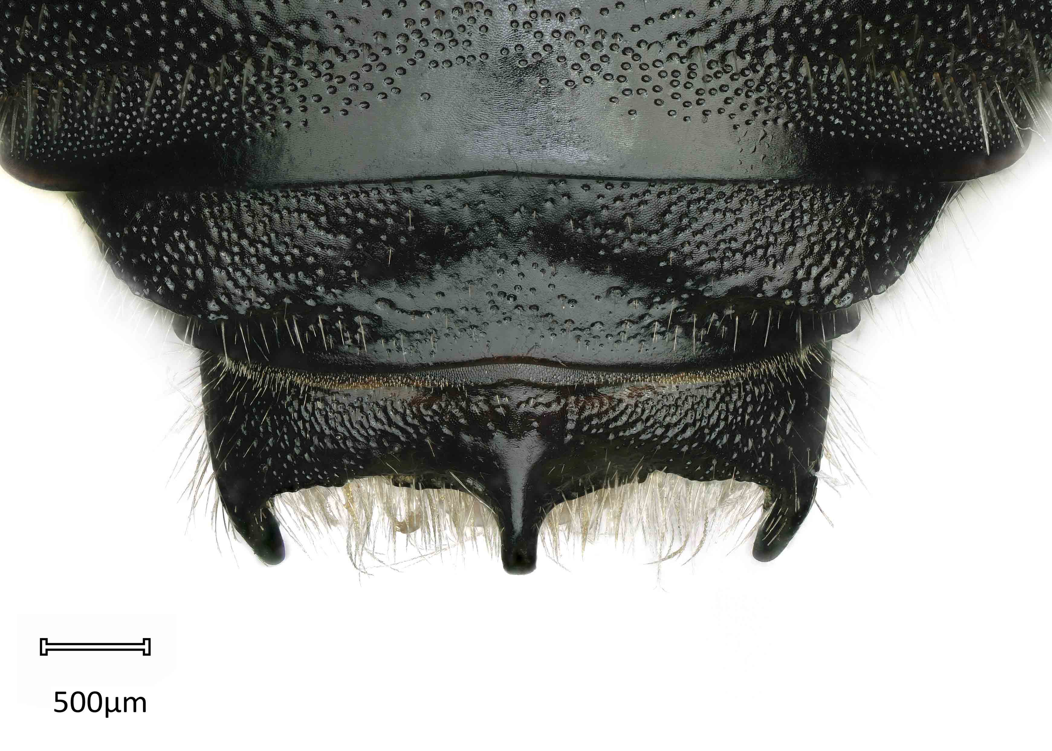
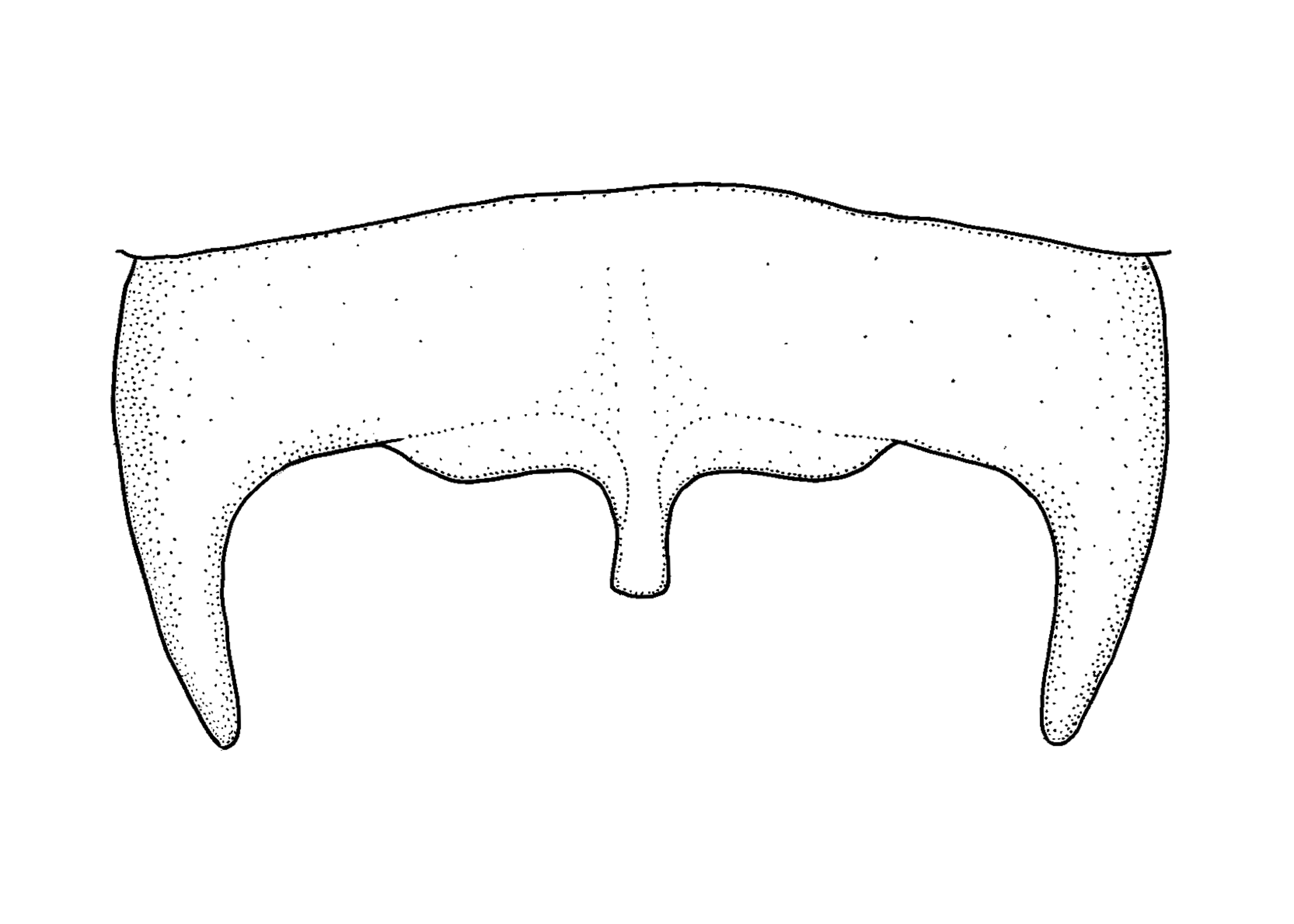
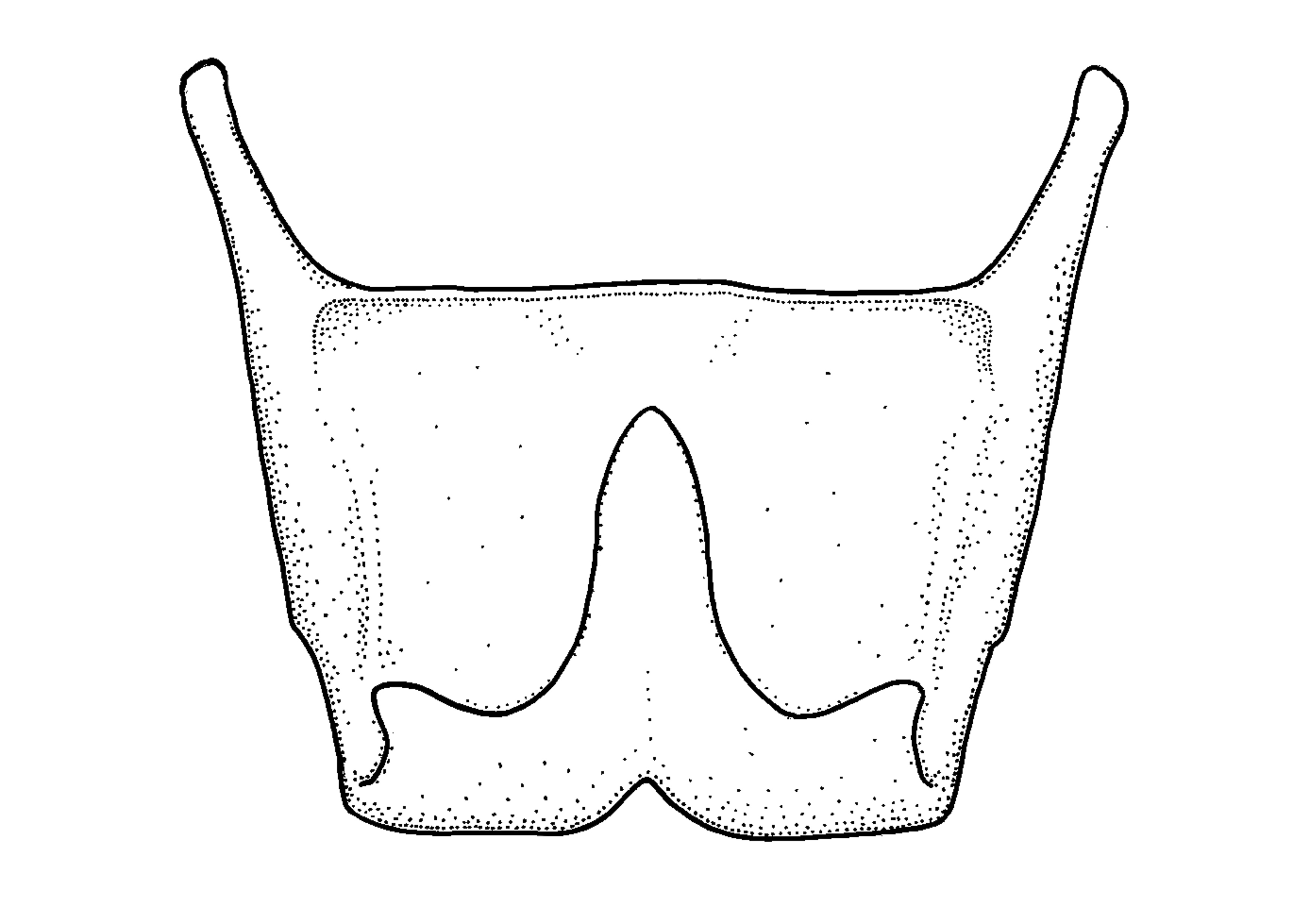
Anthidium chamelense may be confused with A. rodriguezi based on similar body coloration (black abdomen with a yellow scutellumscutellum:
shield shaped plate behind scutum and axillae) and a body length greater than 16 mm (Gonzalez and Griswold 2013Gonzalez and Griswold 2013:
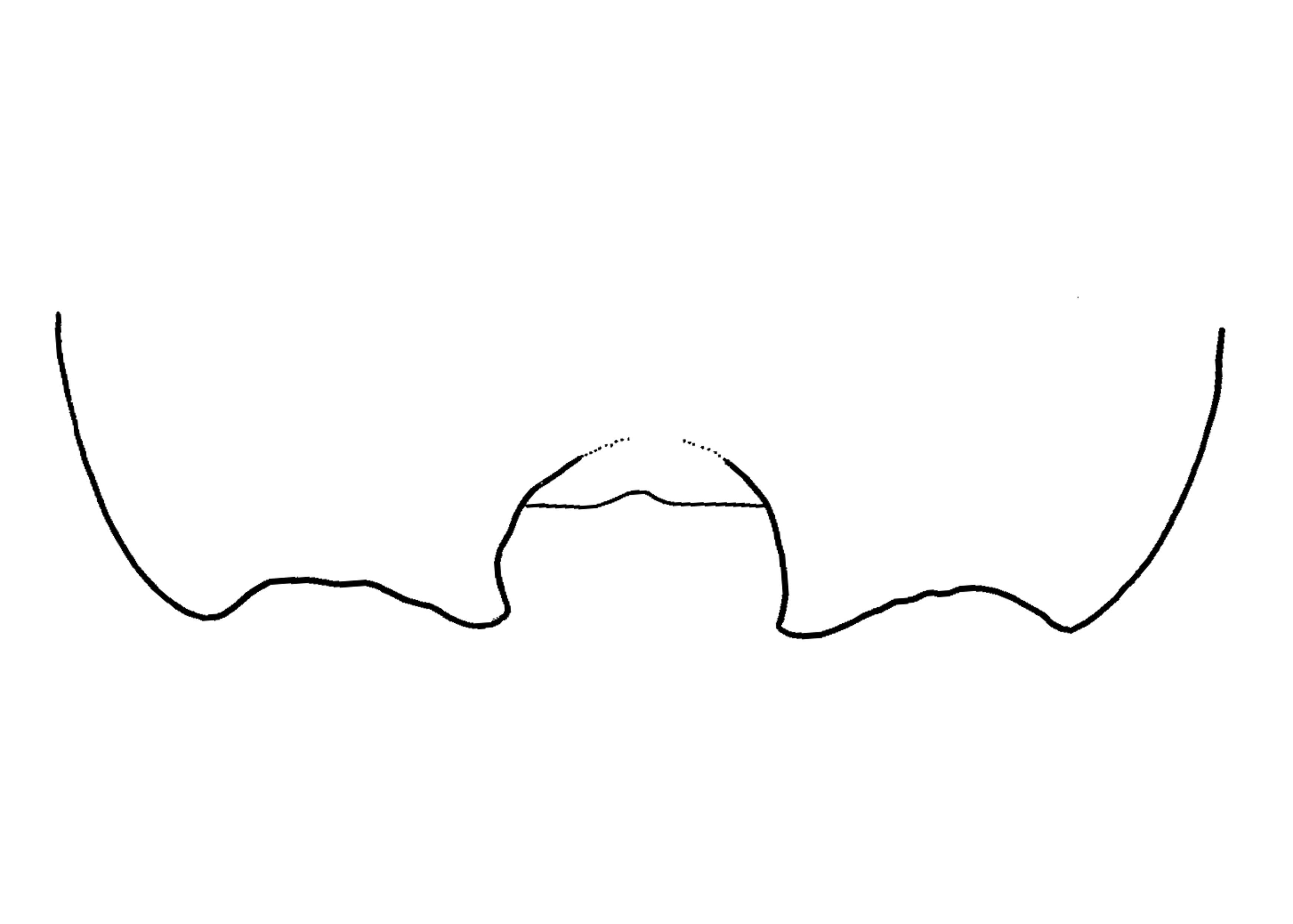
Gonzalez, V.H. and T.L. Griswold. 2013. Wool carder bees of the genus Anthidium in the Western Hemisphere (Hymenoptera: Megachilidae): diversity, host plant associations, phylogeny, and biogeography. Zoological Journal 168: 221ndash;425.). Female A. chamelense can be differentiated from A. rodriguezi based on the presence of a rounded laterallateral:
relating, pertaining, or attached to the side
angle on T6T6:
the segments on the top side of the abdomen, often abbreviated when referring to a specific segment to T1, T2, T3, T4, T5, T6, or T7 and preapicalpreapical:
referring to a section of a bee that is physically found just before the outermost (or apical) end of the section or segment
carinacarina:
a clearly defined ridge or keel, not necessarily high or acute; usually appears on bees as simply a raised line
with a semicircular emarginationemargination:
a notched or cut out place in an edge or margin, can be dramatic or simply a subtle inward departure from the general curve or line of the margin or structure being described
. Male A. chamelense can be differentiated from A. rodriguezi based on an acute laterallateral:
relating, pertaining, or attached to the side
spine on T6T6:
the segments on the top side of the abdomen, often abbreviated when referring to a specific segment to T1, T2, T3, T4, T5, T6, or T7 and a blunt median spine on T7T7:
the segments on the top side of the abdomen, often abbreviated when referring to a specific segment to T1, T2, T3, T4, T5, T6, or T7 (Gonzalez and Griswold 2013Gonzalez and Griswold 2013:
Gonzalez, V.H. and T.L. Griswold. 2013. Wool carder bees of the genus Anthidium in the Western Hemisphere (Hymenoptera: Megachilidae): diversity, host plant associations, phylogeny, and biogeography. Zoological Journal 168: 221ndash;425.).
Anthidium chamelense adults have been recorded in flight from July to November (Gonzalez and Griswold 2013Gonzalez and Griswold 2013:
Gonzalez, V.H. and T.L. Griswold. 2013. Wool carder bees of the genus Anthidium in the Western Hemisphere (Hymenoptera: Megachilidae): diversity, host plant associations, phylogeny, and biogeography. Zoological Journal 168: 221ndash;425.).
Floral associations are unknown.
Nesting behavior is unknown.
Anthidium chamelense occur in Guerrero, Jalisco, and Oaxaca, Mexico. They are primarily found in dry and pine-oak forests (Gonzalez and Griswold 2013Gonzalez and Griswold 2013:
Gonzalez, V.H. and T.L. Griswold. 2013. Wool carder bees of the genus Anthidium in the Western Hemisphere (Hymenoptera: Megachilidae): diversity, host plant associations, phylogeny, and biogeography. Zoological Journal 168: 221ndash;425.). None are known to occur in the U.S. or Canada.
Distribution map generated by Discover Life -- click on map for details, credits, and terms of use.
Gonzalez, V.H. and T.L. Griswold. 2013. Wool carder bees of the genus Anthidium in the Western Hemisphere (Hymenoptera: Megachilidae): diversity, host plant associations, phylogeny, and biogeography. Zoological Journal of the Linnean Society 168: 221-425.
Authors: S. Burrows, C. Ritner, M. Christman, L. Spears, A. Smith-Pardo, S. Price, R. Ramirez, T. Griswold, A. Redford
Edition 3 - last updated November 2021
tool images at ITP Node
idtools.org