
Phytophthora medicaginis
|
Phytophthora spp. in subclade 8a: portion of the seven-loci ML phylogeny featuring the type cultures of 212 described species (by T. Bourret). Notice the position of P. medicaginis Ex-type CBS 119902 = S&T BL 83. Gloria Abad, USDA S&T.
|
|
Phytophthora spp. in subclade 8a: Morphological Tabular key (PDF) and Tabular key legends (PDF) in IDphy2 KEY SECTION. Notice the data of P. medicaginis Ex-type CBS 119902 = S&T BL 83. Gloria Abad, USDA S&T.
|
|
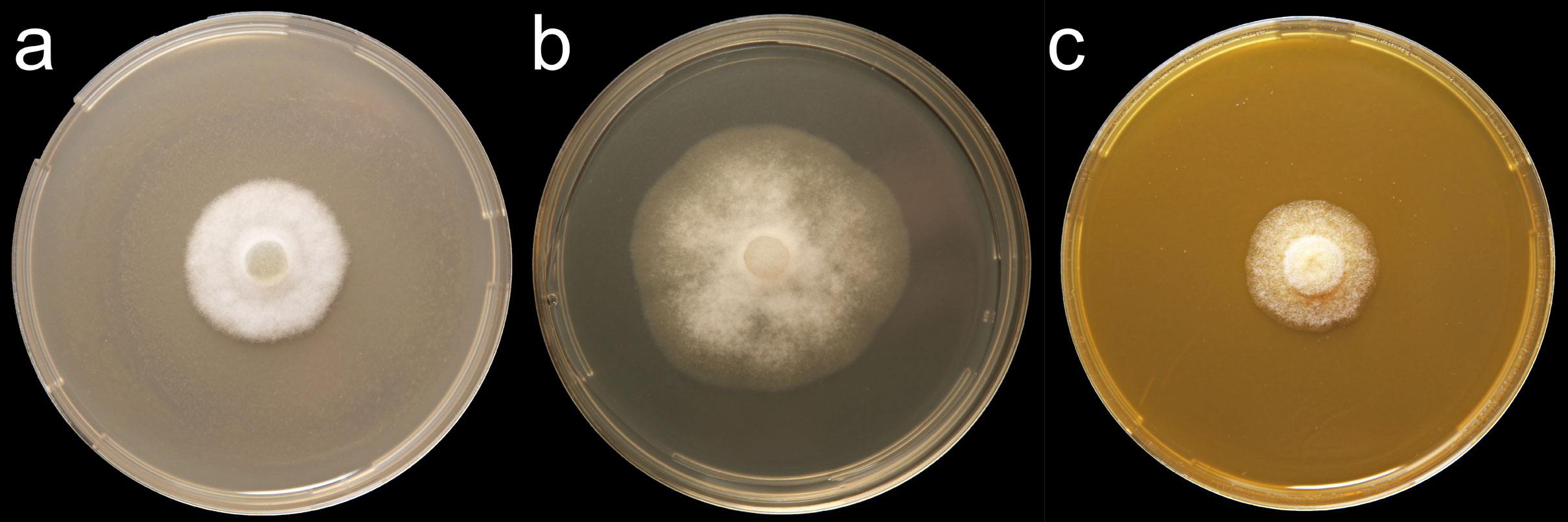
Phytophthora medicaginis (CPHST BL 83) colonies of the ex-type grown for 7 days on (a) V8®Agar, (b) potato dextrose agar, and (c) malt extract agar; photo by Krysta Jennings and Leandra Knight, USDA-APHIS-PPQ |
|
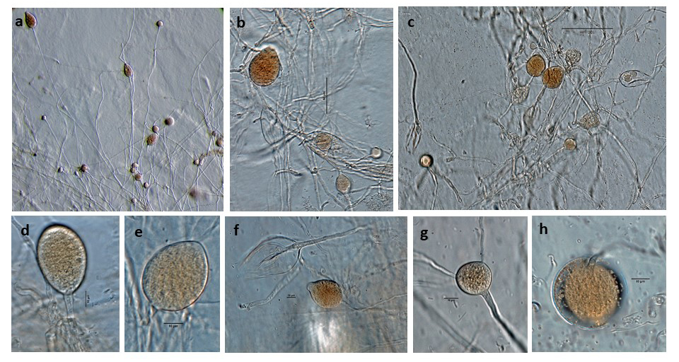
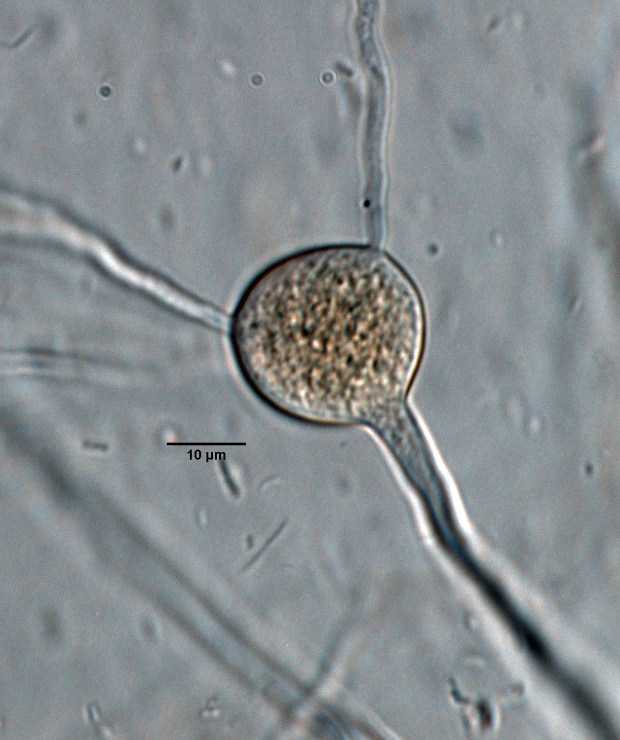
Phytophthora medicaginis (selected specimen P7029) asexual phase (a–h): (a–f) different shape of sporangia, (a–c) sporangia and hyphal swellings, (f) sporangium with internal proliferation, (g) radiating hyphal swelling, (h) chlamydospore; photos by G. Abad, USDA-APHIS-PPQ. |
|
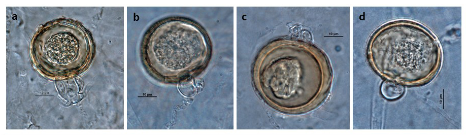
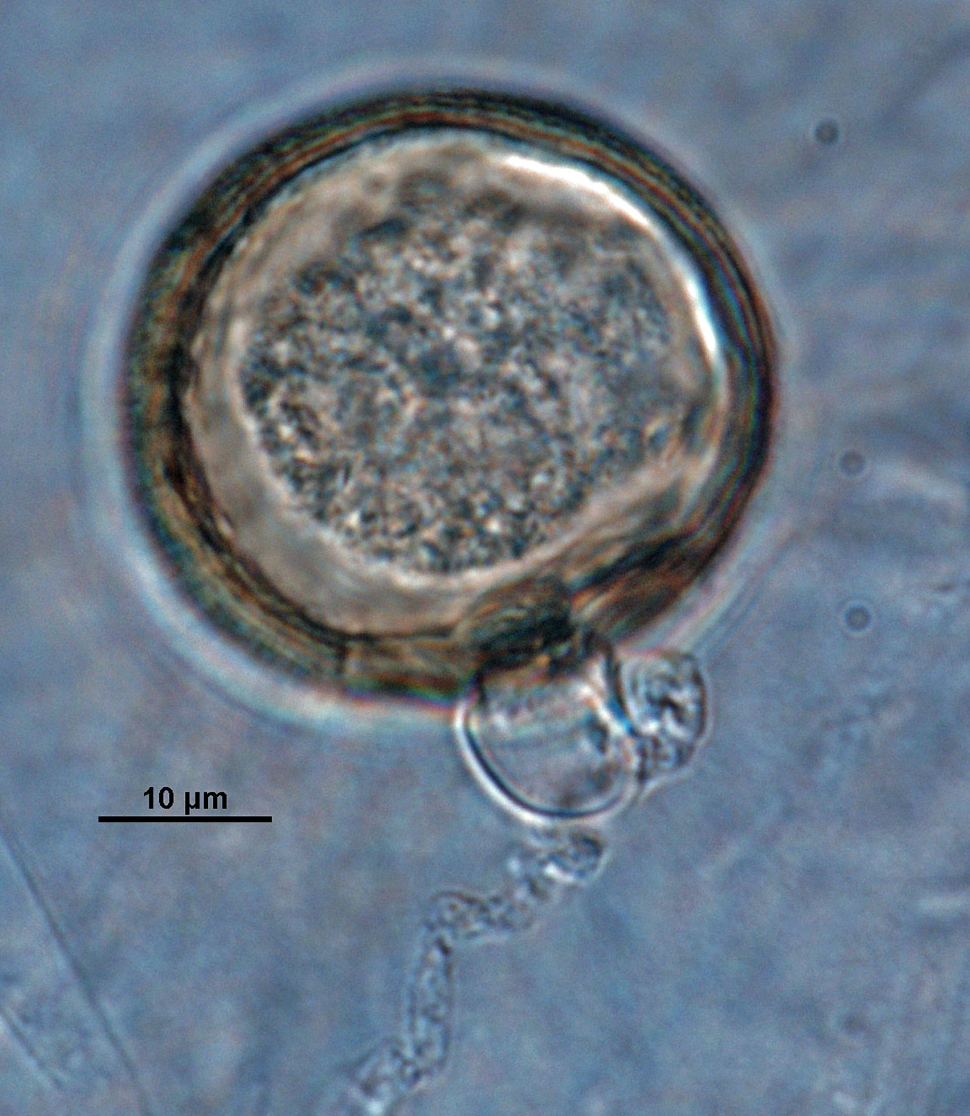
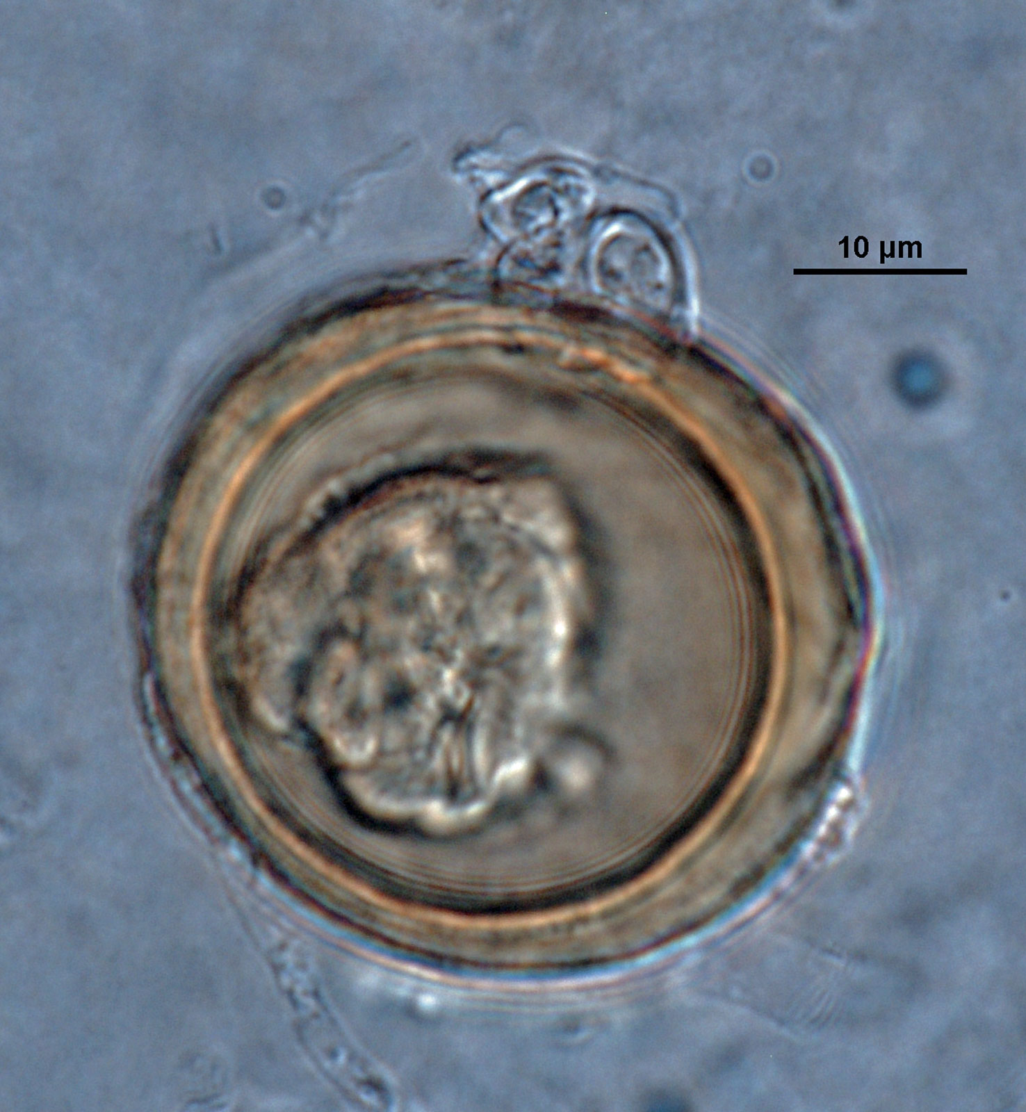
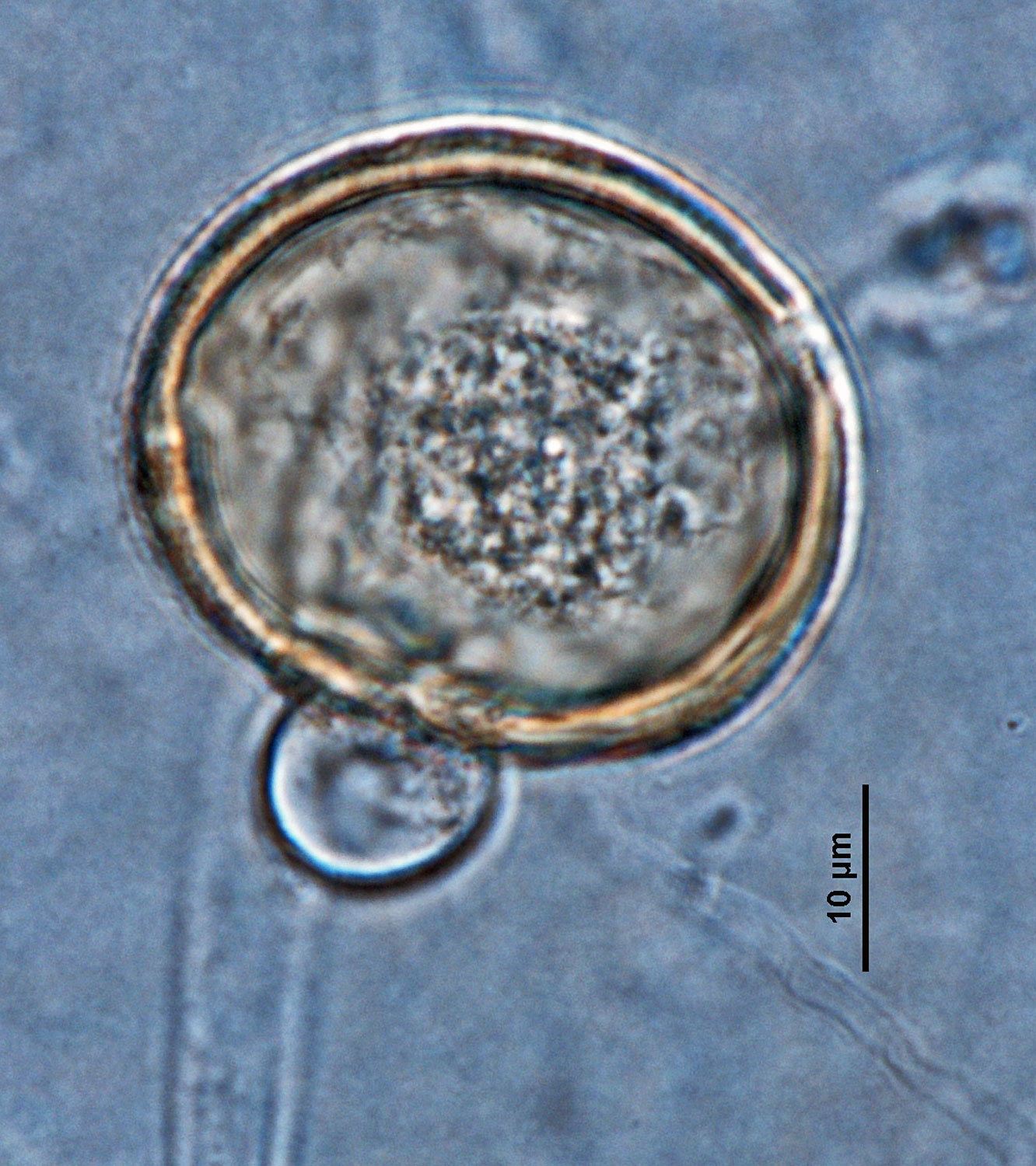
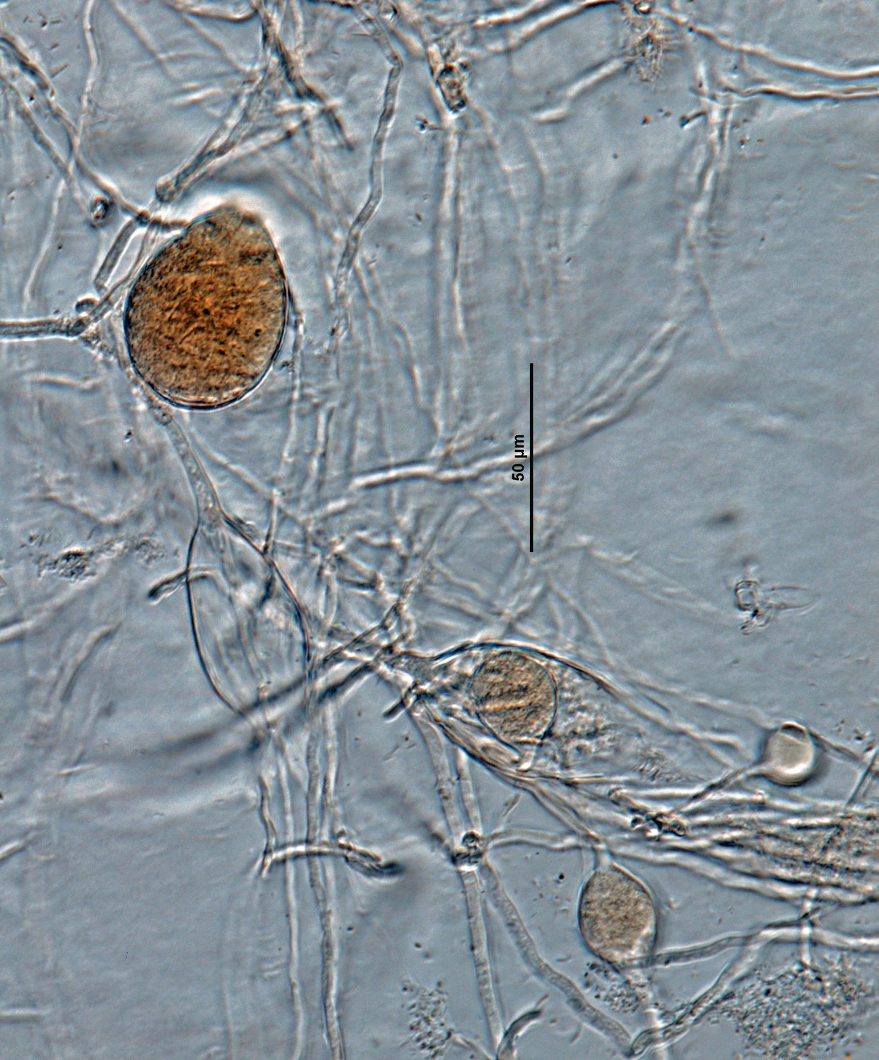
Phytophthora medicaginis (ex-type CPHST BL 83) sexual phase (a–d): (a–c) oogonia with amphigynous antheridia, (d) oogonium with paragynous antheridium; photos by G. Abad, USDA-APHIS-PPQ. |
|
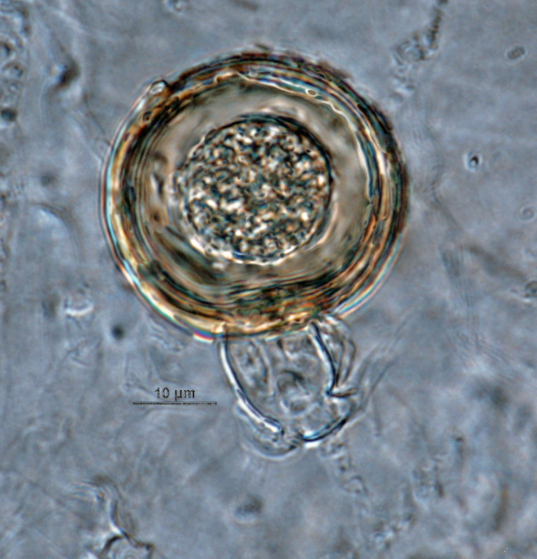
Phytophthora medicaginis (ex-type CPHST BL 83) sexual phase: oogonium with amphigynous antheridium; photo by G. Abad, USDA-APHIS-PPQ. |
|
Phytophthora medicaginis (ex-type CPHST BL 83) sexual phase: oogonium with amphigynous antheridium; photo by G. Abad, USDA-APHIS-PPQ. |
|
Phytophthora medicaginis (ex-type CPHST BL 83) sexual phase: oogonium with amphigynous antheridium; photo by G. Abad, USDA-APHIS-PPQ. |
|
Phytophthora medicaginis (ex-type CPHST BL 83) sexual phase: oogonium with paragynous antheridium; photo by G. Abad, USDA-APHIS-PPQ. |
|
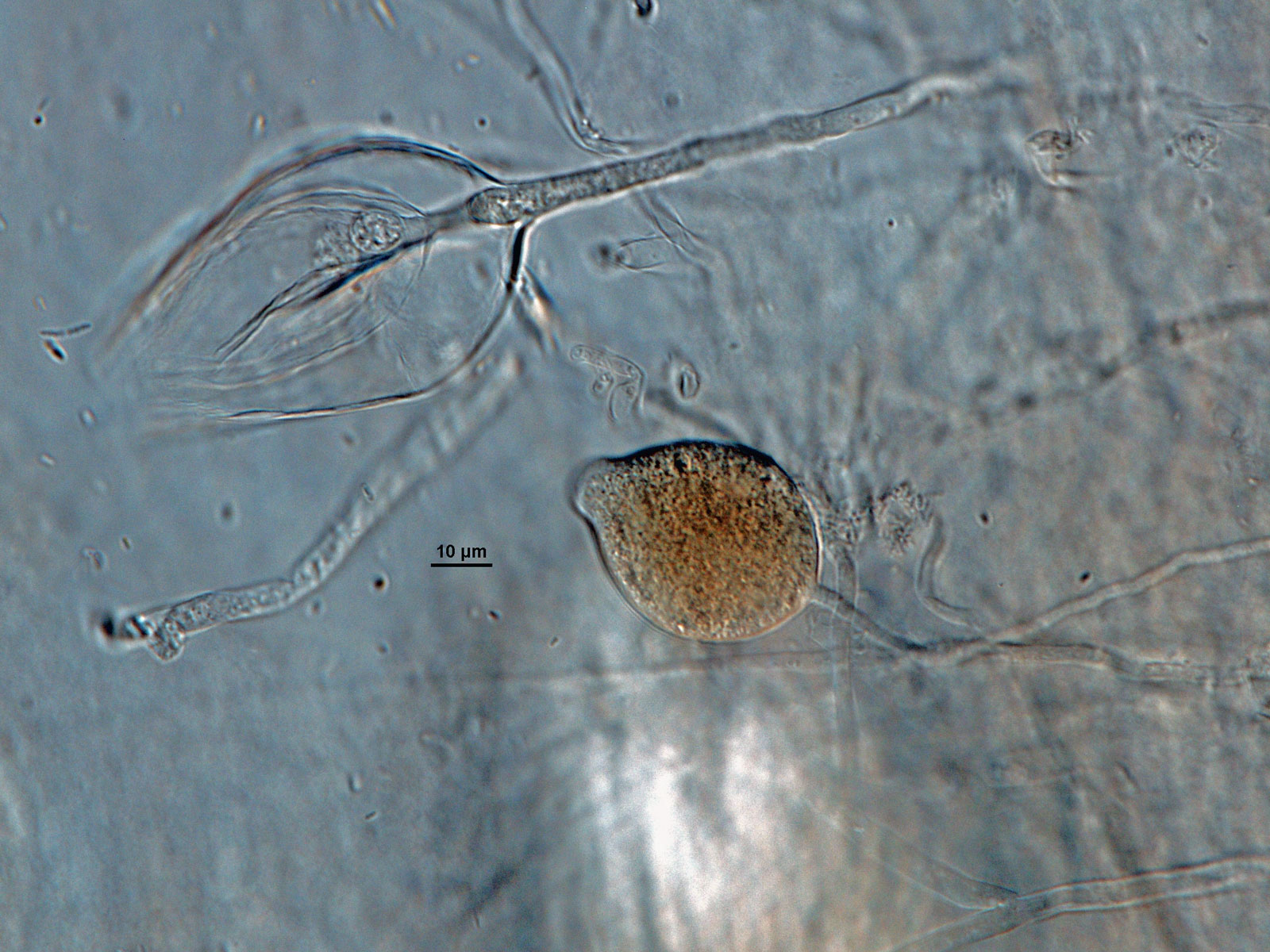
Phytophthora medicaginis (selected specimen P7029) asexual phase: sporangia and hyphal swellings; photo by G. Abad, USDA-APHIS-PPQ. |
|
Phytophthora medicaginis (selected specimen P7029) asexual phase: radiating hyphal swelling; photo by G. Abad, USDA-APHIS-PPQ. |
|
Phytophthora medicaginis (selected specimen P7029) asexual phase: nonpapillate sporangia and hyphal swellings; photo by G. Abad, USDA-APHIS-PPQ. |
|
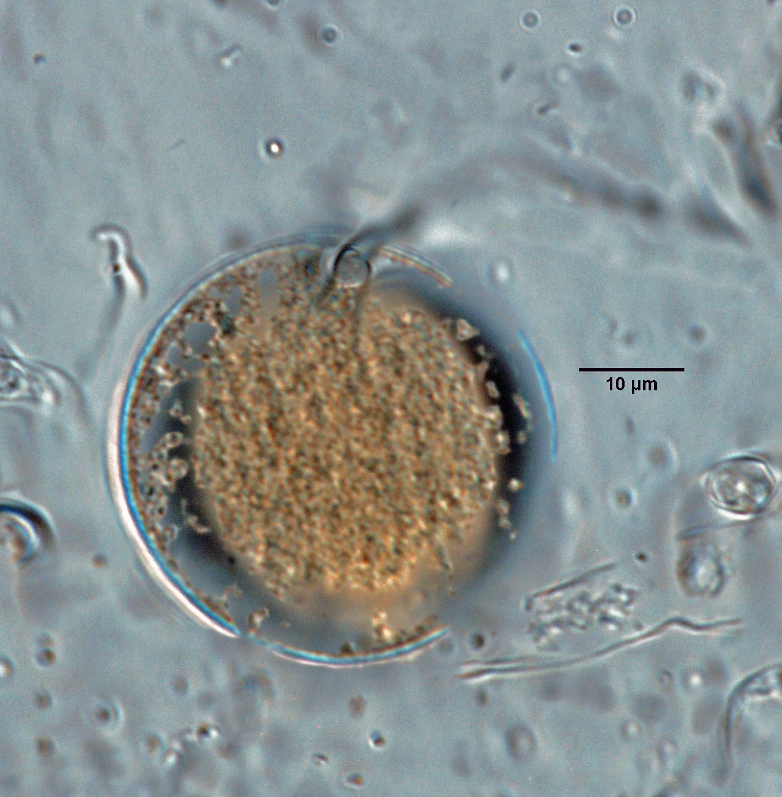
Phytophthora medicaginis (selected specimen P7029) asexual phase: chlamydospore; photo by G. Abad, USDA-APHIS-PPQ. |
|
Phytophthora medicaginis (selected specimen P7029) asexual phase: sporangia and hyphal swellings; photo by G. Abad, USDA-APHIS-PPQ. |
|
Phytophthora medicaginis (selected specimen P7029) asexual phase: nonpapillate sporangium; photo by G. Abad, USDA-APHIS-PPQ. |
|
Phytophthora medicaginis (selected specimen P7029) asexual phase: sporangium with internal proliferation; photo by G. Abad, USDA-APHIS-PPQ. |
|
Phytophthora medicaginis (selected specimen P7029) asexual phase: nonpapillate sporangium; photo by G. Abad, USDA-APHIS-PPQ. |
.JPG)
.JPG)


Name and publication
Phytophthora medicaginis E.M. Hansen & D.P. Maxwell (1991)
Hansen EM and Maxwell DP. 1991. Species of the Phytophthora megasperma-complex. Mycologia 83: 376–381 (pg. 377).
Nomenclature
from Hansen and Maxwell (1991)
Mycobank
Etymology
named for the principal host
Typification
Type: UNITED STATES, Oregon, Salem, from Medicago sativa, collected by P. B. Hamm in 1978 [no. AL1 S1 et no. 4 in herbarium at Oregon State University (OSC)]
Ex-type: CBS 119902 (duplicate deposited by Hansen on 12 Oct 1985)
NOTE: According to the original manuscript, cultures of the type were deposited in the American Type Culture Collection. No isolate for AL1 S1 et no. 4 is found in ATCC. E. Hansen confirmed that CBS 119902 is the authentic ex-holotype in e-mail to G. Abad on 7-25-13.
Ex-type in other collections
(ET) CBS 119902, NRRL 64260, WPC P19830, S&T BL 83 (Abad)
Molecular identification
Voucher sequences for barcoding genes (ITS rDNA and COI) of the ex-type (see Molecular protocols page)
Phytophthora medicaginis isolate CPHST BL 83 (= P19830 WPC) = ITS rDNA MG865532, COI MH136927
Voucher sequences for Molecular Toolbox with seven genes (ITS, β-tub, COI, EF1α, HSP90, L10, and YPT1
(see Molecular protocols page) (In Progress)
Voucher sequences for Metabarcoding High-throughput Sequencing (HTS) Technologies [Molecular Operational Taxonomic Unit (MOTU)]
(see Molecular protocols page) (In Progress)
Sequences with multiple genes for ex-type in other sources
- NCBI: Phytophthora medicaginis CPHST BL 83
- EPPO-Q-bank: Phytophthora medicaginis CBS 119902
- BOLDSYSTEMS: Phytophthora medicaginis (barcoding COI & ITS)
Position in multigenic phylogeny with 7 genes (ITS, β-tub, COI, EF1α, HSP90, L10, and YPT1)
Clade 8a
Morphological identification
Colonies and cardinal temperatures
Colony colony:
assemblage of hyphae which usually develops form a single source and grows in a coordinated way
morphology after 7 days of growth on potato dextrose agar, V8 agar, and malt extract agar with no distinct pattern. Minimum temperature for growth is 3°C, optimum 27°C, and maximum 33°C.
Conditions for growth and sporulation
Sporangia and hyphal swellings produced in V-8 agar flooded with 10% soil solution.
Asexual phase
SporangiaSporangia:
sac within which zoospores form, especially when water is cooled to about 10°C below ambient temperature; in solid substrates, sporangia usually germinate by germ tubes
nonpapillated; persistentpersistent:
pertaining to sporangia that remain attached to the sporangiophore and do not separate or detach easily (cf. caducous)
; ellipsoidellipsoid:
refers to a solid body that forms an ellipse in the longitudinal plane and a circle in cross section; many fungal spores are ellipsoidal or elliptic
, ovoidovoid:
egg-shaped, with the widest part at the base of the sporangium and the narrow part at the apex
, obpyriformobpyriform:
inversely pear-shaped, i.e. with the widest part at the point of attachment (cf. pyriform)
(35–69 L x 20–41 W μm); showing internal proliferationinternal proliferation:
internal proliferation occurs when the sporangiophore continues to grow through an empty sporangium
; originated in unbranched sporangiophores; in some cases the sporangiophoresporangiophore:
the hyphal strand on which the sporangium is formed; may be branched or unbranched to form compound sympodia or simple sympodia
broadens towards the sporangiumsporangium:
sac within which zoospores form, especially when water is cooled to about 10°C below ambient temperature; in solid substrates, sporangia usually germinate by germ tubes
. Hyphal swellings mostly individual subglobose, elongate, irregular or coralloid. ChlamydosporesChlamydospores:
an asexual spore with a thickened inner wall that is delimited from the mycelium by a septum; may be terminal or intercalary, and survives for long periods in soil
rarely produced; globoseglobose:
having a rounded form resembling that of a sphere
or subglobose (20–33 μm diam), intercalaryintercalary:
positioned within a hypha (cf. terminal)
.
Sexual phase
Homothallic. OogoniaOogonia:
the female gametangium in which the oospore forms after fertilization by the antheridium
smooth-walled, globoseglobose:
having a rounded form resembling that of a sphere
(19–40 μm diam), sometimes with tapered bases; antheridia predominantly amphigynousamphigynous:
pertaining to the sexual stage in which the antheridium completely surrounds the stalk of the oogonium (cf. paragynous)
, sometimes with two antheridiaantheridia:
the male gametangium; a multinucleate, swollen hyphal tip affixed firmly to the wall of the female gametangium (the oogonium)
, one amphyginous and one paragynousparagynous:
pertaining to the sexual stage in which the antheridium is attached to the side of the oogonium (cf. amphigynous)
, sometimes with digitate projections; oosporesoospores:
zygote or thick-walled spore that forms within the oogonium after fertilization by the antheridium; may be long-lived
globoseglobose:
having a rounded form resembling that of a sphere
(14–32 μm diam) pleroticplerotic:
pertaining to an oospore that fills the oogonium (cf. aplerotic)
, apleroticaplerotic:
pertaining to a mature oospore that does not fill the oogonium; i.e. there is room left between the oospore wall and oogonium wall (cf. plerotic)
, or slightly apleroticaplerotic:
pertaining to a mature oospore that does not fill the oogonium; i.e. there is room left between the oospore wall and oogonium wall (cf. plerotic)
.
Most typical characters
Phytophthora medicaginis is characterized by the presence of predominantly amphigynousamphigynous:
pertaining to the sexual stage in which the antheridium completely surrounds the stalk of the oogonium (cf. paragynous)
antheridia in conjunction with paragynousparagynous:
pertaining to the sexual stage in which the antheridium is attached to the side of the oogonium (cf. amphigynous)
antheridiaantheridia:
the male gametangium; a multinucleate, swollen hyphal tip affixed firmly to the wall of the female gametangium (the oogonium)
.
Specimen(s) evaluated
Phytophthora medicaginis ex-type CPHST BL 83, duplicate of P19830 (World Phytophthora Collection), which is a duplicate of CBS 119902
Phytophthora medicaginis selected specimen P7029 (World Phytophthora Collection) (ITSrDNA 100% alignment with ex-type CPHST BL 83)
Hosts and distribution
Distribution: cosmopolitan, throughout the range of the host
Substrate: roots
Disease note: root rot, damping-off of seedlings
Host: Medicago sativa (alfalfa), Onobrychis viciifolia (sainfoin), Cicer arietinum (chickpea) (Fabaceae); also reported on Prunus mahaleb (cherry, Rosaceae)
Retrieved January 31, 2018 from U.S. National Fungus Collections Nomenclature Database.
Additional references and links
- SMML USDA-ARS: Phytophthora medicaginis
- EPPO Global Database: Phytophthora medicaginis
- Forest Phytophthoras of the world: Phytophthora medicaginis
- CABI Digital Library: Phytophthora medicaginis
- Encyclopedia of Life (EOL): Phytophthora medicaginis
- Index Fungorum (IF): Phytophthora medicaginis
- Google All Phytophthora medicaginis
- Google Images Phytophthora medicaginis
- Google Scholar Phytophthora medicaginis
Fact sheet author
Z. Gloria Abad, Ph.D., USDA-APHIS-PPQ-S&T Plant Pathogen Confirmatory Diagnostics Laboratory (PPCDL), United States of America.
