
Phytophthora gibbosa
|
Phytophthora spp. in subclade 2b: portion of the seven-loci ML phylogeny featuring the type cultures of 212 described species (by T. Bourret). Notice the position of P. gloveri Ex-type CBS 121969= S&T BL 36. Gloria Abad, USDA S&T.
|
|
Phytophthora spp. in subclade 2b: Morphological Tabular key (PDF) and Tabular key legends (PDF) in IDphy2 KEY SECTION. Notice the data of P. gloveri Ex-type CBS 121969= S&T BL 36. Gloria Abad, USDA S&T.
|
|
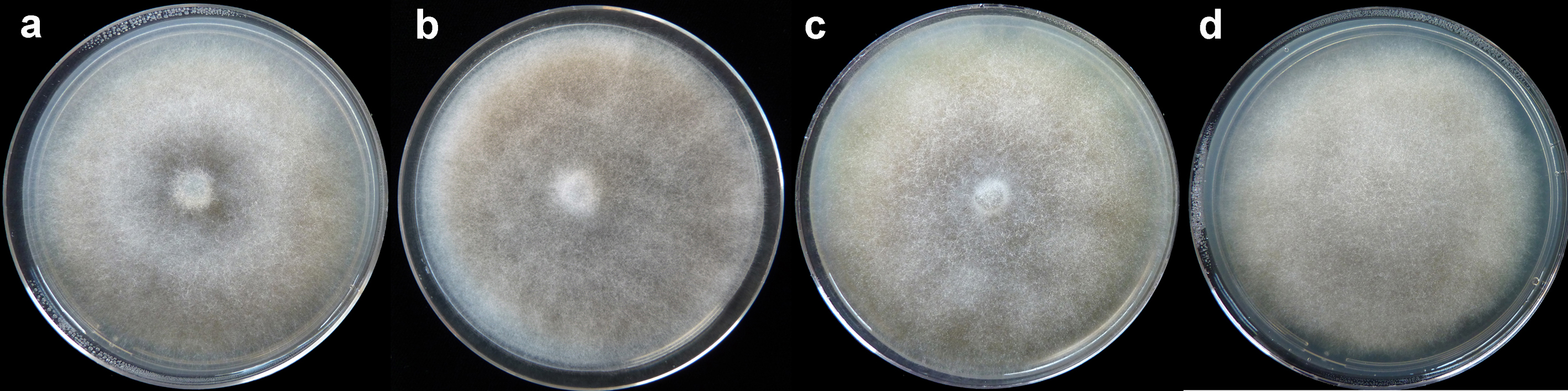
Phytophthora gibbosa colonies grown for 7 days at 20°C on: (a) V8® agar (b) carrot agar (c) malt extract agar (d) potato-dextrose agar |
|
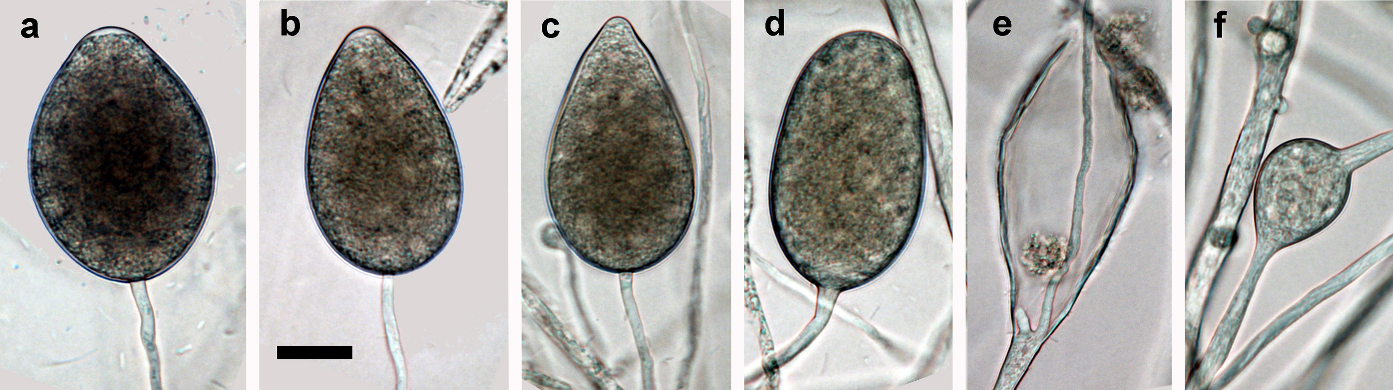
mature sporangia formed on V8 agar flooded with soil extract: (a-b) ovoid semipapillate sporangia, (c) obpyriform sporangium with nonpapillate pointed apex, (d) nonpapillate ellipsoid sporangium, (e) empty limoniform sporangium showing internal extended proliferation, (f) intercalary hyphal swellings originating from undeveloped sporangia; scale bar = 25 µm |
|
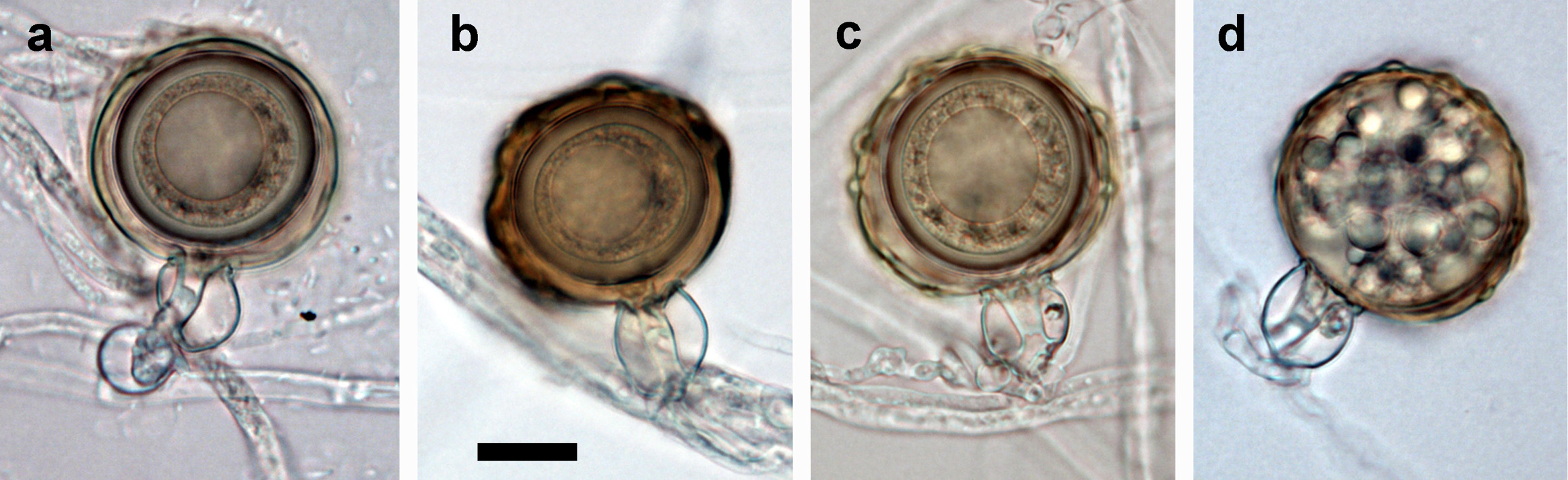
(a-d) mature often bronze-brown oogonia with amphigynous antheridia and thick-walled aplerotic oospores each containing a large ooplast: (a) smooth-walled, (b-c) ornamented gibbose oogonia, (d) gibbose golden-brown oogonium aborted before oospore formation; scale bar = 25 μm |
.JPG)
.JPG)
Name and publication
Phytophthora gibbosa T. Jung, M.J.C. Stukely & T.I. Burgess (2011)
Jung T, Stukely MJC, Hardy GE StJ, White MD, Paap T, Dunstan WA, and Burgess TI. 2011. Multiple new Phytophthora species from ITS Cladeclade:
a taxonomic group of organisms classified together on the basis of homologous features traced to a common ancestor
6 associated with natural ecosystems in Australia: evolutionary and ecological implications. Persoonia 13: 13–39.
Nomenclature
from Jung et al. (2011)
Mycobank
Etymology
refers to the gibbous ornamented surface of the oogoniaoogonia:
the female gametangium in which the oospore forms after fertilization by the antheridium
(gibbosa Latin = gibbous, knaggy)
Typification
Type: WESTERN AUSTRALIA, Scott River ironstones, from rhizosphere soil of dying Acacia pycnantha, 2009, VHS, holotype MURU 461 (dried culture on V8A, Herbarium of Murdoch University, Western Australia)
Ex-type: CBS 127951 and VHS 21998
Sequences for ex-type in original manuscript: CBS 127951 = ITS rDNA HQ012933, HSP90 HQ012892, cox1 HQ012846
Ex-type in other collections
(ET) CBS 127951, NRRL 64012, VHS21998, WPC P19586, S&T BL 65 (Abad), 62B8 (Hong), TJ345 (Jung)
Molecular identification
Voucher sequences for barcoding genes (ITS rDNA and COI) of the ex-type (see Molecular protocols page)
Phytophthora gibbosa isolate CPHST BL 65 (= P19586 WPC) = ITS rDNA MG865499, COI MH136894
Voucher sequences for Molecular Toolbox with seven genes (ITS, β-tub, COI, EF1α, HSP90, L10, and YPT1
(see Molecular protocols page) (In Progress)
Voucher sequences for Metabarcoding High-throughput Sequencing (HTS) Technologies [Molecular Operational Taxonomic Unit (MOTU)]
(see Molecular protocols page) (In Progress)
Sequences with multiple genes for ex-type in other sources
- NCBI: Phytophthora gibbosa CPHST BL 65
- NCBI: Phytophthora gibbosa CBS 127951
- EPPO-Q-bank: Phytophthora gibbosa CBS 127951
- BOLDSYSTEMS: Phytophthora gibbosa (barcoding COI & ITS)
Position in multigenic phylogeny with 7 genes (ITS, β-tub, COI, EF1α, HSP90, L10, and YPT1)
Clade clade:
a taxonomic group of organisms classified together on the basis of homologous features traced to a common ancestor
6b
Morphological identification
Colonies and cardinal temperatures
Colony colony:
assemblage of hyphae which usually develops form a single source and grows in a coordinated way
morphology is uniform on V8A, CA, MEA, and PDA. Minimum growth temperature 7.5°C, optimum 30°C, and maximum 32.5°C.
Conditions for growth and sporulation
SporangiaSporangia:
sac within which zoospores form, especially when water is cooled to about 10°C below ambient temperature; in solid substrates, sporangia usually germinate by germ tubes
are produced in water cultures (soil extract or river water) and not observed in solid media. OogoniaOogonia:
the female gametangium in which the oospore forms after fertilization by the antheridium
are formed readily in single-strain culture on CA and V8A after about 20 d.
Asexual phase
SporangiaSporangia:
sac within which zoospores form, especially when water is cooled to about 10°C below ambient temperature; in solid substrates, sporangia usually germinate by germ tubes
are non-papillate, persistentpersistent:
pertaining to sporangia that remain attached to the sporangiophore and do not separate or detach easily (cf. caducous)
, and ovoidovoid:
egg-shaped, with the widest part at the base of the sporangium and the narrow part at the apex
or ellipsoidellipsoid:
refers to a solid body that forms an ellipse in the longitudinal plane and a circle in cross section; many fungal spores are ellipsoidal or elliptic
in shape. SporangiaSporangia:
sac within which zoospores form, especially when water is cooled to about 10°C below ambient temperature; in solid substrates, sporangia usually germinate by germ tubes
average 48.8 ± 9.6 × 30.8 ± 5.4 μm (overall range 24.8–71.1 × 17.4–48.0 μm). Sporangiophores usually in simple sympodiasympodia:
a type of sporangiophore which appears simple, but where each successive sporangium develops on a branch behind and to one side of the previous apex, where growth has already ceased
with internal extended proliferationextended proliferation:
a type of internal proliferation in which the sporangiophore originates from inside of an empty sporangium, and continues to grow through and out of the old sporangium
. External proliferationexternal proliferation:
formation of a sporangium after a sporangiophore has emerged from beneath and external to an empty sporangium that has previously emitted its zoospores (cf. internal proliferation)
also observed leading to lax sympodiasympodia:
a type of sporangiophore which appears simple, but where each successive sporangium develops on a branch behind and to one side of the previous apex, where growth has already ceased
. Hyphal swellings are common. ChlamydosporesChlamydospores:
an asexual spore with a thickened inner wall that is delimited from the mycelium by a septum; may be terminal or intercalary, and survives for long periods in soil
absent.
Sexual phase
Homothallic. OogoniaOogonia:
the female gametangium in which the oospore forms after fertilization by the antheridium
are globoseglobose:
having a rounded form resembling that of a sphere
with wavy to ornamented gibbous walls turning golden-brown on maturity, average size 38.1 ± 5.4 mm (27.0–49.9 µm). OosporesOospores:
zygote or thick-walled spore that forms within the oogonium after fertilization by the antheridium; may be long-lived
are apleroticaplerotic:
pertaining to a mature oospore that does not fill the oogonium; i.e. there is room left between the oospore wall and oogonium wall (cf. plerotic)
, globoseglobose:
having a rounded form resembling that of a sphere
, with a large ooplast and thick walls, average size 31.4± 4.6 µm (18.9–39.4). AntheridiaAntheridia:
the male gametangium; a multinucleate, swollen hyphal tip affixed firmly to the wall of the female gametangium (the oogonium)
amphigynous.
Most typical characters
Phytophthora gibbosa can be easily differentiated from all other species from ITS Cladeclade:
a taxonomic group of organisms classified together on the basis of homologous features traced to a common ancestor
6 (except Phytophthora ornamentata) by the production of ornamented (gibbous) oogoniaoogonia:
the female gametangium in which the oospore forms after fertilization by the antheridium
in single culture. In addition, it can be separated from Phytophthora gregata and P. taxon raspberry by the lack of nested proliferationnested proliferation:
a type of internal proliferation where a new sporangium develops successively inside the old sporangium after it has emptied
of sporangiasporangia:
sac within which zoospores form, especially when water is cooled to about 10°C below ambient temperature; in solid substrates, sporangia usually germinate by germ tubes
.
Specimen(s) evaluated
Australia, Western Australia, Scott River ironstones, from rhizosphere soil of dying Acacia pycnantha, 2009, CBS 127951 = VHS 21998; VHS 22007; from dying Xanthorrhoea gracilis, VHS 21999; from a dying Grevillea sp., VHS 22008
CPHST BL 65 = P19586 WPC
Hosts and distribution
Distribution: Australia
Substrate: soil associated with dying plants, roots
Retrieved January 30, 2018 from U.S. National Fungus Collections Nomenclature Database.
Additional info:
Distribution: Western Australia
Substrate: roots, collars, and rhizosphere soil
Disease note: no pathogenicity trials have been conducted
Hosts: Acacia pycnantha, Xanthorrhoea gracilis, Grevillea sp.
Additional references and links
Burgess TI, Webster JL, Ciampini JA, White DW, Hardy GESJ, Stukely MJC. 2009. Re-evaluation of Phytophthora species isolated during 30 years of vegetation health surveys in Western Australia using molecular techniques. Plant Disease 93: 215–223.
- SMML USDA-ARS: Phytophthora gibbosa
- EPPO Global Database: Phytophthora gibbosa
- Forest Phytophthoras of the world: Phytophthora gibbosa
- CABI Digital Library: Phytophthora gibbosa
- Encyclopedia of Life (EOL): Phytophthora gibbosa
- Index Fungorum (IF): Phytophthora gibbosa
- Google All Phytophthora gibbosa
- Google Images Phytophthora gibbosa
- Google Scholar Phytophthora gibbosa
Fact sheet authors
Treena Burgess, Ph.D., Phytophthora Science and Management, Harry Butler Institute, Murdoch University, Australia
Z. Gloria Abad, Ph.D., USDA-APHIS-PPQ-S&T Plant Pathogen Confirmatory Diagnostics Laboratory (PPCDL), United States of America.
