
Phytophthora capsici
|
Phytophthora spp. in subclade 2b: portion of the seven-loci ML phylogeny featuring the type cultures of 212 described species (by T. Bourret). Notice the position of P. capsici Ex-type CBS 128.23 = S&T BL 33G. Gloria Abad, USDA S&T.
|
|
Phytophthora spp. in subclade 2b: Morphological Tabular key (PDF) and Tabular key legends (PDF) in IDphy2 KEY SECTION. Notice the data of P. capsici Ex-type CBS 128.23 = S&T BL 33G. Gloria Abad, USDA S&T.
|
|
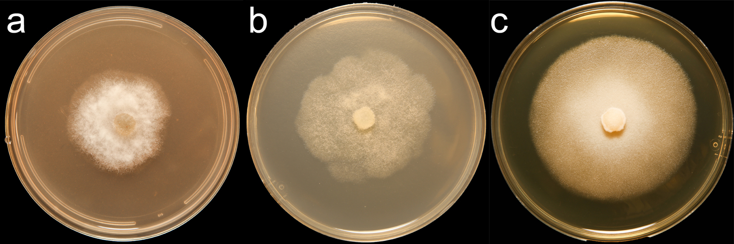
Phytophthora capsici (CPHST BL 33) colonies of the ex-type grown for 7 days on (a) V8® Agar, (b) potato dextrose agar, and (c) malt extract agar; photo by Krysta Jennings and Leandra Knight, USDA-APHIS-PPQ |
|
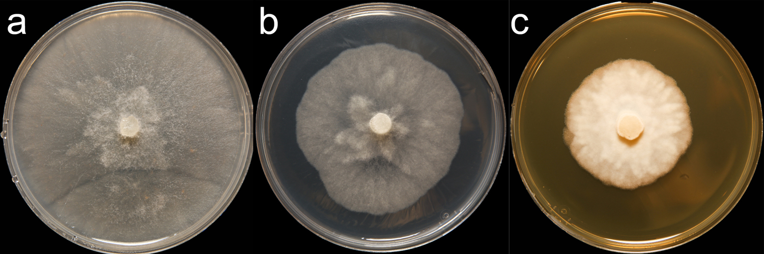
Phytophthora capsici (CPHST BL 136) colonies of a selected specimen grown for 7 days on (a) V8® Agar, (b) potato dextrose agar, and (c) malt extract agar; photo by Krysta Jennings and Leandra Knight, USDA-APHIS-PPQ |
|
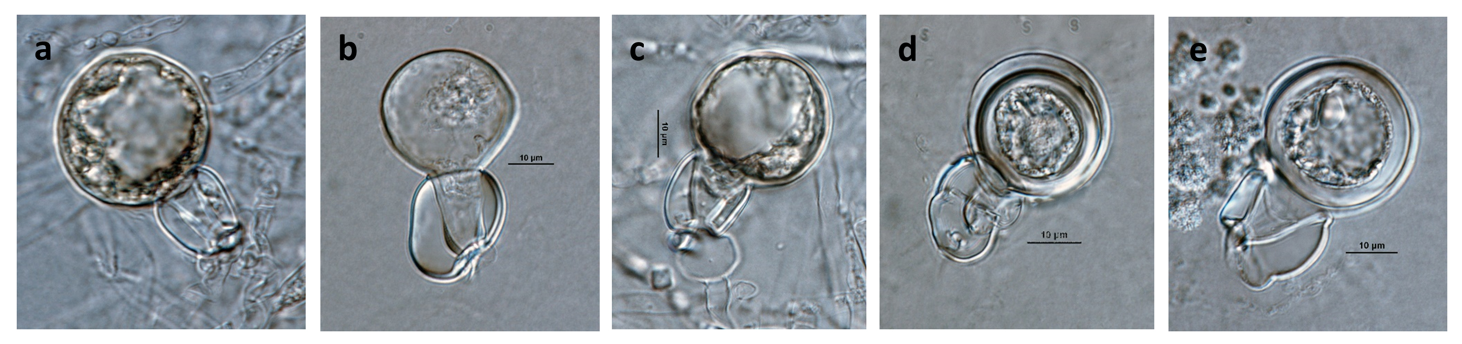
Phytophthora capsici (ex-type CPHST BL 33) heterothallic sexual phase: (a, c) young oogonia with amphigynous antheridia, (d, e) smooth-walled oogonia with amphigynous antheridia (d, e), aplerotic oospore (d), plerotic oospore (e); photos by Gloria Abad, USDA-APHIS-PPQ. |
|
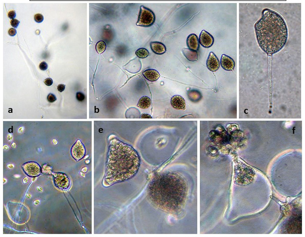
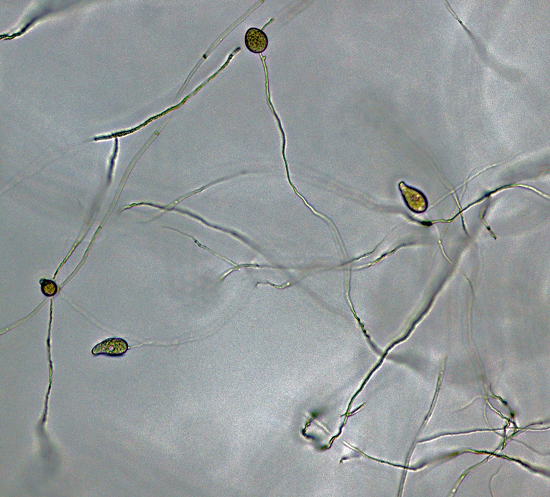
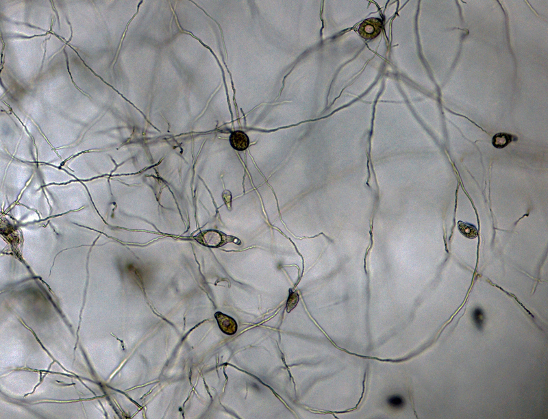
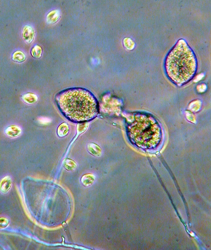
Phytophthora capsici (selected specimen P223): (a) sporangia originated in unbranched and simple sympodial sporangiophores, (b) sporangia originated in unbranched and irregular branched sporangiophores, (2) semipapillate ellipsoid caducous sporangium with long pedicel, (d-f) zoospores produced from semipapillated or bipapillate sporangia; photos by Gloria Abad, USDA-APHIS-PPQ |
|
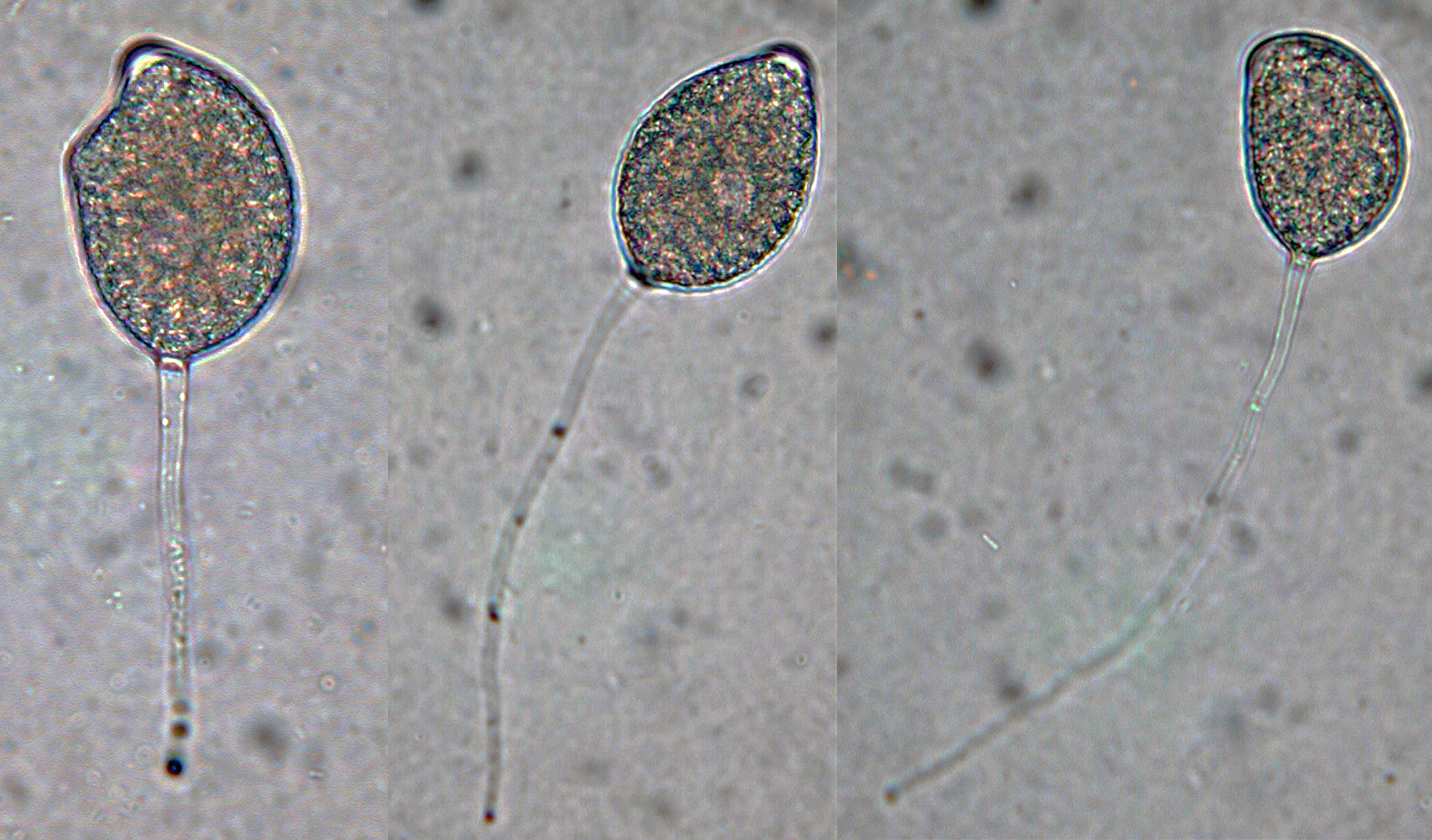
P. capsici (ex-type CPHST BL 33): (a, b) sporangia formed in unbranched and irregular branched sporangiophores, (c) semipapillate ellipsoid sporangium with short pedicel, (d) papillate subglobose sporangium with short pedicel, (e) semipapillate pyriform sporangium with medium pedicel, (f) semipapillate distorted shape sporangium with medium pedicel, (g) semipapillate distorted shape sporangium with long pedicel; photos by Gloria Abad, USDA-APHIS-PPQ. |
|
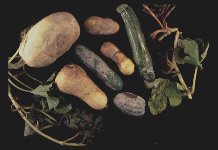
Phytophthora blight of cucurbits caused by Phytophthora capsici; photo by Gloria Abad at the former Plant Pathogen Identification Laboratory - North Carolina State University (present institution: USDA-APHIS-PPQ) |
|
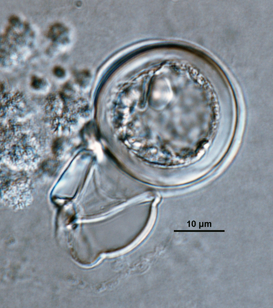
Phytophthora capsici (ex-type CPHST BL 33) heterothallic sexual phase: smooth-walled oogonium with amphigynous antheridium and plerotic oospore; photo by Gloria Abad, USDA-APHIS-PPQ. |
|
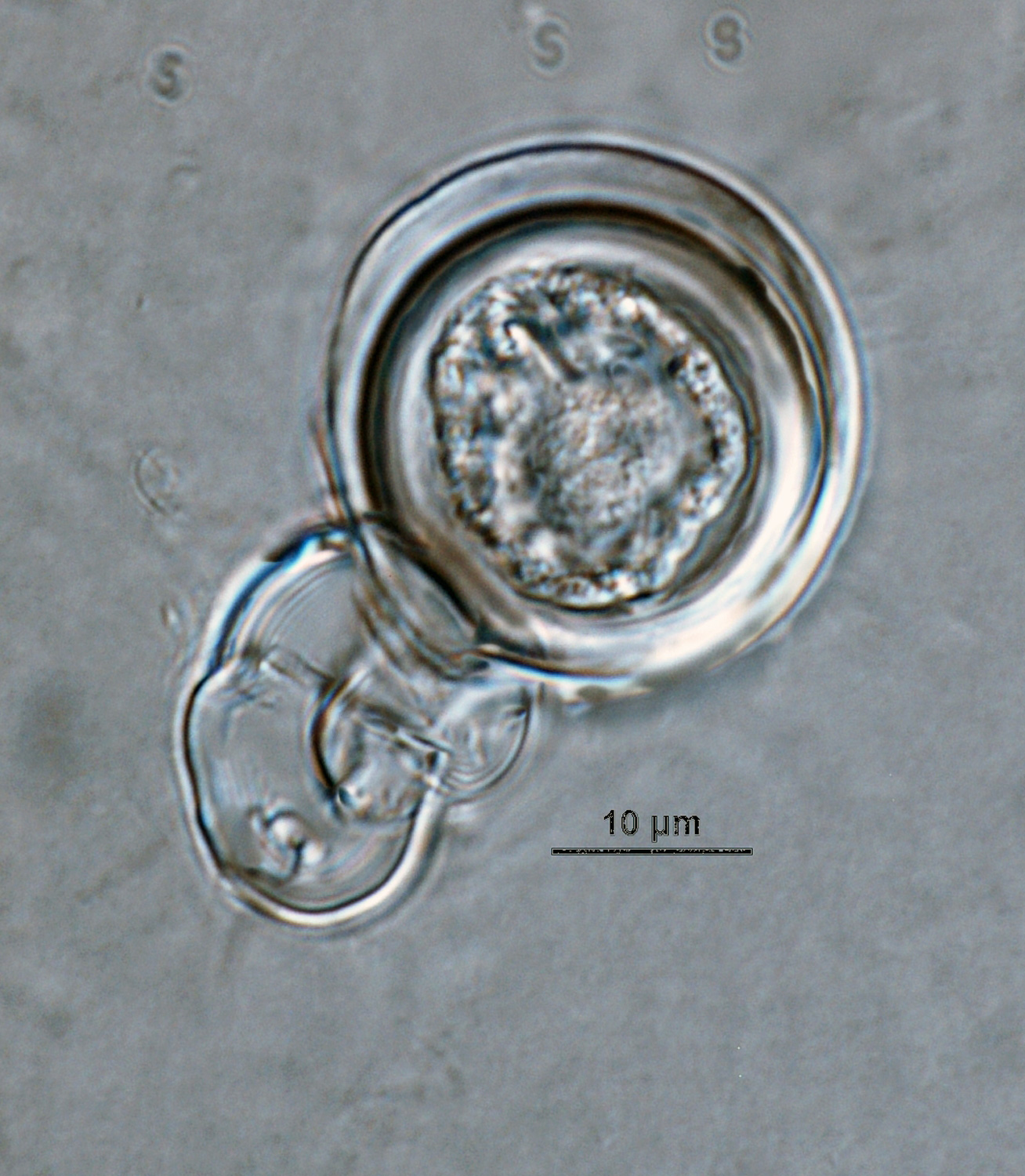
Phytophthora capsici (ex-type CPHST BL 33) heterothallic sexual phase: young oogonium with amphigynous antheridium; photo by Gloria Abad, USDA-APHIS-PPQ. |
|
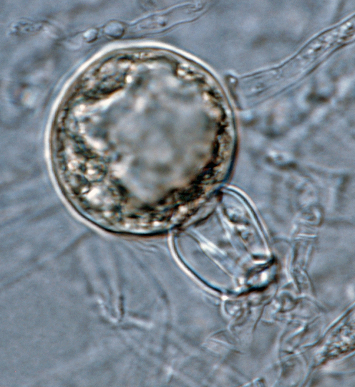
Phytophthora capsici (ex-type CPHST BL 33) heterothallic sexual phase: young oogonium with amphigynous antheridium and undifferentiated oospore; photo by Gloria Abad, USDA-APHIS-PPQ.
|
|
Phytophthora capsici (ex-type CPHST BL 33) heterothallic sexual phase: smooth-walled oogonium with amphigynous antheridium and aplerotic oospore; photo by Gloria Abad, USDA-APHIS-PPQ. |
|
Phytophthora capsici (ex-type CPHST BL 33) heterothallic sexual phase: young oogonium with amphigynous antheridium; photo by Gloria Abad, USDA-APHIS-PPQ. |
|
P. capsici (ex-type CPHST BL 33) asexual phase: sporangia formed in unbranched and irregular branched sporangiophores; photo by Gloria Abad, USDA-APHIS-PPQ. |
|
P. capsici (ex-type CPHST BL 33) asexual phase: sporangia formed in unbranched and irregular branched sporangiophores; photo by Gloria Abad, USDA-APHIS-PPQ. |
|
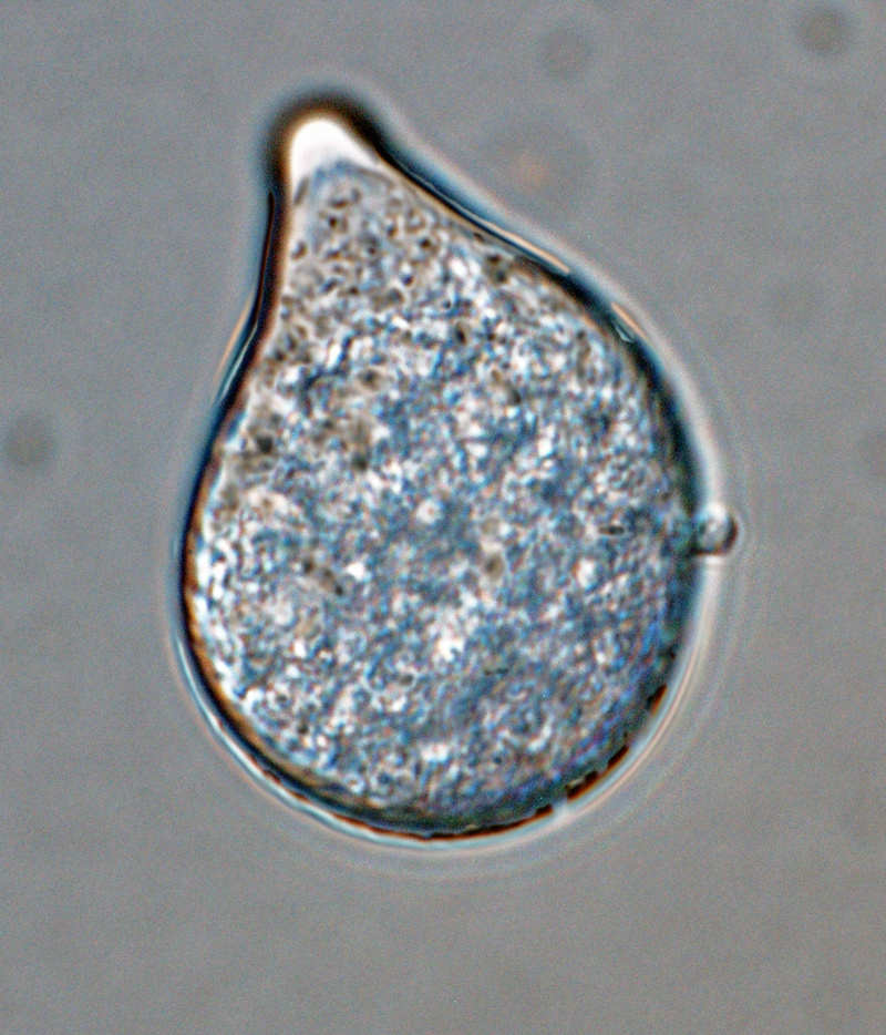
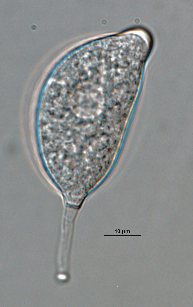
P. capsici (ex-type CPHST BL 33) asexual phase: papillate subglobose sporangium with short pedicel; photo by Gloria Abad, USDA-APHIS-PPQ. |
|
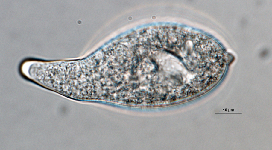
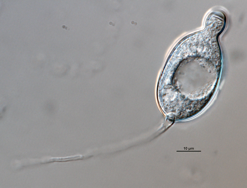
P. capsici (ex-type CPHST BL 33) asexual phase: semipapillate pyriform sporangium with medium pedicel; photo by Gloria Abad, USDA-APHIS-PPQ. |
|
P. capsici (ex-type CPHST BL 33) asexual phase: semipapillate distorted shape sporangium with medium pedicel; photo by Gloria Abad, USDA-APHIS-PPQ. |
|
P. capsici (ex-type CPHST BL 33) asexual phase: semipapillate pyriform sporangium with medium pedicel; photo by Gloria Abad, USDA-APHIS-PPQ. |
|
P. capsici (ex-type CPHST BL 33) asexual phase: semipapillate distorted shape sporangium with long pedicel; photo by Gloria Abad, USDA-APHIS-PPQ. |
|
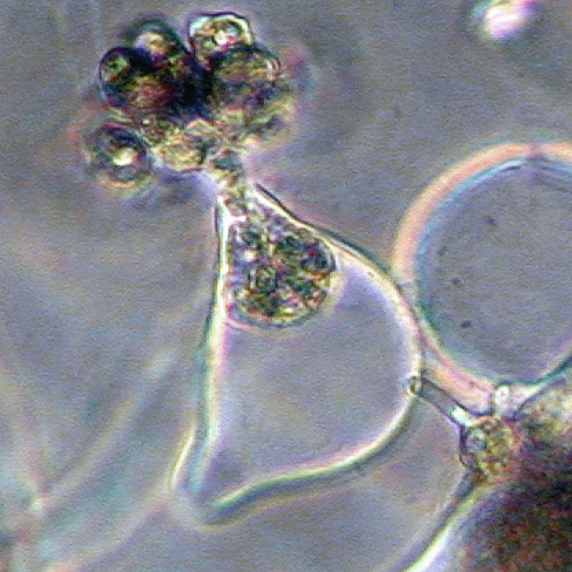
Phytophthora capsici (selected specimen P223) asexual phase: zoospores produced from bipapillate sporangium; photo by Gloria Abad, USDA-APHIS-PPQ. |
|
Phytophthora capsici (selected specimen P223) asexual phase: sporangia originated in unbranched and irregular branched sporangiophores; photo by Gloria Abad, USDA-APHIS-PPQ. |
|
Phytophthora capsici (selected specimen P223) asexual phase: zoospores produced from semipapillated or bipapillate sporangia; photo by Gloria Abad, USDA-APHIS-PPQ. |
|
Phytophthora capsici (selected specimen P223) asexual phase: zoospores produced from semipapillated or bipapillate sporangia; photo by Gloria Abad, USDA-APHIS-PPQ. |
|
Phytophthora capsici (selected specimen P223) asexual phase: sporangia originated in unbranched and simple sympodial sporangiophores; photo by Gloria Abad, USDA-APHIS-PPQ. |
|
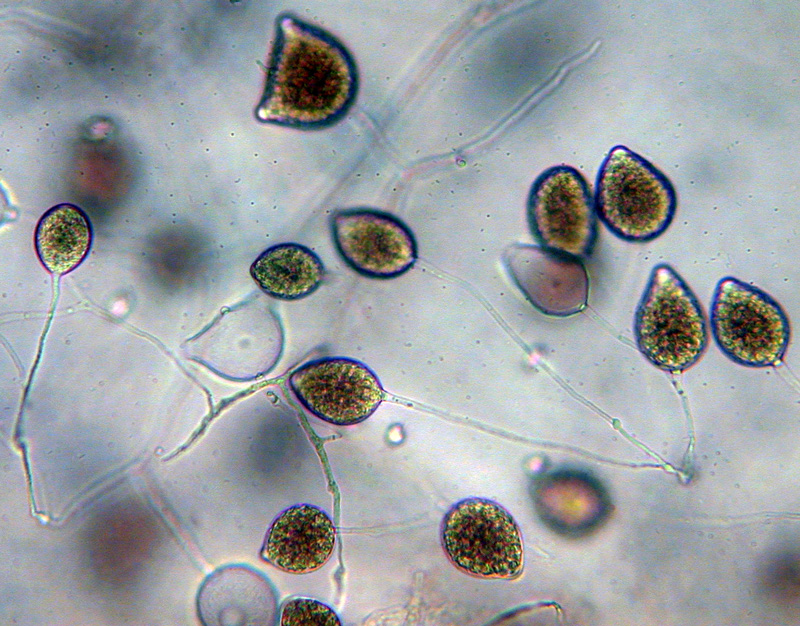
Phytophthora capsici (selected specimen P223) asexual phase: semipapillate variably shaped caducous sporangia with long pedicels; photo by Gloria Abad, USDA-APHIS-PPQ. |
.JPG)
.JPG)


Name and publication
Phytophthora capsici Leonian (1922)
Leonian LH. 1922. Stem and fruit blight of pepper caused by Phytophthora capsici species nov. Phytopathology 12: 401–408.
Nomenclature
from Leonian (1922)
Mycobank
Synonymy
≡ Phytophthora parasitica var. capsici (Leonian) Sarej., Annales de l'Institut Phytopathologique Benaki 2: 44 (1936) [MB353070]
= Phytophthora hydrophila Curzi, Riv. Patol. veg.: 1 (1927) [MB267501]
Typification
Type: UNITED STATES OF AMERICA, parasitic on stems and fruit of chile pepper (Capsicum annuum), State College, New Mexico. L. H. Leonian 1919
Ex-type: CBS 128.23 (deposited by Leonian in 1923) (A2 Type)
Ex-type in other collections
(ET) CBS 128.23 (A2), NRRL 64133, ATCC 52771, CABI IMI40502 (AVA), WPC P1091 P3605, S&T BL 33G (Abad)
CBS 128.23 = P3605 (WPC), P1091 (WPC), ATCC 52771 A2, IMI 40502, CPHST BL 33 (Abad)
NOTE: IMI 40502 The Netherlands??
Molecular identification
Voucher sequences for barcoding genes (ITS rDNA and COI) of the ex-type (see Molecular protocols page)
Phytophthora capsici isolate CPHST BL 33G (= P1091 WPC) = ITS rDNA MG865467, COI MH136863
Selected specimen
Phytophthora capsici isolate CPHST BL 136 (= P1314 WPC) = ITS rDNA MG865468, COI MH136864
Voucher sequences for Molecular Toolbox with seven genes (ITS, β-tub, COI, EF1α, HSP90, L10, and YPT1
(see Molecular protocols page) (In Progress)
Voucher sequences for Metabarcoding High-throughput Sequencing (HTS) Technologies [Molecular Operational Taxonomic Unit (MOTU)]
(see Molecular protocols page) (In Progress)
Sequences with multiple genes for ex-type in other sources
- NCBI: Phytophthora capsici CPHST BL 33G
- NCBI: Phytophthora capsici P3605
- NCBI: Phytophthora capsici P1091
- NCBI: Phytophthora capsici CBS 128.23
- EPPO-Q-bank: Phytophthora capsici CBS 128.23
- BOLDSYSTEMS: Phytophthora capsici OOMYA012-07 (= CBS 128.23), OOMYA490-08 (= CBS 128.23), PHYTO172-10 (= P3605) (barcoding COI & ITS)
Position in multigenic phylogeny with 7 genes (ITS, β-tub, COI, EF1α, HSP90, L10, and YPT1)
Clade clade:
a taxonomic group of organisms classified together on the basis of homologous features traced to a common ancestor
2b
Morphological identification
Colonies and cardinal temperatures
Colonies in V8-A, PDA, and MEA with no distinct pattern to light chrysanthemum pattern. Minimum temperature for growth is 6°C, optimum is 27°C, and maximum 33°C.
Asexual phase
Sporangia are papillatepapillate:
pertaining to the production of a distinct papilla at the distal end of the sporangium (cf. nonpapillate and semipapillate)
and semipapillatesemipapillate:
pertaining to the production of shallow having papilla that are not well developed, shallow and less nipple-like than fully papillate structures
, occasionally with two or three apices; caducouscaducous:
pertaining to sporangia that become dislodged readily (i.e. deciduous) and separate from the sporangiophore (cf. persistent)
with short, medium, and long pedicels (3 to 138 µm); subglobose, ovoidovoid:
egg-shaped, with the widest part at the base of the sporangium and the narrow part at the apex
, obovoidobovoid:
inversely egg-shaped; ovoid, but with the widest part at the apex
, ellipsoidellipsoid:
refers to a solid body that forms an ellipse in the longitudinal plane and a circle in cross section; many fungal spores are ellipsoidal or elliptic
, fusiformfusiform:
spindle-shaped; wide in the middle while narrowing or tapering at each end
, pyriformpyriform:
pear-shaped, with the narrowest part at the base (cf. obpyriform)
, to distorted shapes sometimes with tapered bases (9–41 x 12–47 µm). Sporangiophores unbranched, irregularly branched, and occasionally simple sympodial. Hyphal swellings are occasionally produced by some isolates in aqueous cultures and are globoseglobose:
having a rounded form resembling that of a sphere
, subglobose, produced individually or catenulatecatenulate:
having a chain-like form
. ChlamydosporesChlamydospores:
an asexual spore with a thickened inner wall that is delimited from the mycelium by a septum; may be terminal or intercalary, and survives for long periods in soil
absent.
Sexual phase
Heterothallic. OogoniaOogonia:
the female gametangium in which the oospore forms after fertilization by the antheridium
are spherical or subspherical, smooth-walled, and hyaline to brown (22–40 µm diam); antheridia are amphigynousamphigynous:
pertaining to the sexual stage in which the antheridium completely surrounds the stalk of the oogonium (cf. paragynous)
; oosporesoospores:
zygote or thick-walled spore that forms within the oogonium after fertilization by the antheridium; may be long-lived
are apleroticaplerotic:
pertaining to a mature oospore that does not fill the oogonium; i.e. there is room left between the oospore wall and oogonium wall (cf. plerotic)
and pleroticplerotic:
pertaining to an oospore that fills the oogonium (cf. aplerotic)
(19–32 µm diam).
Most typical characters
Phytophthora capsici is characterized by its production of caducouscaducous:
pertaining to sporangia that become dislodged readily (i.e. deciduous) and separate from the sporangiophore (cf. persistent)
, short, medium, and long pedicelpedicel:
the hyphal base of a sporangium that remains attached after the sporangium separates, or is shed, from the sporangiophore; the pedicel may be short (< 5 µm), medium (5–20 µm), or long (> 20 µm)
sporangia which are subglobose to ellipsoidellipsoid:
refers to a solid body that forms an ellipse in the longitudinal plane and a circle in cross section; many fungal spores are ellipsoidal or elliptic
, and the absence of chlamydosporeschlamydospores:
an asexual spore with a thickened inner wall that is delimited from the mycelium by a septum; may be terminal or intercalary, and survives for long periods in soil
. It differs from closely related Phytophthora tropicalis which produces caducouscaducous:
pertaining to sporangia that become dislodged readily (i.e. deciduous) and separate from the sporangiophore (cf. persistent)
, long pedicelpedicel:
the hyphal base of a sporangium that remains attached after the sporangium separates, or is shed, from the sporangiophore; the pedicel may be short (< 5 µm), medium (5–20 µm), or long (> 20 µm)
, elongated-narrow sporangiasporangia:
sac within which zoospores form, especially when water is cooled to about 10°C below ambient temperature; in solid substrates, sporangia usually germinate by germ tubes
with a tapering base, and chlamydosporeschlamydospores:
an asexual spore with a thickened inner wall that is delimited from the mycelium by a septum; may be terminal or intercalary, and survives for long periods in soil
frequently produced.
Specimen(s) evaluated
Phytophthora capsici ex-type CPHST BL 33 (A2) duplicate of P1091 (World Phytophthora Collection), a duplicate of ATCC 52771 (= ex-type CBS 128.23)
Additional isolates with 100% alignment in ITS rDNA with ex-type CPHST BL 33
CPHST BL 136 (A1) duplicate of P1314 (World Phytophthora Collection) from green bell pepper (Capsicum annuum) in California, USA
P223 (A2) from former NCSU - Plant Pathogen Identification Laboratory, Director Gloria Abad, collected from "chile jalapeño" (Capsicum annum) in Guatemala
Hosts and distribution
Distribution: cosmopolitan; tropical specimens may be Phytophthora tropicalis
Substrate: roots, fruits, stems, seedlings, pods, cotton bolls
Disease note: fruit, stem, and root rot; also seedling damping-off, leaf wilt
Hosts: Phytophthora capsici sensu lato infects 51 genera in 28 families, including Capsicum annuum (peppers), Solanum lycopersicum (tomato), and other Solanaceae. Aragaki & Uchida (2001) refers to isolates from non-Capsicum hosts to P. tropicalis, including isolates from Macadamia spp., Theobroma cacao (cacao), and other tropical crop species.
Retrieved January 29, 2018 from U.S. National Fungus Collections Nomenclature Database.
Additional references and links
Bowers JH, Martin FN, Tooley PW, and Luz ED. 2007. Genetic and morphological diversity of temperate and tropical isolates of Phytophthora capsici. Phytopathology 97: 492-503.
- SMML USDA-ARS: Phytophthora capsici
- EPPO Global Database: Phytophthora capsici
- Forest Phytophthoras of the world: Phytophthora capsici
- CABI Digital Library: Phytophthora capsici
- Encyclopedia of Life (EOL): Phytophthora capsici
- Index Fungorum (IF): Phytophthora capsici
- Google All Phytophthora capsici
- Google Images Phytophthora capsici
- Google Scholar Phytophthora capsici
Fact sheet author
Z. Gloria Abad, Ph.D., USDA-APHIS-PPQ-S&T Plant Pathogen Confirmatory Diagnostics Laboratory (PPCDL), United States of America.
