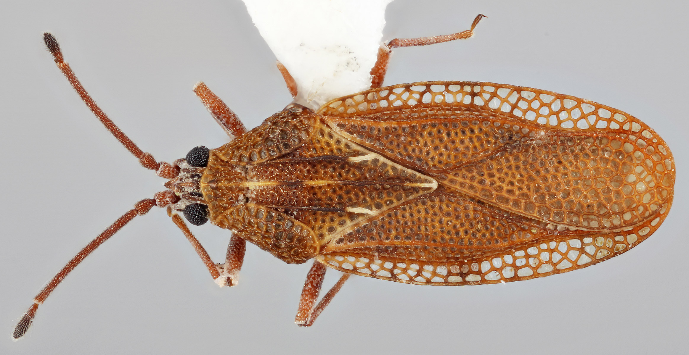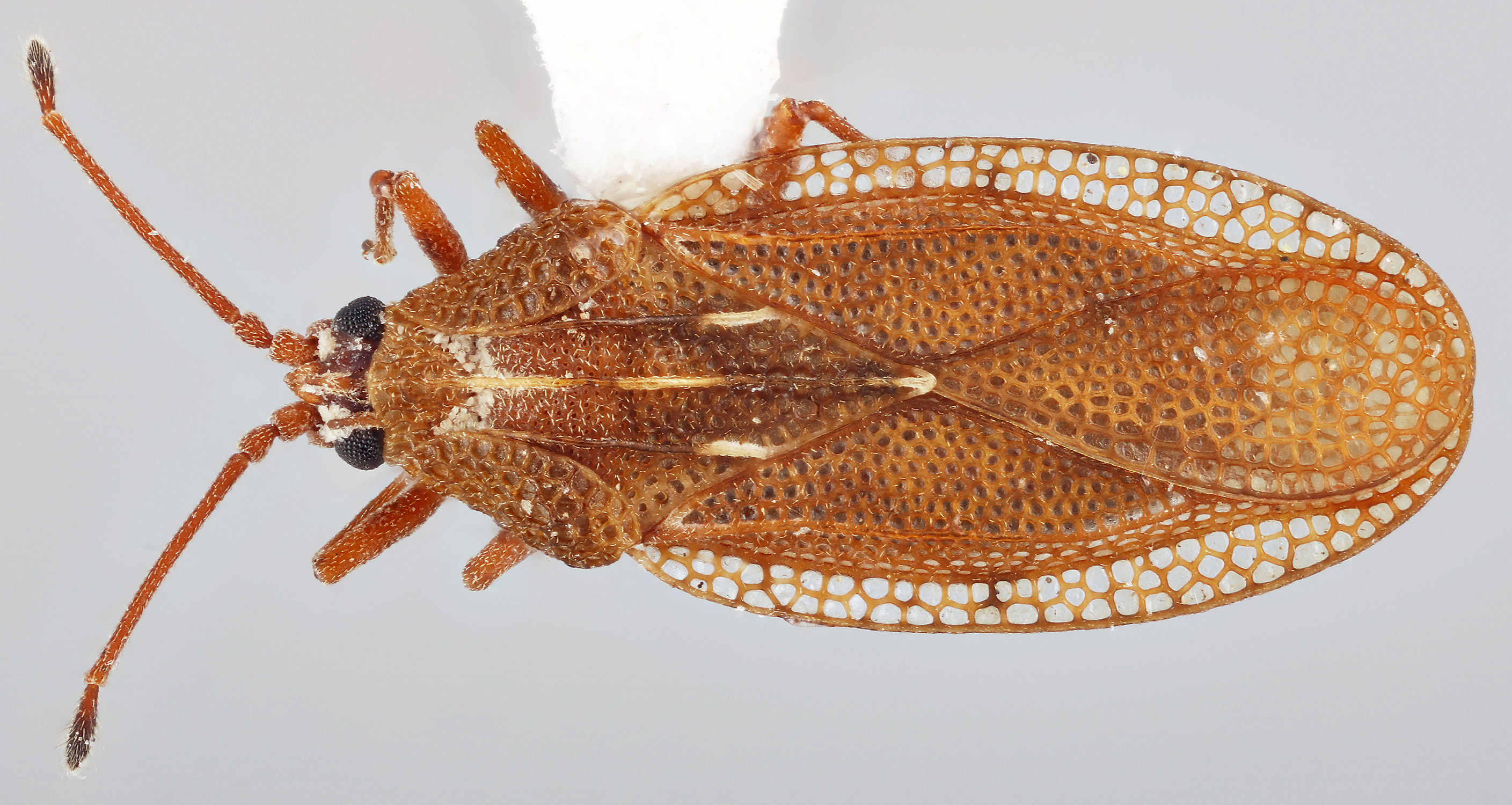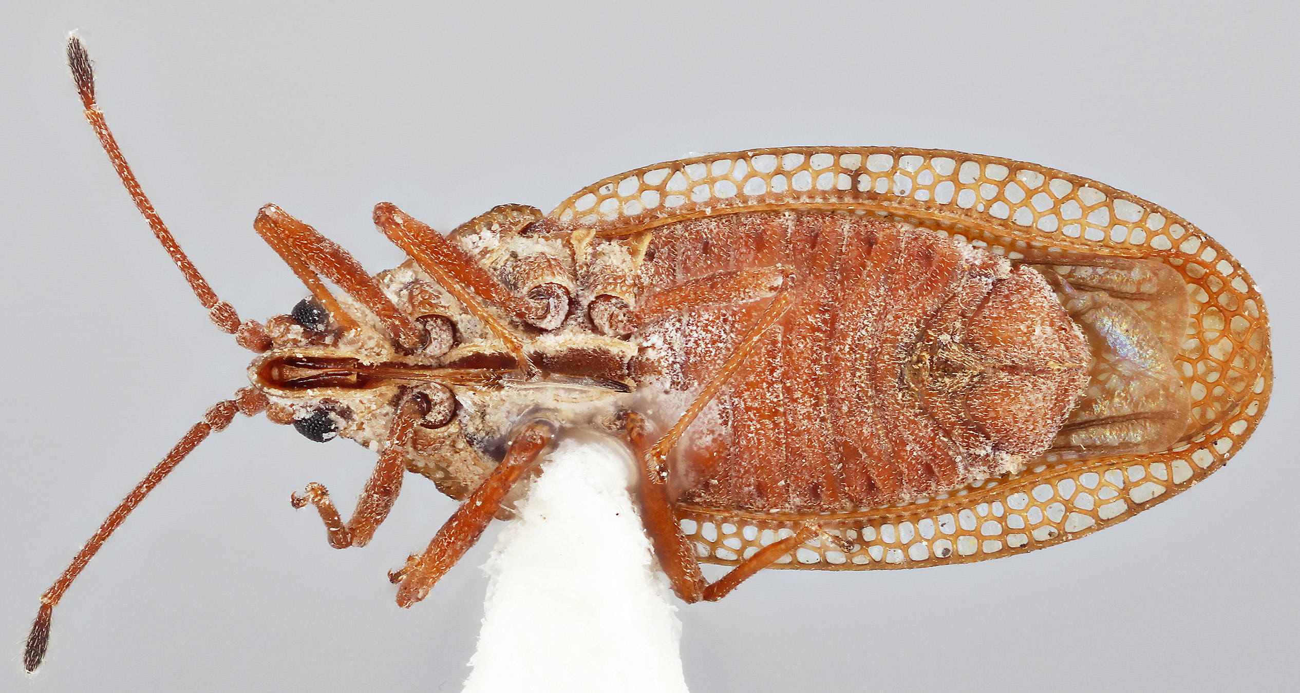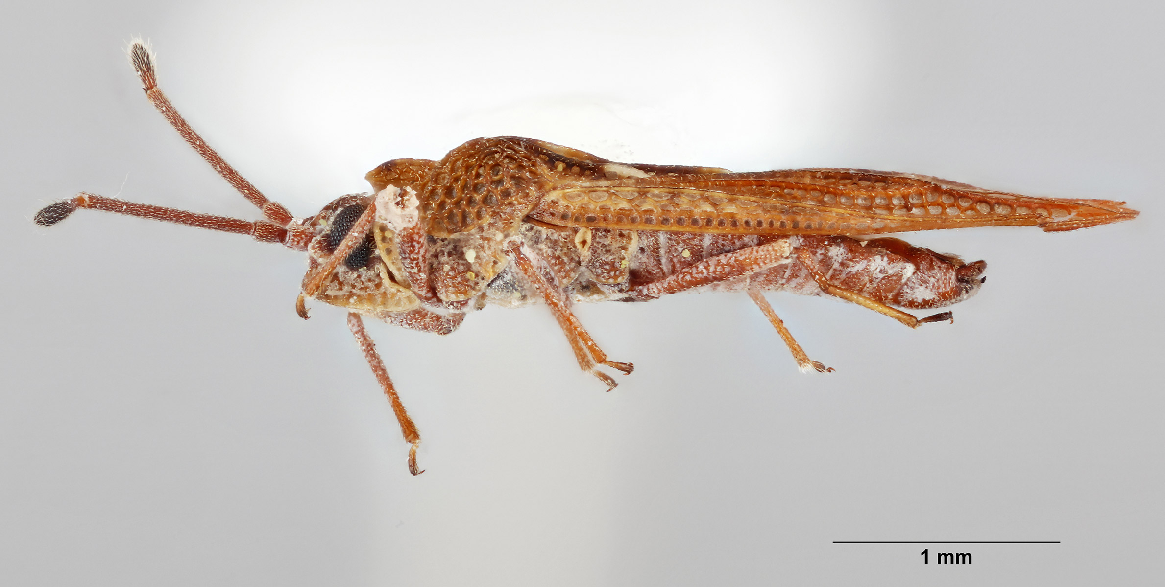
Monanthia (Physatocheila) Fieber, 1844
Physatocheila: Stål, 1973
Type species: Acanthia quadrimaculata Wolff = Acanthia costata Fabricius
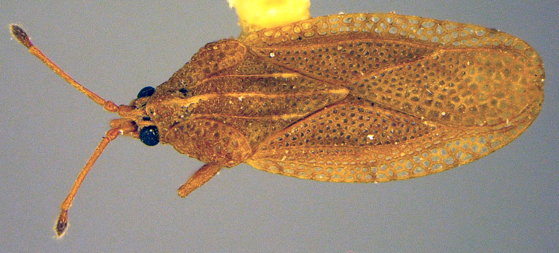
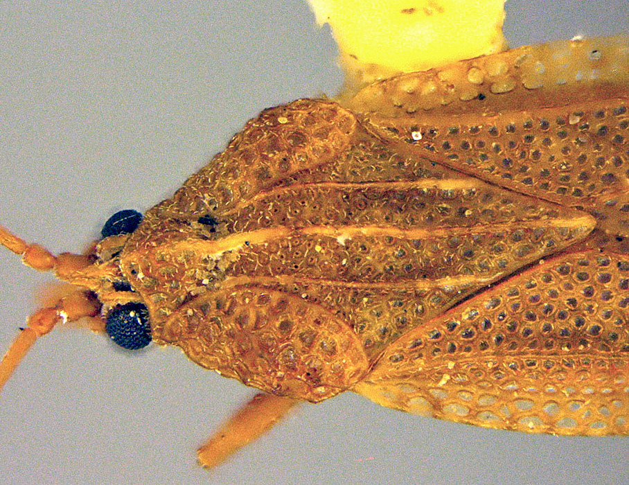
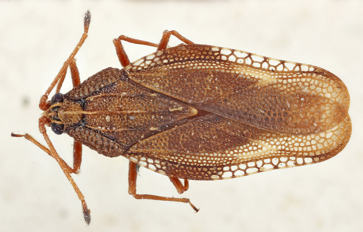
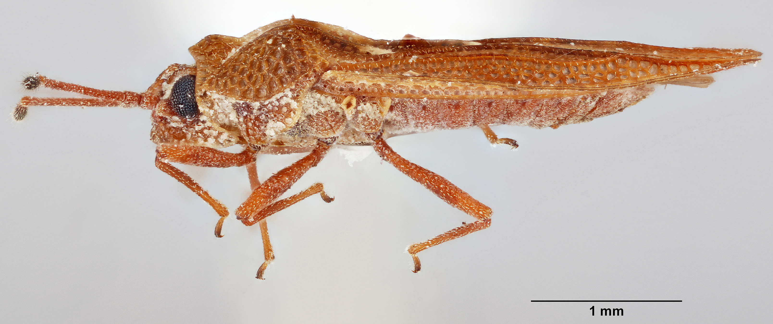
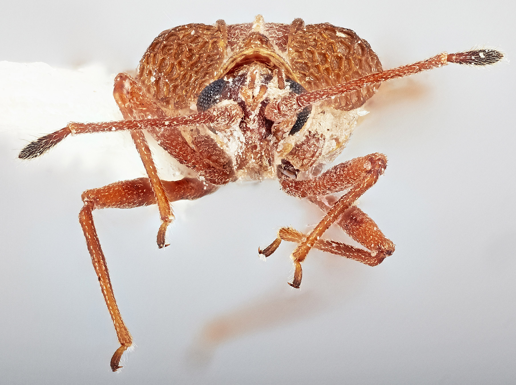
Head short, clypeus not surpassing apical half of first antennal segment; antennae shorter than the length of pronotumpronotum:
dorsal sclerite of the first thoracic segment
; cephalic spinescephalic spines:
a spine on the head
 present; bucculaebucculae:
an elevated ridge on either side of the first labial segment
 closed anteriorly; pronotumpronotum:
dorsal sclerite of the first thoracic segment
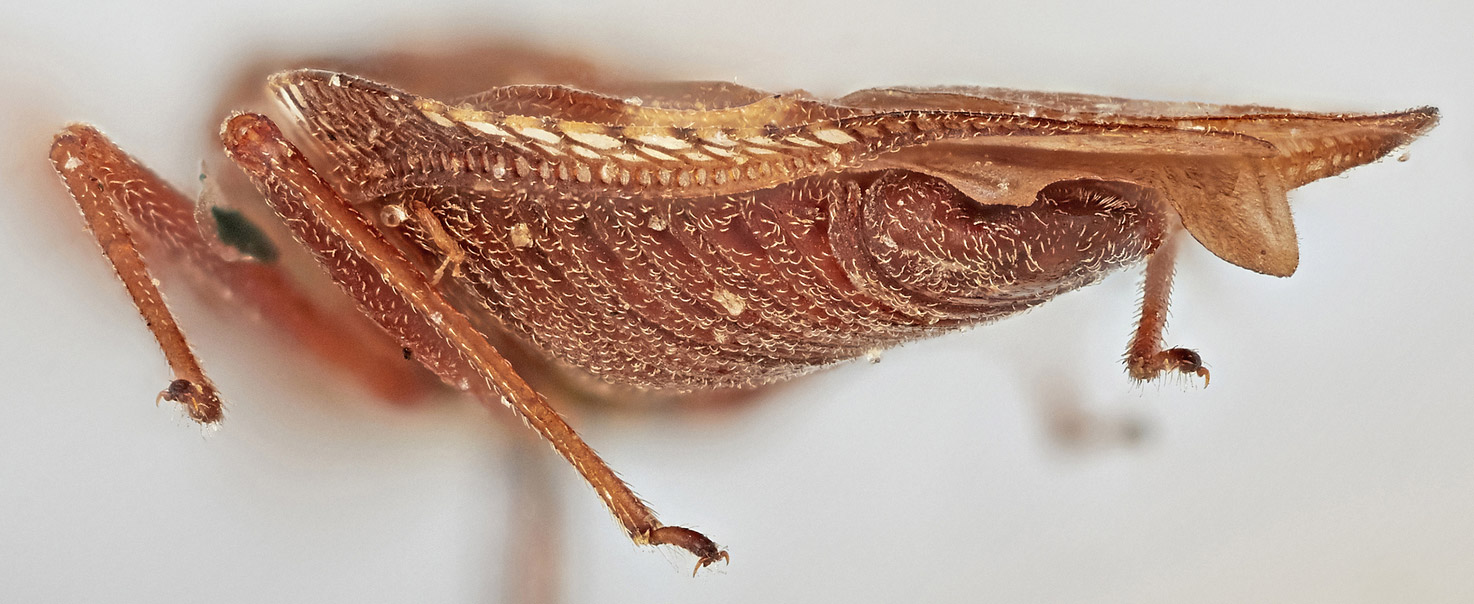
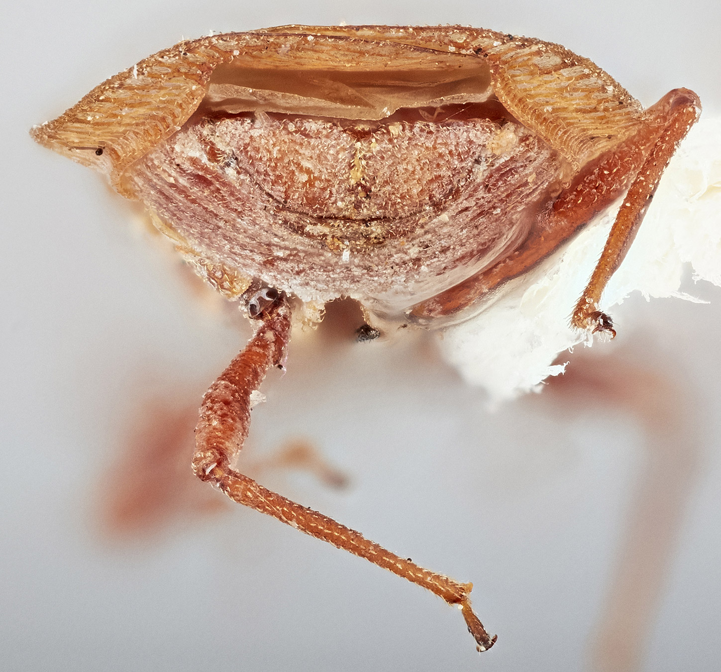
tricarinate; pronotal hoodhood:
term used to describe the modified anterior area of the pronotum, which is sometimes tectiform and sometimes bulbous, with numerous intermediate conditions.
 tectiform, not bulbous; paranotumparanotum:
lateral extension of pronotum; may be carinate, explanate, or reflexed
 expanded widely, reflexedreflexed:
bent or curved backwards, as in a reflexed paranotum
 and appressed to pronotal disc, not forming elevated cyst, in some species, anterior part of lateral carinaecarinae:
elevated ridge or keel
hidden under reflexedreflexed:
bent or curved backwards, as in a reflexed paranotum
pronotumpronotum:
dorsal sclerite of the first thoracic segment
; anterior margin not projecting anterad; hemelytrahemelytra:
one of the basally thickened forewings of Hemiptera
with claval areaclaval area:
parallel-sided and sharply pointed anal area of hemelytron
 weakly developed, almost entirely covered by posterior margin of pronotumpronotum:
dorsal sclerite of the first thoracic segment
; costal areacostal area:
area of the costa delineated by the first longitudinal vein of the wing, usually running along the anterior margin
 narrow, with one to two rows of areolaeareolae:
a small enclosed space on a surface, such as an area enclosed by veinlets on wings
 , not reflexedreflexed:
bent or curved backwards, as in a reflexed paranotum
near its base; discoidal areadiscoidal area:
area of the forewing posterior to the subcostal area
level, discoidal cell long, extending beyond the middle of hemelytra; hypocostal laminae extending beyond the apex of abdomen.
Afrotropical, Australasian, Nearctic, Oriental, Palearctic (Drake and Ruhoff 1960Drake and Ruhoff 1960:
Drake, C. J., and F. Ruhoff, A. 1960. Lace-bug genera of the world (Hemiptera: Tingidae). Proceedings of the United States National Museum 112., Drake and Ruhoff 1965Drake and Ruhoff 1965:
Drake, C. J., and F. A. Ruhoff. 1965. Lacebugs of the World: A Catalog (Hemiptera: Tingidae) Bulletin of the United States National Museum: 1ndash;634., Guilbert 2019Guilbert 2019:
Guilbert, E. 2019. Lace bugs database - http://www.hemiptera-databases.com/tingidae)
| Intercepted species | Shipment origin(s) | Inspected commodities |
|---|---|---|
| Physatocheila brevirostris Osborn & Drake | Italy | Stone product- Ceramic tiles |
| Physatocheila costata (Fabricius) | Unknown | Rosmarinus sp. |
| Physatocheila sp. | Lebanon | Prunus sp. |
Drake and Ruhoff 1960Drake and Ruhoff 1960:
Drake, C. J., and F. Ruhoff, A. 1960. Lace-bug genera of the world (Hemiptera: Tingidae). Proceedings of the United States National Museum 112., Drake and Ruhoff 1965Drake and Ruhoff 1965:
Drake, C. J., and F. A. Ruhoff. 1965. Lacebugs of the World: A Catalog (Hemiptera: Tingidae) Bulletin of the United States National Museum: 1ndash;634., Guilbert 2019Guilbert 2019:
Guilbert, E. 2019. Lace bugs database - http://www.hemiptera-databases.com/tingidae
Physatocheila images at the Smithsonian
Authors: S.M. Krishnankutty, J.L. Scher
Last updated May 2020
idtools.org