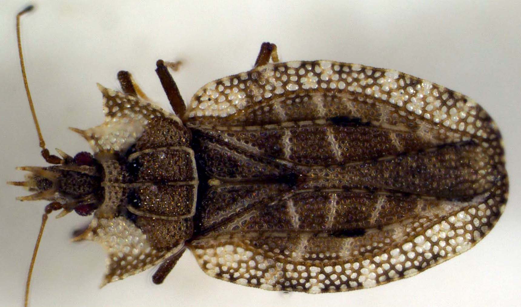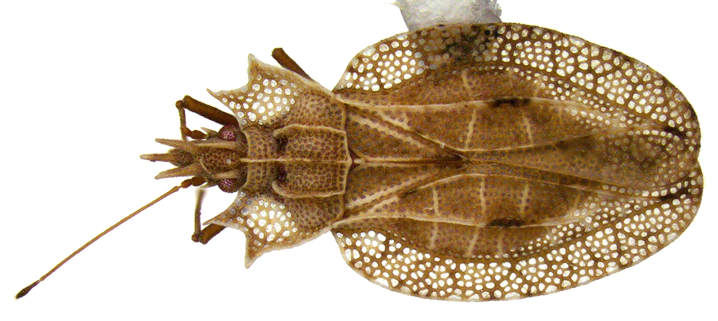
Phatnoma Fieber, 1844
Type species: Phatnoma laciniata Fieber
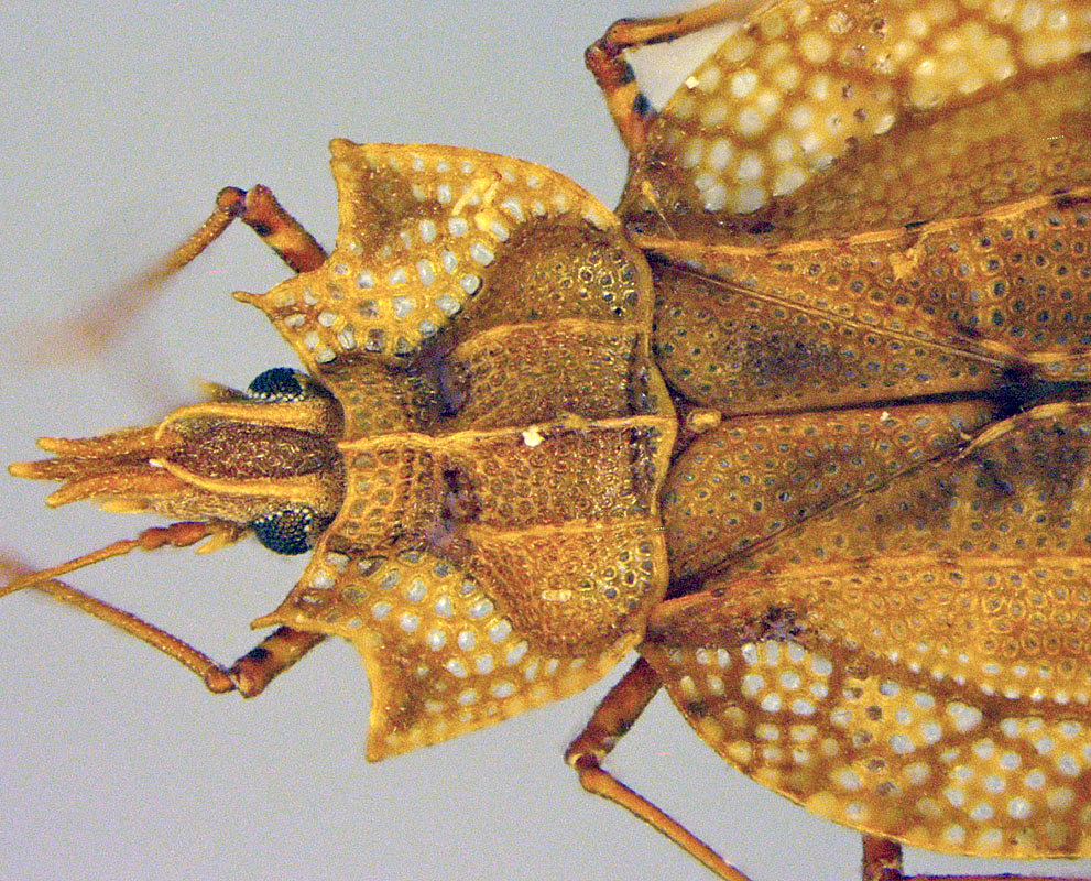
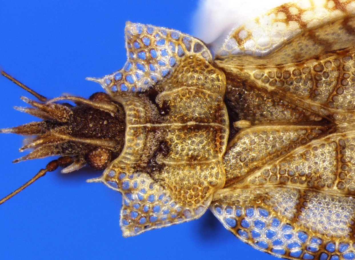
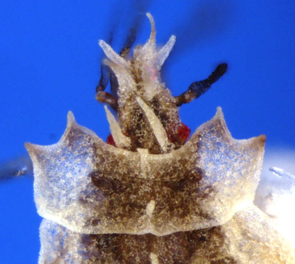
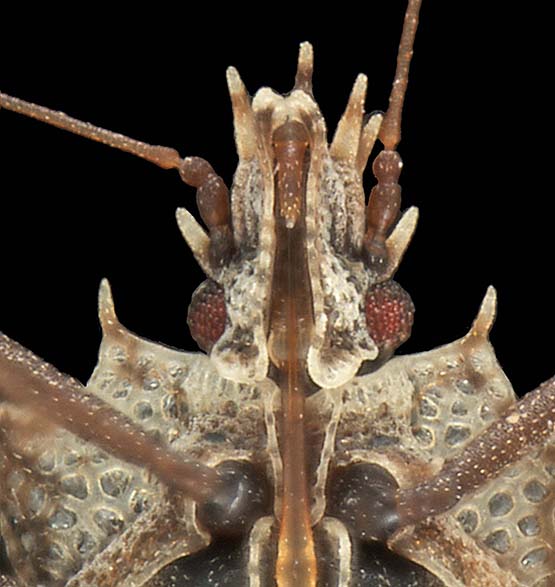
Head long, clypeus surpassing apical half of first antennal segment; cephalic spinescephalic spines:
a spine on the head
 present; bucculaebucculae:
an elevated ridge on either side of the first labial segment
 closed anteriorly; interocular areainterocular area:
area between compound eyes
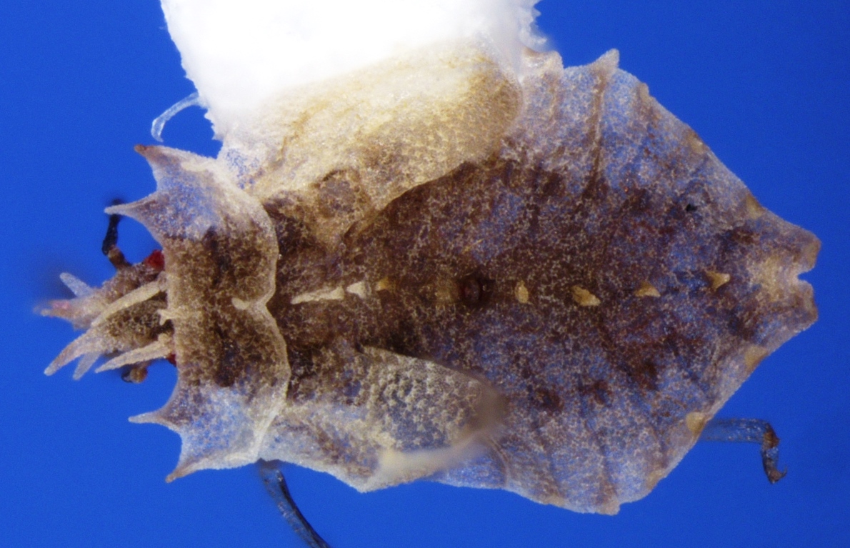
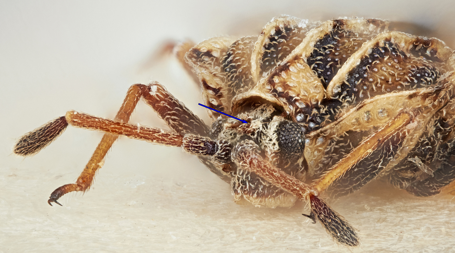 not depressed; rostrumrostrum:
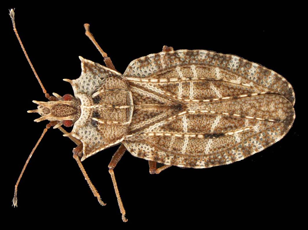
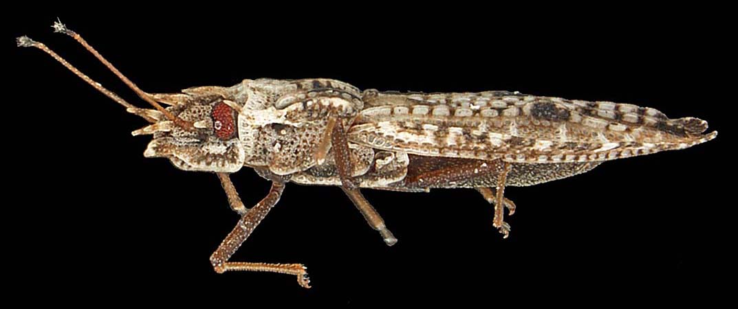
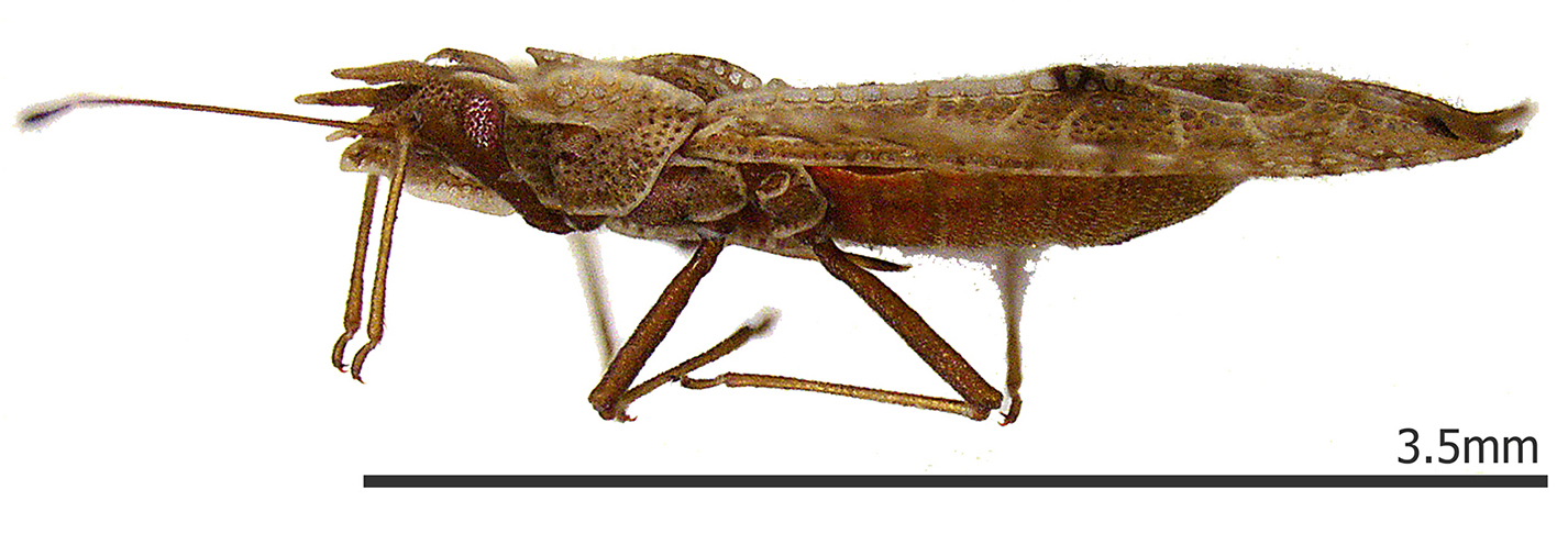
a beak or snout, applied especially to the piercing mouth-parts of bugs and elongated snouts of weevils long, reaching abdominal segment III, ventral side of abdomen with mediolongitudinal groove; pronotumpronotum:
dorsal sclerite of the first thoracic segment
tricarinate, lateral carinaecarinae:
elevated ridge or keel
obsolete on anterior regions of pronotal disc; pronotal hoodhood:
term used to describe the modified anterior area of the pronotum, which is sometimes tectiform and sometimes bulbous, with numerous intermediate conditions.
 absent; paranotumparanotum:
lateral extension of pronotum; may be carinate, explanate, or reflexed
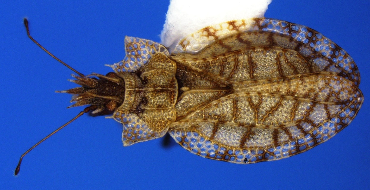
 widely expanded with three or more rows of areolaeareolae:
a small enclosed space on a surface, such as an area enclosed by veinlets on wings
 , anterior margin projecting anterad; scutellumscutellum:
a small triangular plate behind the pronotum and between the forewing bases
 exposed; hemelytrahemelytra:
one of the basally thickened forewings of Hemiptera
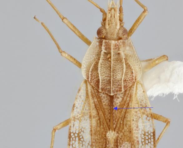
with claval areaclaval area:
parallel-sided and sharply pointed anal area of hemelytron
 well developed, meeting in a straight line to form claval commissureclaval commissure:
junction of hemelytra along clavus in middle of dorsum, posterior to scutellum
 , not covered by posterior margin of pronotumpronotum:
dorsal sclerite of the first thoracic segment
; discoidal areadiscoidal area:
area of the forewing posterior to the subcostal area
 level, extending beyond the middle of hemelytra; hypocostal laminae extending to the apex of abdomen, stenocostal areastenocostal area:
area of hemelytra along the outer margin of costal area
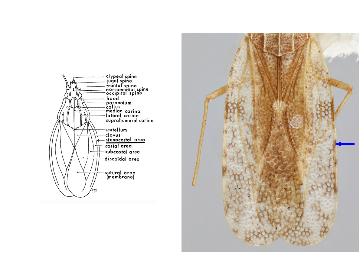 absent (Froeschner 1996Froeschner 1996:
Froeschner, R. C. 1996. Lace bug genera of the World, I: Introduction, Subfamily Cantacaderinae (Heteroptera: Tingidae). Smithsonian Contributions to Zoology. 1ndash;43.).
Afrotropical, Australasian, Neotropical, Oceanian, Oriental (Drake and Ruhoff 1960Drake and Ruhoff 1960:
Drake, C. J., and F. Ruhoff, A. 1960. Lace-bug genera of the world (Hemiptera: Tingidae). Proceedings of the United States National Museum 112., Froeschner 1996Froeschner 1996:
Froeschner, R. C. 1996. Lace bug genera of the World, I: Introduction, Subfamily Cantacaderinae (Heteroptera: Tingidae). Smithsonian Contributions to Zoology. 1ndash;43., Guilbert 2019Guilbert 2019:
Guilbert, E. 2019. Lace bugs database - http://www.hemiptera-databases.com/tingidae)
| Intercepted species | Shipment origin(s) | Inspected host(s) |
|---|---|---|
| Phatnoma annulipes (Champion) | Costa Rica, Protugal, Mexico | Ananas comosus, Murraya paniculata, Rubus sp. |
| Phatnoma marmorata Champion | Costa Rica, Ecuador | Ananas comosus |
| Phatnoma sp. | Costa Rica, Jamaica, Malaysia, Mexico, Thailand, Viet Nam | Alocasia sp., Ananas comosus, Annona sp., Cupressus sp., Dimocarpus longan, Lansium domesticum |
Drake and Ruhoff 1960Drake and Ruhoff 1960:
Drake, C. J., and F. Ruhoff, A. 1960. Lace-bug genera of the world (Hemiptera: Tingidae). Proceedings of the United States National Museum 112., Froeschner 1996Froeschner 1996:
Froeschner, R. C. 1996. Lace bug genera of the World, I: Introduction, Subfamily Cantacaderinae (Heteroptera: Tingidae). Smithsonian Contributions to Zoology. 1ndash;43., Guilbert 2019Guilbert 2019:
Guilbert, E. 2019. Lace bugs database - http://www.hemiptera-databases.com/tingidae
Authors: S.M. Krishnankutty, J.L. Scher
Last updated May 2020
idtools.org