
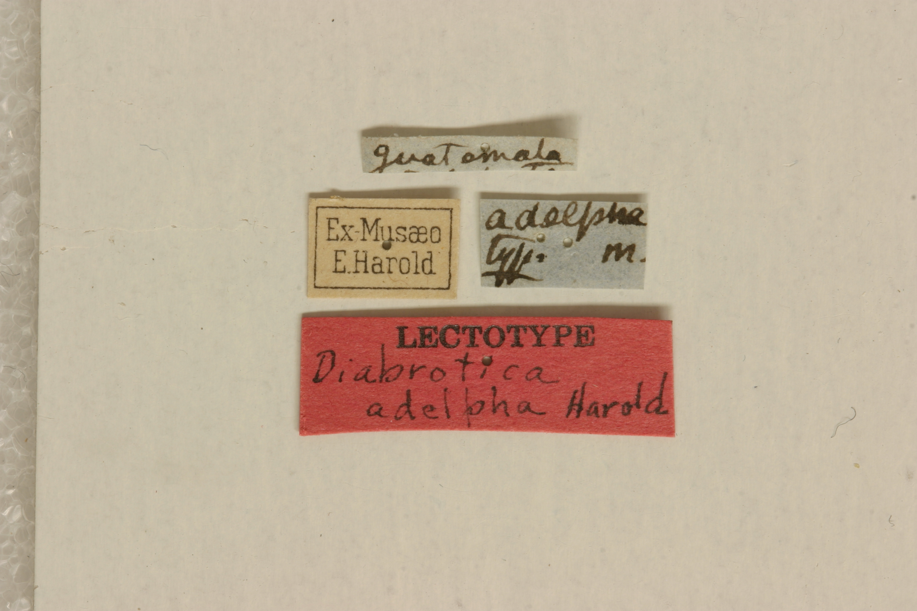
Guatemala
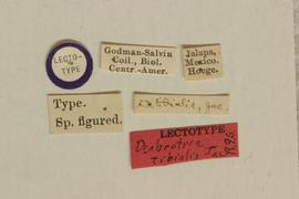
MNHN, lectotype, female, verified
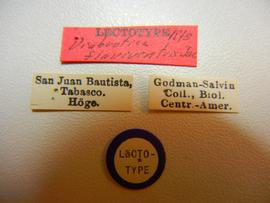
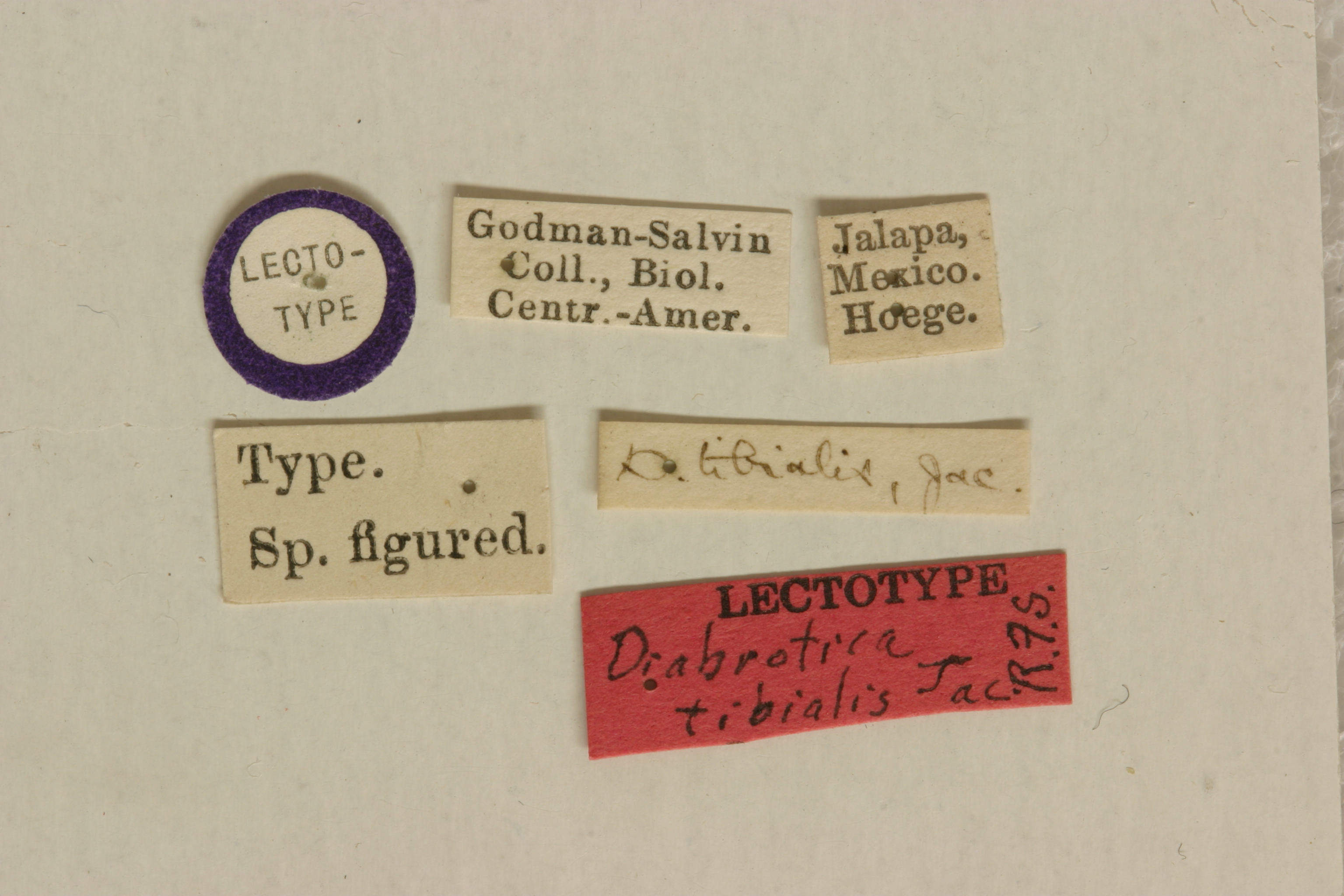
flaviventris Jacoby 1887: 517 (type locality: Mexico, San Juan Bautista; type depository: BMNH, lectotype, female, verified) new synonym
picticornis Horn 1893: 90 (type locality: Texas, USA; type depository: ANSP, holotype, female) synonym of Diabrotica tibialis Jacoby (Smith, 1966)
tibialis Jacoby 1887: 512 (type locality: Mexico, Jalapa; type depository: BMNH, lectotype, female, verified) new synonym
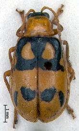
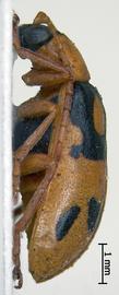
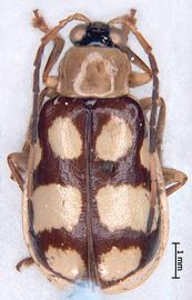
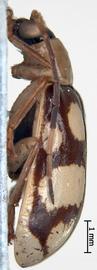
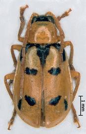
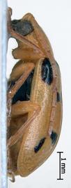
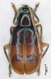
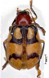
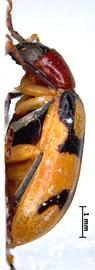
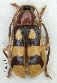
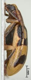
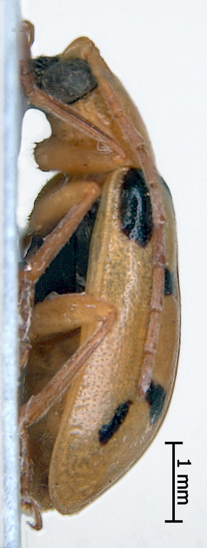
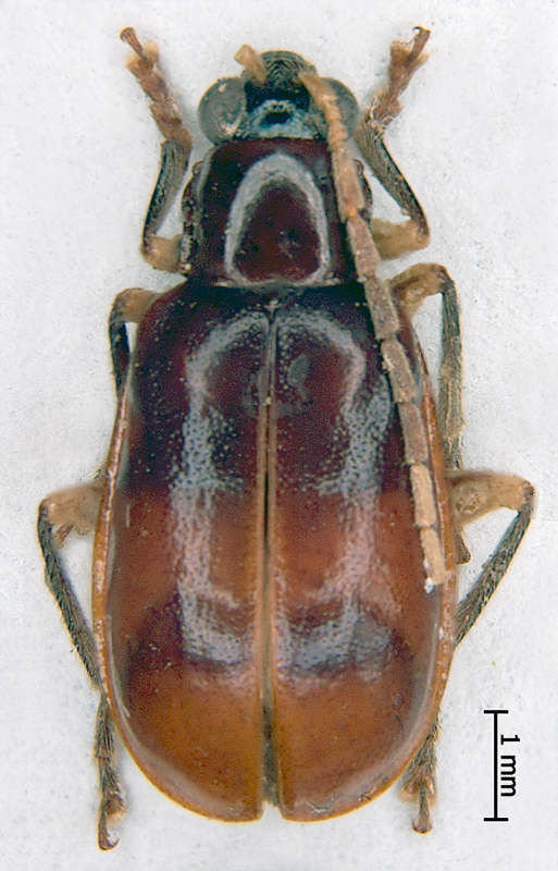
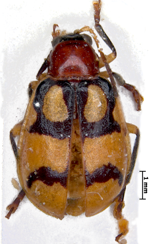
Body length 5.8-7.4. Body width 3.0-4.2. Head basic coloration black. Male antennae serrateserrate:
in reference to antennae, saw-like, often with a sharp ridge on the outer margin
, uniformly yellow, bi- or tricolored, antennomeres 1-3, and 9-10 uniformly yellow or yellow ocher, antennomeres 4 - 8 uniformly cinnamon brown or gradually infuscated by brown, antennomereantennomere:
"segment" of antenna, more or less clearly separated
11 completely dark (brussels brown) or dark apically, Female antennae filiformfiliform:
slender antennae with antennomeres of similar shape
or serrateserrate:
in reference to antennae, saw-like, often with a sharp ridge on the outer margin
(sometimes only slightly so). Maxillary palpi yellow, yellow ocher or amber brown, labrumlabrum:
the "upper lip" of beetles, a movable sclerite joined under clypeus
black. Pronotumpronotum:
the notum of the prothorax with highly sclerotized pronotal disc
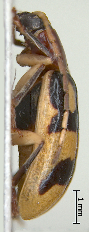
yellow, sulphur yellow, yellow ocher or testaceous, subquadrate, nonfoveate, shagreen absent. Scutellumscutellum:
small, usually triangular shield between the bases of elytra
piceous, black or amber brown. Basic color of elytra yellow or rufous, with 2 black bandsbands:
(here) transverse maculae on the beetle elytra
. One yellow macula of the irregular triangular shape is in center of basalbasal:
of or pertaining to the base, as in the first, or basal segment of an appendage; opposite of apical
band on each elytronelytron:
<em>(pl. elytra)</em> the fore highly sclerotized wing of beetle
. Posteriorposterior:
the region of the body parts of the beetle furthest from the head
margin of the band is produced laterally and slightly distally like a handle. Distal band is narrow, arcuate, may be reduced to two round or drop-shaped spots. Elytrae of D. flaviventris are almost entirely testaceous with appeared outlines of the bandsbands:
(here) transverse maculae on the beetle elytra
. Color of bandsbands:
(here) transverse maculae on the beetle elytra
black, humeral calli black, epipleura completely yellow or completely rufous, not sulcatesulcate:
bearing the carinae or ridges on the elytron
. Sutural anglesutural angle:
the posterior angle or apex of the elytron near the suture
of elytra obtuse-angled or dentiform, punctation dense, fine. Abdomen yellow or yellow ocher. Tarsi coloration black, chestnut, yellow ocher or ochraceous-orange. Tibiatibia:
<em>(pl. tibiae)</em> the forth part of the beetle leg articulated with femur on the one side and with tarsus on the other side
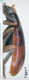
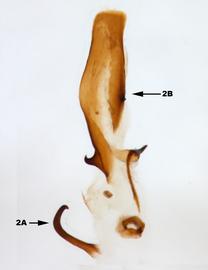
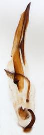
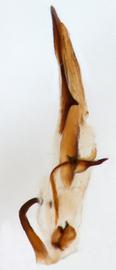
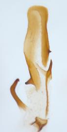
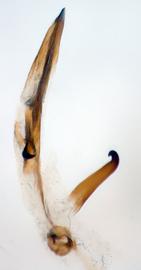
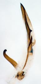
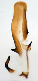
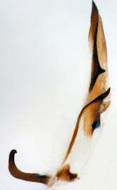
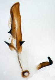
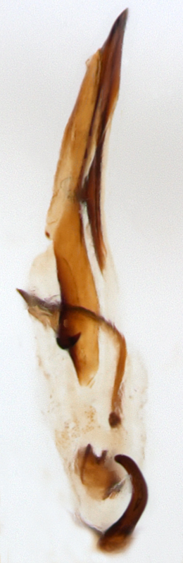
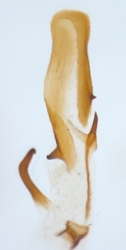
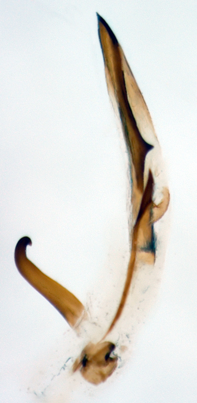
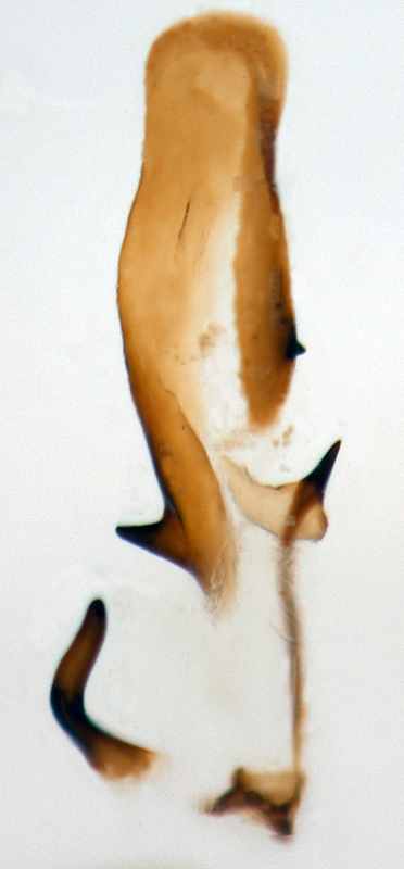
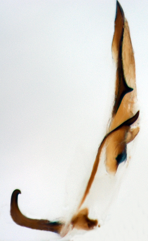
uniformly black, yellow ocher or bicolored: yellow, outer edge with piceous or testaceous line, sometimes extensively darkened. Femora uniform yellow or yellow ocher. Aedeagusaedeagus:
the main sclerotized part of the male genitalia; "aedeagus" is used here instead of "median lobe of aedeagus"
asymmetric. Number of internal sac scleritessclerites:
(here) the sclerotized hooks, spines or plates in the internal sac
2.
Mexico, Honduras, Guatemala, Costa Rica, Nicaragua, Panama. One female in the USNM collection was collected in the Texas, USA, in 1993. The only female could be found occasionally introduced with feeding substrate. Therefore this record need to be confirmed by the new data.
There are the records of Diabrotica tibialis Jacoby on Verbesina L. (Asteraceae), Beta vulgaris L., cucumber, different species of Cucurbita L., horse bean (Vicia faba L.), okra (Abelmoschus esculentus (L.) Moench), cotton, Hibiscus rosa-sinensis L., Zea mays L., tomato, potato (Clark et al., 2004). One specimen of D. adelpha in the USNM collection was collected on Asparagus officinalis L. and one on Sphaeralcea A.St.-Hil.
Smith and Lawrence (1967) could recognize the only specimen (female) of D. adelpha in the Allard collection in the MNHN as part of Harold's original series. They designated it as the lectotype. The male in Allard collection in the MNHN, bearing the labels (1) [Diabrotica adelpha Harold] handwritten and (2) [Ex Musaeo Quedenfeldt] white machine-printed, is morphologically identical with the type female. We based our concept of male of D. adelpha on this specimen. The male genitalia have been examined in this male, in 6 paralectotype males of D. flaviventris in BMNH and MCZ collections, and in 4 paralectotype males of D. tibialis in BMNH and MCZ collections. Male genitalia of additional specimens of all three species have been examined from the USNM collection. The armament of the internal sac of the male aedeagusaedeagus:
the main sclerotized part of the male genitalia; "aedeagus" is used here instead of "median lobe of aedeagus"
is identical in all three species. The species are very similar in coloration patterns. Jacoby wrote (1887): "This [D. adelpha] and several of the following species [D. tibialis, D. brunneosignata, etc.] are extremely closely allied, their characters of distinction being slight and variable". Only two substantial differences in coloration are recognizable on the type females of D. adelpha and D. tibialis: in D. adelpha the pronotumpronotum:
the notum of the prothorax with highly sclerotized pronotal disc
is yellow or yellow ocher, in D. tibialis - testaceous; in D. adelpha tibiatibia:
<em>(pl. tibiae)</em> the forth part of the beetle leg articulated with femur on the one side and with tarsus on the other side
are yellow or yellow ocher, in D. tibialis - black or chestnut. After study of the large series of these two species we could recognize a few transitional coloration patterns of tibiatibia:
<em>(pl. tibiae)</em> the forth part of the beetle leg articulated with femur on the one side and with tarsus on the other side
from uniformly yellow through bicolored (yellow, outer edge with piceous or testaceous line, sometimes extensively darkened) to uniformly black or chestnut. The outlines of the bandsbands:
(here) transverse maculae on the beetle elytra
on elytrae of all three species are very similar. The D. flaviventris with almost entirely testaceous elytrae is probable melanistic color morph of the same species. No substantial differences were recognized in the body size or habitus of three species. Hereby we place D. tibialis and D. flaviventris as synonyms of D. adelpha .
Authors: A. Derunkov, A. Konstantinov, A. Tishechkin, L. Hartje, and A.J. Redford
Last updated Feb. 12, 2015
idtools.org | tool images at ITP Node

