
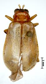
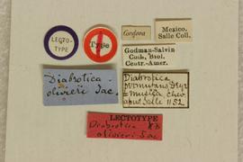
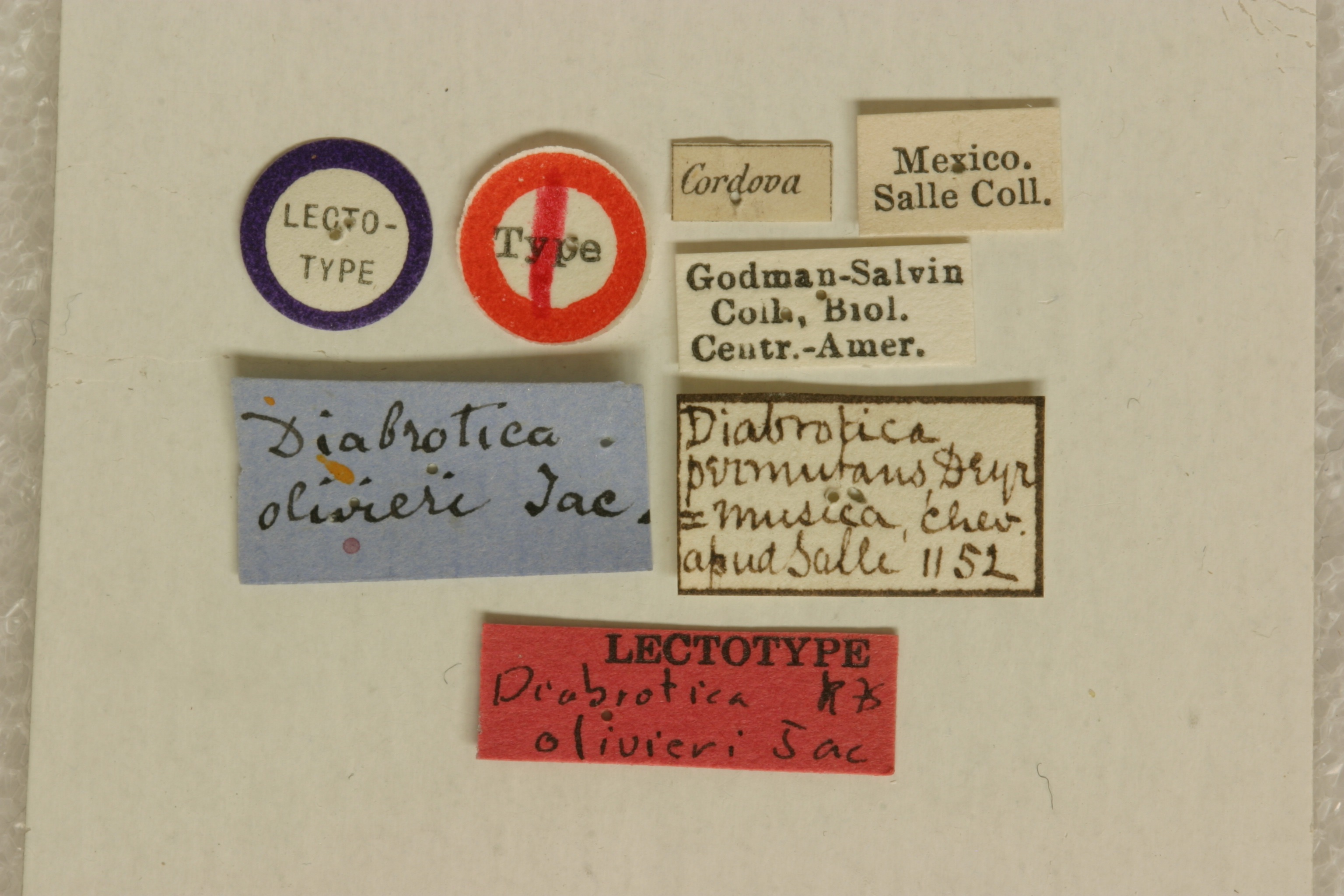
Diabrotica olivieri Jacoby 1887: 526
Cordova, Mexico
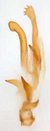
BMNH, lectotype, female, verified
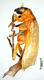
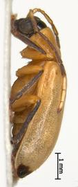
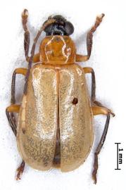
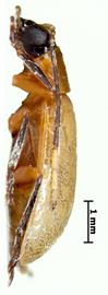
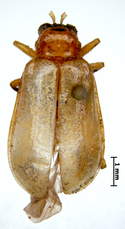
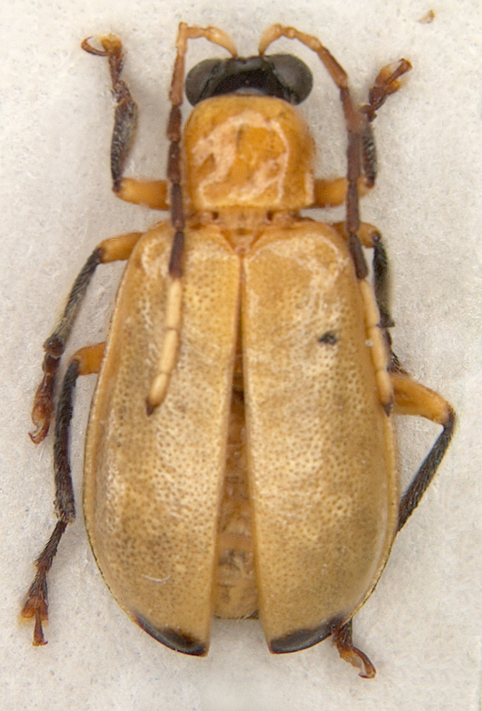
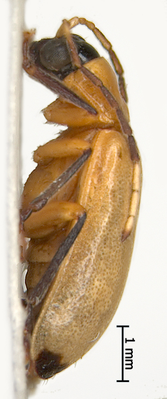
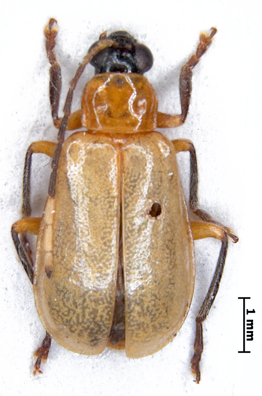
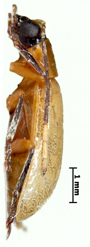
Body length 5.7-6.1 mm. Body width 2.9-3.0 mm. Head basic color black. Antennae filiformfiliform:
slender antennae with antennomeres of similar shape
, bi- or tricolored, antennomeres 1-3 and 9-10 sulphur yellow, antennomeres 4-8 cinnamon brown, antennomereantennomere:
"segment" of antenna, more or less clearly separated
11 dark apically. Pronotumpronotum:
the notum of the prothorax with highly sclerotized pronotal disc
yellow or mustard yellow, subquadrate, weakly bifoveate, with wide shallow foveae, not shagreened. Scutellumscutellum:
small, usually triangular shield between the bases of elytra
yellow. Elytra yellow or rufous, maculatemaculate:
(here) marked by maculae or patches of a different shape and size, usually clearly separated from each other
, with one black macula on the apexapex:
<em>(pl. apices)</em> the far distal end of a structure; opposite of base
of each elytronelytron:
<em>(pl. elytra)</em> the fore highly sclerotized wing of beetle
. Elytral epipleura completely yellow, sutural anglesutural angle:
the posterior angle or apex of the elytron near the suture
of elytra obtuse-angled, punctation dense, fine. Abdomen yellow or pygidium (at least the apexapex:
<em>(pl. apices)</em> the far distal end of a structure; opposite of base
) black. Tarsi black or chestnut, tibiatibia:
<em>(pl. tibiae)</em> the forth part of the beetle leg articulated with femur on the one side and with tarsus on the other side
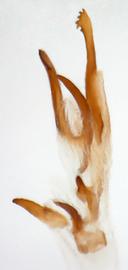
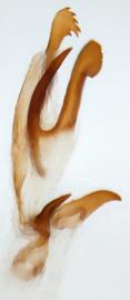
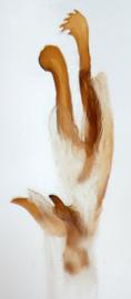
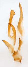
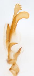
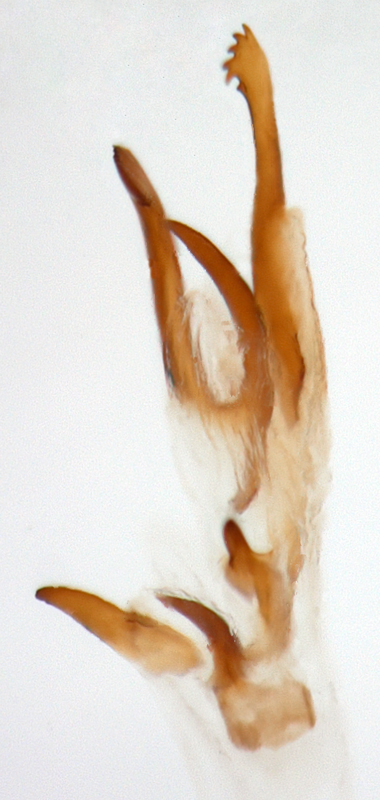
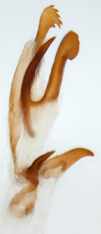
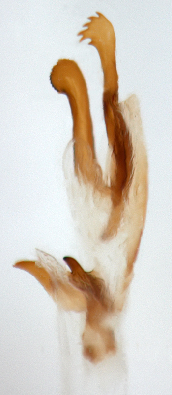
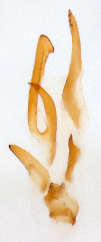
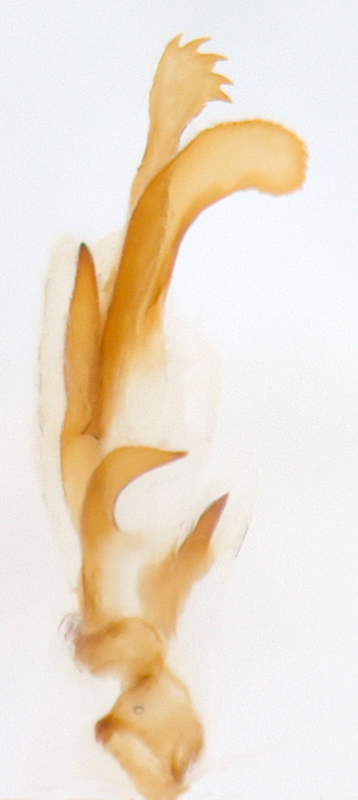
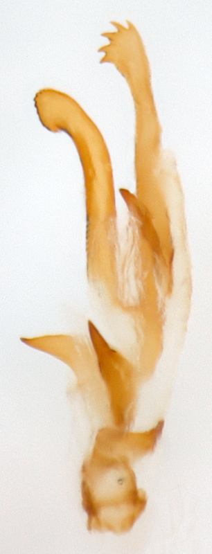
black or piceous. Femora yellow or yellow ocher. Aedeagusaedeagus:
the main sclerotized part of the male genitalia; "aedeagus" is used here instead of "median lobe of aedeagus"
symmetric, with five internal sac scleritessclerites:
(here) the sclerotized hooks, spines or plates in the internal sac
.
Mexico, Belize, Nicaragua, Costa Rica
Unknown
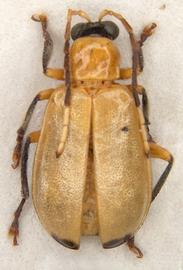
Diabrotica olivieri Jacoby is very similar to D. apicalis Baly and D. obscura Jacoby. They can be separated by the following features: elytra with apicalapical:
of or pertaining to the apex; opposite of basal
black macula on each elytronelytron:
<em>(pl. elytra)</em> the fore highly sclerotized wing of beetle
in D. olivieri, but completely yellow in D. obscura; D. apicalis is larger than D. olivieri; elytrla sides are almost parallel in D. apicalis, while the body shape in D. olivieri is roundish; elytra punctuation is stronger in D. olivieri than in D. apicalis, so elytra in D. olivieri have dull luster, whereas they are shining in D. apicalis.
Authors: A. Derunkov, A. Konstantinov, A. Tishechkin, L. Hartje, and A.J. Redford
Last updated Feb. 12, 2015
idtools.org | tool images at ITP Node
